. 2023 Sep 1;435(17):168192.
doi: 10.1016/j.jmb.2023.168192. Epub 2023 Jul 1.

Conformation-specific Synthetic Antibodies Discriminate Multiple Functional States of the Ion Channel CorA

Affiliations

Conformation-specific Synthetic Antibodies Discriminate Multiple Functional States of the Ion Channel CorA

Satchal K Erramilli et al. J Mol Biol. .

Abstract

CorA, the primary magnesium ion channel in prokaryotes and archaea, is a prototypical homopentameric ion channel that undergoes ion-dependent conformational transitions. CorA adopts five-fold symmetric non-conductive states in the presence of high concentrations of Mg2+, and highly asymmetric flexible states in its complete absence. However, the latter were of insufficient resolution to be thoroughly characterized. In order to gain additional insights into the relationship between asymmetry and channel activation, we exploited phage display selection strategies to generate conformation-specific synthetic antibodies (sABs) against CorA in the absence of Mg2+. Two sABs from these selections, C12 and C18, showed different degrees of Mg2+-sensitivity. Through structural, biochemical, and biophysical characterization, we found the sABs are both conformation-specific but probe different features of the channel under open-like conditions. C18 is highly specific to the Mg2+-depleted state of CorA and through negative-stain electron microscopy (ns-EM), we show sAB binding reflects the asymmetric arrangement of CorA protomers in Mg2+-depleted conditions. We used X-ray crystallography to determine a structure at 2.0 Å resolution of sAB C12 bound to the soluble N-terminal regulatory domain of CorA. The structure shows C12 is a competitive inhibitor of regulatory magnesium binding through its interaction with the divalent cation sensing site. We subsequently exploited this relationship to capture and visualize asymmetric CorA states in different [Mg2+] using ns-EM. We additionally utilized these sABs to provide insights into the energy landscape that governs the ion-dependent conformational transitions of CorA.

Keywords: CorA; X-ray crystallography; ion channel; membrane proteins; synthetic antibodies.


Conflict of interest statement

Declaration of Competing Interest The authors declare that they have no known competing financial interests or personal relationships that could have appeared to influence the work reported in this paper.

Figures

Figure 1 –
Figure 1 –
Generation and characterization of conformation-specific sABs against CorA. A. Phage display selection strategy for conformation-specific sABs against CorA involved counter selection with CorA D253K, which mimics the magnesium-bound state in the cytoplasmic regulatory domain. B. Affinity estimation ELISA of purified Fabs C12 (EC50 ~192 pM) and C18 (EC50 ~610 pM) shows high affinity binding to CorA in 1 mM EDTA. Assays performed in triplicate. Means and error bars plotted together with sigmoidal dose response curve. C. Single point binding data of purified Fabs C12 and C18 to CorA in biotinylated nanodiscs immobilized in ELISA wells. Fabs tested in ELISA buffer with 20 mM MgCl2 (blue), without (1 mM EDTA, red), or a buffer-only control (gray).
Figure 2 –
Figure 2 –
Dissecting the relationship between environmental Mg2+ concentration and sAB C18 binding. A-F. Double-referenced SPR sensorgrams of a two-fold dilution series of sAB C18 (200–12.5 nM) binding to immobilized CorA in the presence of 0 (A., 11.0 nM KD), 0.25 (B., 28.0 nM KD), 0.5 (C., 65.8 nM KD), 0.75 (D., 1.26 μM KD), 1.0 (E., 11.3 μM KD), 1.5 (F., dark purple), and 2.0 (F., light purple) mM Mg2+. For A-E, experimental curves are overlaid with kinetic fitted curves (black) from a one-to-one binding model. Kinetic fitting was not performed for the last two concentrations (F). G. Plot of log(kon) vs. log(koff) for C18 SPR data from 0–1 mM Mg2+. H. Negative-stain EM two-dimensional class averages of the nanodisc-reconstituted CorA-C18 complex in 1 mM EDTA. Sample micrograph shown in Supplemental Figure 1.
Figure 3 –
Figure 3 –
C12 shows a stepwise binding model to CorA in the presence of increasing [Mg2+]. A. SPR sensorgrams depict 25 nM C12 binding to CorA to determine binding kinetics without Mg2+ (teal, 0.83 nM KD) and with 20 mM Mg2+ (brown, 5.9 nM KD). See Supplemental Table 1 and Supplemental Fig. 2. B. ELISA measuring binding of C12 at constant concentration to CorA in different magnesium concentrations. The EC50 for Mg2+ is 13.03 millimolar, compared to the KD of CorA for Mg2+ without sAB C12 (1–2 mM). C. Overlay of size-exclusion chromatograms of CorA-C12 complexes in different environmental magnesium concentrations. Curves are colored and labeled according to condition. D. Negative stain EM 2D classes of CorA-C12 complex in 1 mM EDTA showing two visible sABs. The distance between the two visible sAB lobes in the side view on the bottom right class was ~150 Å. E. Negative stain EM 2D classes of CorA-C12 in 12.5 mM MgCl2. A mixture of classes with one and two bound sABs are visible. The approximate distribution for each was 50/50 from a set of 6500 particles. F. Negative stain EM 2D classes of CorA-C12 in 40 mM MgCl2. Only one sAB is visible in views. Sample micrograph in Supplemental Figure 2.
Figure 4 –
Figure 4 –
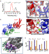
Structural analysis of the sAB C12 epitope. A. A stable complex of CorANTD-C12H12 was isolated by SEC, with the complex chromatogram (red) showing a shift to an earlier retention volume compared with CorANTD alone (black). B. Crystal structure shows a 1:1 complex formed by CorANTD and sAB C12H12. CorANTD is red, sAB C12H12 heavy chain is blue, and sAB C12H12 light chain is green. See Table 1 for data processing statistics. C. Interaction interface of sAB C12 with CorANTD, colored by percentage buried surface area: 10–49%, light purple; 50–70%, medium purple; 71–100% dark purple. Non-interacting areas colored dark gray. C12 CDRs that are part of interface are colored by chain (heavy chain – dark green; light chain – light green). D. Top panel - A closer inspection of the epitope reveals a salt bridge interaction formed by sAB C12H12-R108 and CorANTD-D89. D89 forms part of the magnesium sensor site. Water molecules shown as purple spheres. Bottom panel – In the magnesium-saturated closed crystal structure (PDB ID: 4I0U), D89 and D253 from adjacent monomers (shown as blue and red) form a bidentate interaction to bind a hydrated Mg2+. 2Fc-F0 electron density maps, shown as light gray mesh, were contoured at 2.0σ and 1.5σ, respectively, for the CorANTD-C12 and 4I0U structures. Magnesium ions and water molecules shown as green and red spheres, respectively. E. Binding analysis of sAB C12 to CorA reveals that the binding can be abolished by specific mutation of either CorANTD D89 or sAB C12 R108, indicating that this interaction contributes to magnesium-dependency of sAB binding to CorA.
Figure 5 –
Figure 5 –
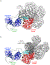
Modeling of the sAB C12 paratope-epitope interaction against the full-length CorA structure. In both figures the sAB light chain is shown as as transparent surface for clarity. A. Superposition of CorANTD (red) and sAB C12 (heavy chain – blue) onto the closed state pentamer (PDB: 4I0U; epitope-adjacent monomer colored dark gray, others light gray) reveals significant steric hindrance of C12 CDR-H3 (cyan) which precludes its binding to closed state of the channel. B. Superposition of CorANTD and sAB C12 (heavy chain colored as in A) onto the open state pentamer (PDB: 3JCG; monomers colored as in A) reveals complete accessibility of the epitope in the Mg-depl condition, which the sAB was selected in.
Figure 6 –
Figure 6 –
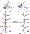
EPR spectroscopy analysis of CorA and CorA-C12 complex. A. Visualization of positions mutated to prepare single-cysteine CorA mutants for site-directed spin-labeling EPR spectroscopy. CorA structure 4I0U was used to visualized Mg2+-bound closed state (left). CorA structure 3JCG was used to visualized Mg2+-free form (right). Sidechains were added to 3JCG using Coot[42] purely for visualization purposes. Residues at indicated positions represented as red spheres and labeled accordingly. Mutated positions shown only for chain A, which undergoes significant conformational changes in the Mg2+-free “open” condition designated “State 1”, for clarity and illustrative purposes. However, all five possible positions for each mutant are labeled. Dotted lines denote boundary of transmembrane region. B. EPR spectra for all five single cysteine mutants of CorA in absence (1 mM EDTA, red) and presence of Mg2+ (black). Spectra are overlaid to show differences between the two conditions. The labeled mutant at V199C (bottom-most spectra) showed the most sensitivity to the two different conditions. C. EPR spectra for all five single cysteine mutants of CorA in absence (1 mM EDTA, red) and presence of Mg2+ (black) after the addition of sAB C12. Spectra are overlaid to show differences. The V199C mutant produced identical spectra (bottom-most spectra) in the two conditions after addition of sAB C12.

Update of

Similar articles

Cited by

References

    1. Paduch M, Koide A, Uysal S, Rizk SS, Koide S, Kossiakoff AA. Generating conformation-specific synthetic antibodies to trap proteins in selected functional states. Methods 2013;60:3–14. 10.1016/j.ymeth.2012.12.010. - DOI - PMC - PubMed
    1. Paduch M, Kossiakoff AA. Generating Conformation and Complex-Specific Synthetic Antibodies. Methods Mol Biol 2017;1575:93–119. 10.1007/978-1-4939-6857-2_6. - DOI - PubMed
    1. Rizk SS, Paduch M, Heithaus JH, Duguid EM, Sandstrom A, Kossiakoff AA. Allosteric control of ligand-binding affinity using engineered conformation-specific effector proteins. Nat Struct Mol Biol 2011;18:437–42. 10.1038/nsmb.2002. - DOI - PMC - PubMed
    1. Dominik PK, Kossiakoff AA. Phage display selections for affinity reagents to membrane proteins in nanodiscs. Methods Enzymol 2015;557:219–45. 10.1016/bs.mie.2014.12.032. - DOI - PubMed
    1. Fellouse FA, Esaki K, Birtalan S, Raptis D, Cancasci VJ, Koide A, et al. High-throughput generation of synthetic antibodies from highly functional minimalist phage-displayed libraries. J Mol Biol 2007;373:924–40. 10.1016/j.jmb.2007.08.005. - DOI - PubMed

Publication types

MeSH terms

LinkOut - more resources