. 2023 Jul 3;8(1):255.
doi: 10.1038/s41392-023-01456-x.

SIRT6 is an epigenetic repressor of thoracic aortic aneurysms via inhibiting inflammation and senescence

Affiliations

SIRT6 is an epigenetic repressor of thoracic aortic aneurysms via inhibiting inflammation and senescence

Yang-Nan Ding et al. Signal Transduct Target Ther. .

Abstract

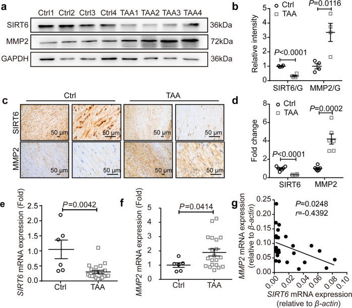
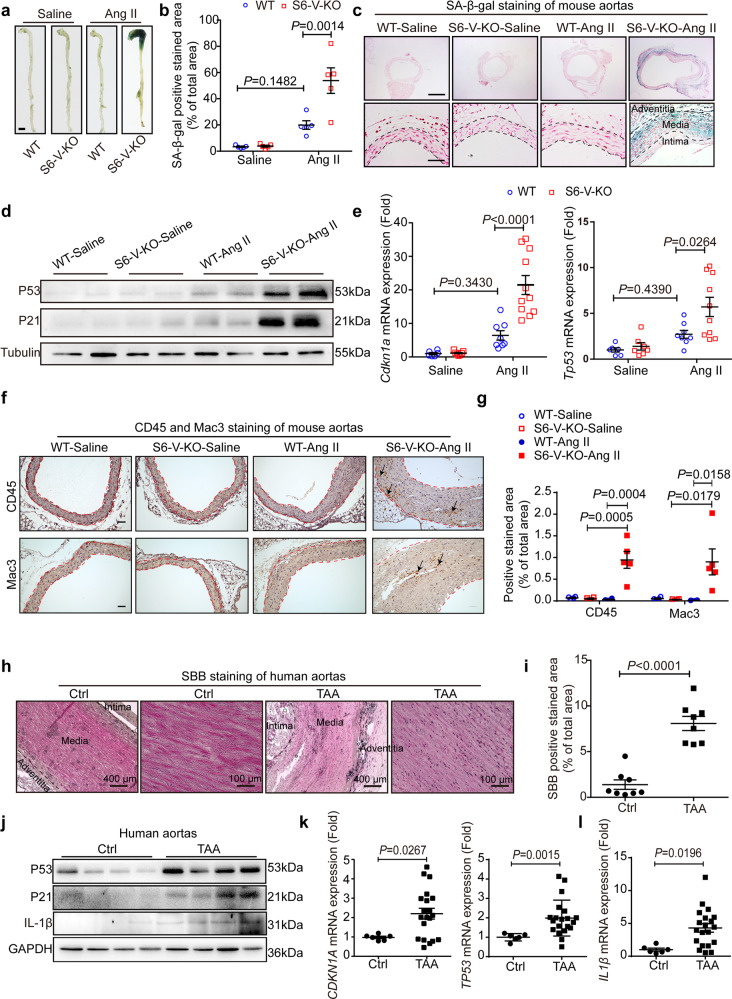
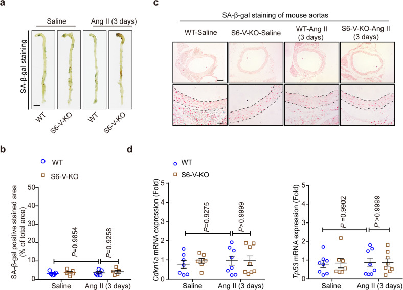
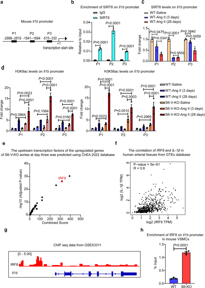
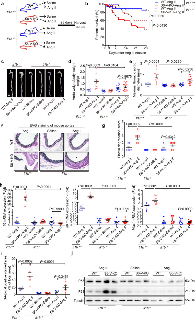
Thoracic aortic aneurysms (TAAs) develop asymptomatically and are characterized by dilatation of the aorta. This is considered a life-threating vascular disease due to the risk of aortic rupture and without effective treatments. The current understanding of the pathogenesis of TAA is still limited, especially for sporadic TAAs without known genetic mutation. Sirtuin 6 (SIRT6) expression was significantly decreased in the tunica media of sporadic human TAA tissues. Genetic knockout of Sirt6 in mouse vascular smooth muscle cells accelerated TAA formation and rupture, reduced survival, and increased vascular inflammation and senescence after angiotensin II infusion. Transcriptome analysis identified interleukin (IL)-1β as a pivotal target of SIRT6, and increased IL-1β levels correlated with vascular inflammation and senescence in human and mouse TAA samples. Chromatin immunoprecipitation revealed that SIRT6 bound to the Il1b promoter to repress expression partly by reducing the H3K9 and H3K56 acetylation. Genetic knockout of Il1b or pharmacological inhibition of IL-1β signaling with the receptor antagonist anakinra rescued Sirt6 deficiency mediated aggravation of vascular inflammation, senescence, TAA formation and survival in mice. The findings reveal that SIRT6 protects against TAA by epigenetically inhibiting vascular inflammation and senescence, providing insight into potential epigenetic strategies for TAA treatment.


Conflict of interest statement

The authors declare no competing interests.

Figures

Fig. 1
Fig. 1
SIRT6 expression is decreased in sporadic human TAA samples. a Representative western blots of SIRT6 and MMP2 in sporadic human TAA and control (Ctrl) samples. b Normalized protein levels of SIRT6 and MMP2 in sporadic human TAA and Ctrl samples (n = 4). c Representative IHC staining images of SIRT6 and MMP2 in sporadic human TAA and Ctrl samples (scale bar: 50 μm). d Densitometric analysis of IHC staining for SIRT6 and MMP2 (n = 6). The results are normalized to the percentage of the stained area in Ctrl samples. e, f mRNA levels of SIRT6 (e) and MMP2 (f) in aortic homogenates from TAA patients (n = 20) and Ctrl samples (n = 6), as determined by quantitative reverse transcription-polymerase chain reaction (qRT-PCR). g Correlation between SIRT6 and MMP2 mRNA levels in 26 thoracic tissues. Each point in the scatter plot represents a single patient sample. Pearson’s correlation coefficient (r) and significant P values are shown
Fig. 2
Fig. 2
Sirt6 deletion in VSMCs promotes TAA formation and rupture after Ang II infusion for 28 days. a Experimental design. S6-V-KO or WT mice were infused with saline or Ang II (0.72 mg/kg/d) for 28 days. b Survival curve after Ang II infusion (n = 31 for WT mice and n = 49 for S6-V-KO mice). c Maximal internal diameter of ascending aorta at days -1, 3, 7, 14, 21 and 28 (n = 12 mice/group). * and #, P < 0.05; ** and ##, P < 0.01; *** and ###, P < 0.001. d Representative images showing macroscopic features of the normal aorta and aneurysms in WT and S6-V-KO mice after infusion with saline or Ang II for 28 days (scale bar: 2 mm). e, f The ratio of aorta weight to body weight (n = 15 mice/group) (e) and the maximal thoracic aortic outer diameter (n = 11–12 mice/group) (f) in saline- and Ang II-infused mice at day 28. g, h. Representative images of Elastica van Gieson (EVG) staining of thoracic aortic sections from mice (g) and semiquantitative analysis of elastin degradation (h). Scale bars: 800 µm (top) and 200 µm (bottom). (n = 10 mice/group). i Representative western blots of MMP2. j Densitometric analysis of the protein levels of MMP2 (n = 3 mice/group)
Fig. 3
Fig. 3
Increased vascular inflammation and senescence in Ang II-infused mice with Sirt6 deficiency and human TAA samples. Mice were infused with saline or Ang II (0.72 mg/kg/d) for 28 days. a Representative images of SA-β-gal-stained aorta (scale bar: 2 mm). b Densitometric analysis of SA-β-gal staining in the whole aorta from WT and S6-V-KO mice infused with saline or Ang II for 28 days (n = 5 mice/group). c Representative images of SA-β-gal-stained transverse sections of thoracic aorta from WT and S6-V-KO mice infused with saline or Ang II. The blue regions are positively stained, and the nuclei were counterstained using nuclear Fast Red. Scale bars: 800 µm (top) and 100 µm (bottom). d Western blots of P21 and P53 in the mouse aorta. e mRNA levels of Cdkn1a and Tp53 in the mouse aorta (n = 6–11 mice/group). f Representative images of IHC staining of leukocytes (CD45, top) and macrophages (Mac3, bottom) in the thoracic aortas of mice (scale bar: 50 µm). Arrows represent positively stained areas. g Statistical analysis of the CD45 and Mac3 positive area percentage in mouse aorta (n = 4–5 mice/group). h Representative staining with SBB as another indicator of cell senescence in human TAA and the control aorta. Scale bars: 400 µm (left) and 100 µm (right). i Statistical analysis of the SBB-positive area percentage in clinical TAA and control samples (n = 8). j Representative western blots of P21, P53 and IL-1β in human TAA and the control aorta. k The mRNA levels of CDKN1A and TP53 in the human control thoracic aorta (n = 6) and TAA samples (n = 20). l The mRNA levels of IL1β in the human control thoracic aorta (n = 6) and TAA samples (n = 20)
Fig. 4
Fig. 4
Sirt6 deficiency does not induce vascular senescence in mouse aortas after Ang II infusion for 3 days. All mice were infused with saline or Ang II (0.72 mg/kg/d) for 3 days. a Representative images showing the SA-β-gal-stained aorta from the indicated groups (scale bars: 2 mm). b Densitometric analysis of SA-β-gal staining in whole aorta from WT and S6-V-KO mice infused with saline or Ang II (n = 5 mice/group). c Representative images of SA-β-gal-stained transverse sections of thoracic aortas from WT and S6-V-KO mice infused with saline or Ang II. Nuclei were counterstained using nuclear Fast Red. Scale bars: 400 µm (top) and 100 µm (bottom). d mRNA levels of Cdkn1a and Tp53 in mouse aorta (n = 7–8 mice/group)
Fig. 5
Fig. 5
IL-1β expression is increased early in S6-V-KO mouse aorta after Ang II infusion for 3 days. a KEGG pathway analysis revealed that cytokine-cytokine receptor interaction was strongly affected in aorta of S6-V-KO mice compared with that in the aorta of WT littermates after Ang II-treated for 3 days (n = 3 mice/group). b Volcano plot of differentially expressed genes (DEGs) in the cytokine-cytokine receptor interaction pathway between S6-V-KO and WT mouse aortas after Ang II infusion for 3 days. The thresholds used for differential genes were a false discovery rate (FDR) of 0.05 and 1.5-fold up- or downregulation with Sirt6 deficiency. c qRT-PCR analysis of Il1b expression in the mouse aorta (n = 7–11 mice/group). d Representative images of IHC staining of IL-1β in the mouse thoracic aorta (scale bar: 200 µm), with brown representing positive staining areas. e Densitometric analysis of IHC staining for IL-1β. The results were normalized to the percentage of the stained area in saline-treated WT mice. f Representative images of IF staining of leukocytes (CD45, top), macrophages (Mac3, bottom), VSMCs (αSMA) and nuclei (Hoechst) in the mouse thoracic aorta (scale bar: 100 µm). g Densitometric analysis of IF staining for CD45 and Mac3 (n = 3–6 mice/group). h qRT-PCR analysis of the expression of inflammation-related genes (Il6, Il8, and Mcp-1) in mouse aorta (n = 7–12 mice/group)
Fig. 6
Fig. 6
Sirt6 deficiency increases H3K9ac and H3K56ac levels on the Il1b promoter. a Diagram showing designed primers for mouse Il1b promoter. b SIRT6 enrichment on the Il1b promoter as assessed by ChIP assays performed with chromatin prepared from aorta of WT mice (n = 3 mice/group). Chromatin was immunoprecipitated with normal rabbit IgG or antibodies against SIRT6, and precipitated genomic DNA was analyzed by real-time PCR using different primers for the different regions of the Il1b promoter. c SIRT6 enrichment on the Il1b promoter as assessed by ChIP assays performed with chromatin prepared from the WT mouse aorta with or without Ang II infusion for 3 days and 28 days (n = 3 mice/group). Procedures were as for (b). d ChIP assay of H3K9ac and H3K56ac at the Il1b promoter in the aorta of WT and S6-V-KO mice after saline or Ang II infusion for 3 days and 28 days (n = 3 mice/group). Chromatin was immunoprecipitated with normal rabbit IgG or antibodies against H3K9ac, H3K56ac and H3, and precipitated genomic DNA was analyzed by real-time PCR. e Upstream transcription factors of the upregulated genes in S6-V-KO aortas at day three were predicted using the ChEA 2022 database (https://maayanlab.cloud/Enrichr/). f The correlation of IRF8 and IL1β in human aorta from the GTEx database. The Spearman analysis was performed using the webtool GEPIA (http://gepia.cancer-pku.cn/). g IRF8-binding to the Il1b promoter in mouse dendritic cells. The ChIP-seq data from GSE53311 was analyzed using IGV. h Sirt6 knockout promotes IRF8 binding to Il1b promoter in VSMCs. The ChIP assay was performed with IRF8 antibody and qPCR of designated promoter region was performed (−1841 to −1648 bp)
Fig. 7
Fig. 7
Genetic inhibition of Il1b alleviates Ang II-induced TAA and vascular inflammation and senescence in S6-V-KO mice. a Schematic of experimental design. All mice were infused with saline or Ang II for 28 days. b Survival curve of mice after Ang II infusion (n = 19 in the WT/Il1b+/+-Ang II group and n = 35 in the S6-V-KO/ Il1b+/+-Ang II group; n = 22 in the WT/ Il1b−/−-Ang II group, and n = 35 in the S6-V-KO/ Il1b/−-Ang II group). c Representative images showing macroscopic features of the aorta (scale bar: 2 mm). d, e The ratio of aortic weight to body weight (n = 8–16 mice/group) (d) and the maximal thoracic aortic outer diameter (n = 8–13 mice/group) (e). f, g Representative images of EVG staining of mouse thoracic aortic sections (f) and semiquantitative analysis of elastin degradation (g). Scale bars: 200 µm (n = 6 mice/group). h mRNA levels of inflammatory genes in the mouse aorta (n = 7–8 mice/group). i Densitometric analysis of SA-β-gal-stained mouse aorta (n = 4–5 mice/group). j Protein levels of P21, P53 and Tubulin in aortic homogenate as determined by western blotting
Fig. 8
Fig. 8
Pharmacological inhibition of IL-1β alleviates Ang II-induced inflammation, senescence and TAA in vitro and in vivo with Sirt6 deficiency. a Representative images of SA-β-gal-stained human VSMCs with or without IL-1β (100 ng/ml) treatment and densitometric analysis of SA-β-gal positive cells. Blue-stained cells were considered senescent. Scale bar, 500 μm. b mRNA levels for inflammatory genes in human VSMCs (n = 3). c Relative mRNA levels of SIRT6 in human VSMCs transfected with negative control siRNA (si NC) and SIRT6 siRNA (si SIRT6) (n = 3). d Densitometric analysis of SA-β-gal-stained WT and SIRT6 knockdown (S6-KD) human VSMCs with or without anakinra treatment following Ang II treatment and densitometric analysis of SA-β-gal- positive cells. (n = 3). e mRNA levels for inflammatory and senescence genes in WT and S6-KD human VSMCs with or without anakinra treatment after Ang II treatment (n = 3). f Survival curves for mice after Ang II infusion (n = 22 for WT-Ang II mice, n = 30 for S6-V-KO-Ang II mice, n = 13 for WT-Ang II-Anakinra mice and n = 29 for S6-V-KO-Ang II-Anakinra mice). g Representative images showing the macroscopic features of aorta (scale bar: 2 mm). h Representative images of EVG staining of the mouse thoracic aortic sections (Scale bars: 200 µm). i mRNA levels of inflammatory genes in the mouse aorta (n = 6–11 mice/group). j Densitometric analysis of SA-β-gal-stained mouse aorta (n = 4–6 mice/group). k Protein levels of P21, P53 and Tubulin in aortic homogenates as measured by western blotting

References

    1. Milewicz DM, Prakash SK, Ramirez F. Therapeutics targeting drivers of thoracic aortic aneurysms and acute aortic dissections: insights from predisposing genes and mouse models. Annu. Rev. Med. 2017;68:51–67. doi: 10.1146/annurev-med-100415-022956. - DOI - PMC - PubMed
    1. Bossone E, Eagle KA. Epidemiology and management of aortic disease: aortic aneurysms and acute aortic syndromes. Nat. Rev. Cardiol. 2021;18:331–348. doi: 10.1038/s41569-020-00472-6. - DOI - PubMed
    1. Quintana RA, Taylor WR. Introduction to the compendium on aortic aneurysms. Circ. Res. 2019;124:470–471. doi: 10.1161/CIRCRESAHA.119.314765. - DOI - PubMed
    1. Fletcher AJ, Syed MBJ, Aitman TJ, Newby DE, Walker NL. Inherited thoracic aortic disease: new insights and translational targets. Circulation. 2020;141:1570–1587. doi: 10.1161/CIRCULATIONAHA.119.043756. - DOI - PMC - PubMed
    1. Isselbacher EM, Lino Cardenas CL, Lindsay ME. Hereditary influence in thoracic aortic aneurysm and dissection. Circulation. 2016;133:2516–2528. doi: 10.1161/CIRCULATIONAHA.116.009762. - DOI - PMC - PubMed

Publication types

LinkOut - more resources