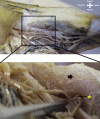
Anatomical study of the distal course of the posterior tibial artery: A cadaver study
- PMID: 37395357
- PMCID: PMC10544252
- DOI: 10.5152/j.aott.2023.22158
Anatomical study of the distal course of the posterior tibial artery: A cadaver study
Abstract
Objective: This study aimed to describe the course, branches, and variances of the posterior tibial artery, which provides the arterial supply of the plantar surface of the foot, starting from the tarsal tunnel level to provide descriptive information for all surgical interventions, diagnostic radiological procedures, and promising endovascular therapies in the tarsal region.
Methods: In this study, a dissection of 48 feet was performed on 25 formalin-fixed cadavers (19 males and 6 females). Surgical instruments and a digital caliper were used for dissection and measurements, and the critical structures were recorded by a Canon 250D camera to be illustrated later.
Results: All parameters were significantly longer in male cadavers compared to females. According to the correlation analysis, while there was a significant and robust correlation between the axial line and pternion-deep plantar arch (R=.830, P .05), a moderate correlation was found between the axial line and sphyrion-bifurcation (R=.575; P < .05), axial line and deep plantar arch-2nd interdigital commissure (R=.457; P < .05), and sphyrion-bifurcation and pternion-deep plantar arch (R=.480; P < .05). Variation in any branch of the posterior tibial artery was observed in 27 of the 48 studied sides.
Conclusion: In our study, the branching and variability of posterior tibial artery on the plantar surface of the foot were described in detail with the determined parameters. In conditions that cause tissue and function loss and require reconstruction, such as diabetes mellitus and atherosclerosis, the most critical factor in increasing treatment success is a better understanding of the region's anatomy.
Figures


References
-
- Mir, Mir L. Follow-up clinic. Functional graft of the heel, by Lorenzo Mir y Mir, M.D. Plast Reconstr Surg. 1954. Plast Reconstr Surg. 1975;55(6):702 703. - PubMed
MeSH terms
Substances
LinkOut - more resources
Full Text Sources
Medical
