. 2023 Jul 27;97(7):e0159622.
doi: 10.1128/jvi.01596-22. Epub 2023 Jul 3.

Targeting the Spike Receptor Binding Domain Class V Cryptic Epitope by an Antibody with Pan-Sarbecovirus Activity

Affiliations

Targeting the Spike Receptor Binding Domain Class V Cryptic Epitope by an Antibody with Pan-Sarbecovirus Activity

Jaime L Jensen et al. J Virol. .

Abstract

Novel therapeutic monoclonal antibodies (MAbs) must accommodate comprehensive breadth of activity against diverse sarbecoviruses and high neutralization potency to overcome emerging variants. Here, we report the crystal structure of the severe acute respiratory syndrome coronavirus 2 (SARS-CoV-2) receptor binding domain (RBD) in complex with MAb WRAIR-2063, a moderate-potency neutralizing antibody with exceptional sarbecovirus breadth, that targets the highly conserved cryptic class V epitope. This epitope overlaps substantially with the spike protein N-terminal domain (NTD) -interacting region and is exposed only when the spike is in the open conformation, with one or more RBDs accessible. WRAIR-2063 binds the RBD of SARS-CoV-2 WA-1, all variants of concern (VoCs), and clade 1 to 4 sarbecoviruses with high affinity, demonstrating the conservation of this epitope and potential resiliency against variation. We compare structural features of additional class V antibodies with their reported neutralization capacity to further explore the utility of the class V epitope as a pan-sarbecovirus vaccine and therapeutic target. IMPORTANCE Characterization of MAbs against SARS-CoV-2, elicited through vaccination or natural infection, has provided vital immunotherapeutic options for curbing the COVID-19 pandemic and has supplied critical insights into SARS-CoV-2 escape, transmissibility, and mechanisms of viral inactivation. Neutralizing MAbs that target the RBD but do not block ACE2 binding are of particular interest because the epitopes are well conserved within sarbecoviruses and MAbs targeting this area demonstrate cross-reactivity. The class V RBD-targeted MAbs localize to an invariant site of vulnerability, provide a range of neutralization potency, and exhibit considerable breadth against divergent sarbecoviruses, with implications for vaccine and therapeutic development.

Keywords: COVID-19; SARS-CoV; SARS-CoV-2; X-ray crystallography; betacoronaviruses; convalescent; cryptic; epitope; neutralizing antibodies; receptor binding domain; sarbecoviruses; spike; structural biology; variants of concern.


Conflict of interest statement

The authors declare a conflict of interest. Patent application number PCT/US 63/140,763, PCT WO 2022/159839 A1 was filed containing the mAbs described in this publication for authors S.J.K., K.M., V.D., and N.L.M. M.G.J. and K.M. are named as inventors on international patent application WO/2021/178971 A1 entitled “Vaccines against SARS-CoV-2 and other coronaviruses.” M.G.J. is named as an inventor on international patent application WO/2018/081318 and U.S. patent 10,960,070 entitled “Prefusion coronavirus spike proteins and their use.” The other authors declare no competing interests.

Figures
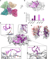
FIG 1
FIG 1
Crystal structure and epitope analysis of the SARS-CoV-2 RBD with WRAIR-2063 Fab. (A) Structure of the SARS-CoV-2 RBD (gray surface), aligned with complexes representative of each canonical RBD-targeted antibody epitope class. Class I, CB6 (pale yellow, PDB 7C01); class II, P2B-2F6 (pale green, PDB 8DCC); class III, S309 (teal, PDB 7SOC); class IV, CR3022 (magenta, PDB 7JN5). The class V epitope (represented by WRAIR-2063) is outlined and shaded with purple. (B) Key antibody contact residues of the RBD are shown as sticks and colored to indicate heavy chain (dark purple) and light chain (light purple). Only residues with a BSA of >20 Å2 are highlighted. The RBD is rotated about the z axis in relation to the RBD displayed in panel A. (C) BSA (Å2) contributions of the heavy and light chain CDR loops. (D) Surface representation of the RBD is highlighted with the WRAIR-2063 in dark and light violet. CDR loops are displayed and labeled, with critical interface residues of the CDR L3, CDR L1, CDR H2, and CDR H3 shown in expanded views. Hydrogen bonds are indicated with dashed lines. Electron density mesh surface is contoured at 1 σ. (E) The RBD (ribbon representation with surface overlay) with the WRAIR-2063 footprint shown in purple. Mutations found in VoCs are displayed as red spheres and labeled in red.
FIG 2
FIG 2
WRAIR-2063 epitope footprint in the context of the SARS-CoV-2 spike trimer. (A) Structural alignment of the WRAIR-2063–RBD with the RBD of the S-2P spike trimer, in the closed conformation (PDB 6ZGE). The aligned RBDs are shown in dark gray, the WRAIR-2063 Fab in violet, and the NTD in pale blue. In this conformation, the WRAIR-2063 epitope is shielded by the NTD. (Lower panel) Butterfly view of the RBD-NTD interface; interacting residues with a BSA of >20 Å2 are indicated. (B) Structural alignment of the WRAIR-2063–RBD with the RBD of the S-2P trimer in the partially open (1 RBD up) conformation (PDB 6X2A). Movement of the RBD allows the NTD to shift, exposing the WRAIR-2063 epitope. (C) Possible interface between the NTD (pale blue; bottom of panel) and the WRAIR-2063 LC (violet; top of panel), based on the superimposition of the WRAIR-2063–RBD complex with the open spike. (D) Footprints of the NTD in the closed spike (left), the overlap (orange) of the NTD and WRAIR-2063 footprints in the closed spike (center), and the NTD and WRAIR-2063 footprints in the open spike. In this orientation, interaction of the RBD with the NTD is minimal, displaying no overlap with the WRAIR-2063 epitope.
FIG 3
FIG 3
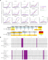
WRAIR-2063 binding to VoCs and knockout mutants. (A) Representative sensorgrams used to determine binding kinetics of WRAIR-2063 with SARS-CoV-2 WA-1, variants of concern, and class V knockout mutants, as measured by BLI. (B) Kinetics constants were determined using a minimum of four dilutions of the RBD (31.3 nM to 500 nM) and fitted using a 1:1 binding model.
FIG 4
FIG 4
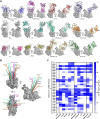
WRAIR-2063 epitope conservation and MAb cross-reactivity. (A) Primary sequence and structural analysis of the conserved WRAIR-2063 epitope across betacoronaviruses. Epitope residues are numbered in relation to the WA-1 (Wuhan-Hu-1) reference. The height and color of the histogram indicate the strength of the interaction between WRAIR-2063 and the RBD. Sequences are ordered according to phylogenetic relationship, based on a maximum likelihood phylogenetic tree derived from RBD primary sequences. The RBD structure shown in surface representation is colored to depict sequence conservation between the SARS-CoV-2 and SARS-CoV-1 RBDs, with the WRAIR-2063 epitope outlined in violet. (B) Heat map of SARS-CoV-2 and SARS-CoV RBD with NTD or WRAIR-2063 interface residues and corresponding percent buried surface area. Key antibody contact residues of the SARS-CoV RBD (gray surface; PDB 2GHV) are shown as sticks and colored by heavy chain (dark purple) and light chain (light purple). Only residues with a BSA of >20 Å2 are highlighted. (C) Binding of WRAIR-2063, WRAIR-2057, WRAIR-2134, WRAIR-2151, and WRAIR-2173 to sarbecovirus and merbecovirus RBDs, as measured by BLI. The heat map displays area under the curve values. RBD molecules were immobilized to the probe, with WRAIR-2063, -2057, -2134, -2151, or -2173 IgGs in solution.
FIG 5
FIG 5
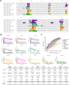
Pseudovirus neutralization of diverse sarbecoviruses. (A) Neutralization curves of WRAIR-2063 MAb from a pseudovirus assay, assessed against multiple sarbeco-pseudoviruses, with S2H97, CR9114, and DH1047 included as comparative controls. Geometric mean IC50 values are presented in the table from experiments outlined in the figure above. (B) Sequence alignment of RBDs from clade 1a and 1b sarbecoviruses, with the WRAIR-2063 epitope highlighted in purple. *, :, . denote identical, similar and less similar residues, respectively.
FIG 6
FIG 6
Structural comparative analysis of available class V RBD-targeted MAbs. (A) Alignment of the WRAIR-2063–RBD complex with structures of additional RBD-class V antibodies. All structures are shown in the same orientation with respect to the RBD. PDB identifiers and references for each structure are listed in Table 2. (B) Three views showing the binding angle between RBD and the antibody center of mass for each class V MAb. (C) Heat map of SARS-CoV-2 RBD–class V RBD-targeted MAb interface residues and corresponding percent buried surface area.
FIG 7
FIG 7
In vitro characterization of class V MAbs. (A) Sequence of the SARS-CoV-2 RBD with NTD interacting and, WRAIR-2063, S2H97, ION-300, 6D6. 7D6, WRAIR-2057, COVOX-45, X17, FD20, WRAIR-2134, and N-612-056 binding sites highlighted. (B) Representative sensorgrams used to determine binding kinetics of class V MAbs with SARS-CoV-2 WA-1, as measured by BLI. Kinetics constants were determined using a minimum of four dilutions of the RBD (31.3 nM to 500 nM), fitted using a 1:1 binding model, and indicated in the table at the bottom of the figure. (C) Neutralization curves of class V RBD-targeted MAbs from a pseudovirus assay, assessed against WA-1 pseudotyped virus. IC50 values for each MAb are indicated.
FIG 8
FIG 8
WRAIR-2063 antibody competition with diverse spike-targeted antibodies. (A) BLI-based binding competition assay in which stabilized, biotinylated S protein (HexaPro) was immobilized to streptavidin biosensors, saturated with WRAIR-2063, and then incubated with a combination of WRAIR-2063 and other S protein-directed MAbs. Values represent the percent residual binding of the indicated second MAb after antigen (S protein) saturation with WRAIR-2063. Shading from dark red to white indicates competition strength, while shading from dark blue to white indicates enhancement of binding in the presence of WRAIR-2063. The BLI sensorgram displays the final association step, after WRAIR-2063 saturation, and is representative of duplicate experiments. The WRAIR-2063 response has been subtracted from that of the MAb combinations response to emphasize binding enhancement. (B) Alignment of the RBDs from the WRAIR-2063–RBD and CR3022-RBD (PDB 6W41) complexes. (C) Superimposition of the WRAIR-2063–RBD and CR3022-RBD complexes onto the 1 RBD up S trimer (left; PDB 6VYB) and the 3 RBD up S protein (right; PDB 7V82). RBDs are displayed in dark gray surface representation, with the NTD, WRAIR-2063, and CR3022 shown in cartoon representation, colored blue, purple, and orange, respectively. Steric clashes of CR3022 with the S2 or RBD are highlighted in red. The NTD is not displayed in the profile view of the 3 RBD up S trimer.

References

    1. Ferdinands JM, Rao S, Dixon BE, Mitchell PK, DeSilva MB, Irving SA, Lewis N, Natarajan K, Stenehjem E, Grannis SJ, Han J, McEvoy C, Ong TC, Naleway AL, Reese SE, Embi PJ, Dascomb K, Klein NP, Griggs EP, Konatham D, Kharbanda AB, Yang DH, Fadel WF, Grisel N, Goddard K, Patel P, Liao IC, Birch R, Valvi NR, Reynolds S, Arndorfer J, Zerbo O, Dickerson M, Murthy K, Williams J, Bozio CH, Blanton L, Verani JR, Schrag SJ, Dalton AF, Wondimu MH, Link-Gelles R, Azziz-Baumgartner E, Barron MA, Gaglani M, Thompson MG, Fireman B. 2022. Waning 2-dose and 3-dose effectiveness of mRNA vaccines against COVID-19-associated emergency department and urgent care encounters and hospitalizations among adults during periods of Delta and Omicron variant predominance—VISION Network, 10 states, August 2021-January 2022. MMWR Morb Mortal Wkly Rep 71:255–263. doi:10.15585/mmwr.mm7107e2. - DOI - PMC - PubMed
    1. Reynolds CJ, Pade C, Gibbons JM, Otter AD, Lin K-M, Muñoz Sandoval D, Pieper FP, Butler DK, Liu S, Joy G, Forooghi N, Treibel TA, Manisty C, Moon JC, Semper A, Brooks T, McKnight Á, Altmann DM, Boyton RJ, Abbass H, Abiodun A, Alfarih M, Alldis Z, Altmann DM, Amin OE, Andiapen M, Artico J, Augusto JB, Baca GL, Bailey SNL, Bhuva AN, Boulter A, Bowles R, Boyton RJ, Bracken OV, O'Brien B, Brooks T, Bullock N, Butler DK, Captur G, Carr O, Champion N, Chan C, Chandran A, Coleman T, Couto de Sousa J, Couto-Parada X, Cross E, Cutino-Moguel T, D'Arcangelo S, COVIDsortium Immune Correlates Network, et al. . 2022. Immune boosting by B.1.1.529 (Omicron) depends on previous SARS-CoV-2 exposure. Science 377:eabq1841. doi:10.1126/science.abq1841. - DOI - PMC - PubMed
    1. Seaman MS, Siedner MJ, Boucau J, Lavine CL, Ghantous F, Liew MY, Mathews J, Singh A, Marino C, Regan J, Uddin R, Choudhary MC, Flynn JP, Chen G, Stuckwisch AM, Lipiner T, Kittilson A, Melberg M, Gilbert RF, Reynolds Z, Iyer SL, Chamberlin GC, Vyas TD, Vyas JM, Goldberg MB, Luban J, Li JZ, Barczak AK, Lemieux JE. 2022. Vaccine breakthrough infection with the SARS-CoV-2 Delta or Omicron (BA.1) variant leads to distinct profiles of neutralizing antibody responses. medRxiv. doi:10.1101/2022.03.02.22271731. - DOI - PMC - PubMed
    1. Thompson MG, Natarajan K, Irving SA, Rowley EA, Griggs EP, Gaglani M, Klein NP, Grannis SJ, DeSilva MB, Stenehjem E, Reese SE, Dickerson M, Naleway AL, Han J, Konatham D, McEvoy C, Rao S, Dixon BE, Dascomb K, Lewis N, Levy ME, Patel P, Liao IC, Kharbanda AB, Barron MA, Fadel WF, Grisel N, Goddard K, Yang DH, Wondimu MH, Murthy K, Valvi NR, Arndorfer J, Fireman B, Dunne MM, Embi P, Azziz-Baumgartner E, Zerbo O, Bozio CH, Reynolds S, Ferdinands J, Williams J, Link-Gelles R, Schrag SJ, Verani JR, Ball S, Ong TC. 2022. Effectiveness of a third dose of mRNA vaccines against COVID-19-associated emergency department and urgent care encounters and hospitalizations among adults during periods of Delta and Omicron variant predominance—VISION Network, 10 states, August 2021-January 2022. MMWR Morb Mortal Wkly Rep 71:139–145. doi:10.15585/mmwr.mm7104e3. - DOI - PMC - PubMed
    1. Morens DM, Fauci AS. 2020. Emerging pandemic diseases: how we got to COVID-19. Cell 183:837. doi:10.1016/j.cell.2020.10.022. - DOI - PMC - PubMed

Publication types

MeSH terms