. 2023 Nov;160(5):391-405.
doi: 10.1007/s00418-023-02214-4. Epub 2023 Jul 3.

Muscle hypertrophy and neuroplasticity in the small bowel in short bowel syndrome

Affiliations

Muscle hypertrophy and neuroplasticity in the small bowel in short bowel syndrome

Rasul Khasanov et al. Histochem Cell Biol. 2023 Nov.

Abstract

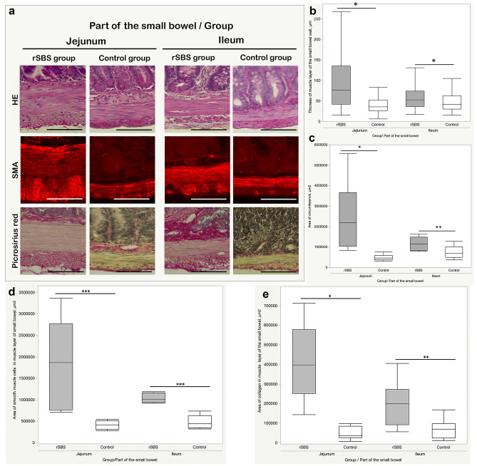
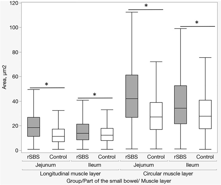
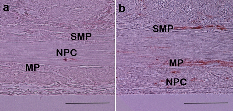
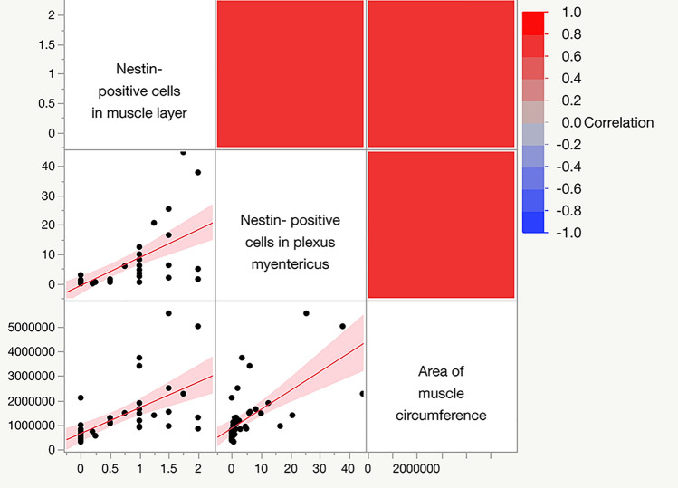
Short bowel syndrome (SBS) is a severe, life-threatening condition and one of the leading causes of intestinal failure in children. Here we were interested in changes in muscle layers and especially in the myenteric plexus of the enteric nervous system (ENS) of the small bowel in the context of intestinal adaptation. Twelve rats underwent a massive resection of the small intestine to induce SBS. Sham laparotomy without small bowel transection was performed in 10 rats. Two weeks after surgery, the remaining jejunum and ileum were harvested and studied. Samples of human small bowel were obtained from patients who underwent resection of small bowel segments due to a medical indication. Morphological changes in the muscle layers and the expression of nestin, a marker for neuronal plasticity, were studied. Following SBS, muscle tissue increases significantly in both parts of the small bowel, i.e., jejunum and ileum. The leading pathophysiological mechanism of these changes is hypertrophy. Additionally, we observed an increased nestin expression in the myenteric plexus in the remaining bowel with SBS. Our human data also showed that in patients with SBS, the proportion of stem cells in the myenteric plexus had risen by more than twofold. Our findings suggest that the ENS is tightly connected to changes in intestinal muscle layers and is critically involved in the process of intestinal adaptation to SBS.

Keywords: Bowel resection; ENS; Enteric neurons; Nestin; PGP 9.5; Short bowel syndrome.


Conflict of interest statement

None to declare.

Figures

Fig. 1
Fig. 1
Process of nuclear analysis in the muscle layers of the small intestinal wall (rat). a DAPI staining of nuclei. Areas of longitudinal (green) and circular (red) muscle were outlined. b Isolated circular muscle layer in 16-bit picture using ImageJ software. c Mapping of the picture in b using ImageJ software based on binary imaging. d Counting and analyzing of the circular muscle layer using ImageJ software. Scale bar is 50 µm
Fig. 2
Fig. 2
Comparison thickness of muscle layer of the small bowel between short bowel syndrome group (rSBS) and control group sections (rat): a Hematoxylin–eosin (HE), Picrosirius Red, and fluorescent smooth muscle actin (SMA) staining of jejunum and ileum; in Picrosirius Red staining, collagen fibers have a red color. In SMA staining, smooth muscle cells present a fluorescence red color, scale bar is 50 µm; b significant increase in the thickness of muscle layer in rSBS compared to control in jejunum and ileum; c area of muscle circumference of the small bowel (rat)—significant increase of the muscle circumference of the small bowel in rSBS compared to control in jejunum and ileum; d area of smooth muscle cells in muscle circumference of the small bowel (rat)—significant increase in rSBS compared to control in jejunum and ileum; e area of collagen in muscle circumference of the small bowel (rat)—significant increase in rSBS compared to control in jejunum and ileum. Line within box represents the median; the top and bottom of the box represent the 75th and 25th percentile, respectively. The whiskers indicate the maximum and minimum (not including outliers). Wilcoxon test: *p < 0.0001, **p = 0.0051, ***p = 0.0004
Fig. 3
Fig. 3
Size of the nuclei (area) (rat). Significant increase of the size of the nuclei in rSBS compared to control in jejunum and ileum. Line within box represents the median; the top and bottom of the box represent the 75th and 25th percentile, respectively. The whiskers indicate the maximum and minimum (not including outliers). Wilcoxon test: *p < 0.0001
Fig. 4
Fig. 4
Number of nuclei in a 1000 μm2 area of muscle layer (rat). The was a significant increase in the number of nuclei in the muscle layer in rSBS compared to control in jejunum and ileum. Line within box represents the median; the top and bottom of the box represent the 75th and 25th percentile, respectively. The whiskers indicate the maximum and minimum (not including outliers). Wilcoxon test: *p < 0.0001
Fig. 5
Fig. 5
Nestin–peroxidase-stained ganglion of myenteric plexus: a control group, b short bowel syndrome (rSBS) group (rat). Sections were stained 2 weeks after surgery. MP myenteric plexus, SMP plexus submucosus, NPC nestin-positive cells in muscle layer. In the rSBS group myenteric plexus, plexus submucosa and some cells in the muscle layer are nestin positive (brown color). Scale bar is 50 µm
Fig. 6
Fig. 6
Nestin fluorescence stained ganglion of myenteric plexus of the small intestine. a and c control group, b and d short bowel syndrome (rSBS) group (rat). Green color represents nestin fluorescence, e ratio of nestin-positive areas within the myenteric plexus compared between control and short bowel syndrome (rSBS). The rSBS small intestine presents more nestin-positive areas within the myenteric plexus. Line within box represents the median; the top and bottom of the box represent the 75th and 25th percentile, respectively. The whiskers indicate the maximum and minimum (not including outliers). Wilcoxon test: *p < 0.0001. Scale bar is 20 µm
Fig. 7
Fig. 7
Spearman’s correlation between the area of muscle circumference and the upregulation of nestin expression in the myenteric plexus and in the muscle layer (rat)
Fig. 8
Fig. 8
Ganglion of myenteric plexus in a biopsy specimen of the human small intestine. Immunofluorescence staining: stem cells, nestin (green); neurons, PGP 9.5 (red); cell nuclei, DAPI (4′,6-diamidino-2-phenylindole dihydrochloride) (blue). a Stem cell research, b study of nerve cells, c analysis of double staining (stem cells as well as nerve cells), d mapping of the myenteric plexus (green—stem cells, red—neurons). e The proportion of the area of stem cells to the area of the nerve cells in a cross section of the myenteric plexus. f Stem cells to nerve cells ratio in the myenteric plexus Line within box represents the median; the top and bottom of the box represent the 75th and 25th percentile, respectively. The whiskers indicate the maximum and minimum. Wilcoxon test: *p < 0.0001. Scale bar is 50 µm

Similar articles

Cited by

  • In focus in HCB.
    Taatjes DJ, Roth J. Taatjes DJ, et al. Histochem Cell Biol. 2023 Nov;160(5):371-373. doi: 10.1007/s00418-023-02246-w. Histochem Cell Biol. 2023. PMID: 37904027 No abstract available.

References

    1. Barth BB, Spencer NJ, Grill WM. Activation of ENS circuits in mouse colon: coordination in the mouse colonic motor complex as a robust, distributed control system. Adv Exp Med Biol. 2022;1383:113–123. doi: 10.1007/978-3-031-05843-1_11. - DOI - PubMed
    1. Beguin PC, Gosselin H, Mamarbachi M, Calderone A. Nestin expression is lost in ventricular fibroblasts during postnatal development of the rat heart and re-expressed in scar myofibroblasts. J Cell Physiol. 2012;227(2):813–820. doi: 10.1002/jcp.22794. - DOI - PubMed
    1. Birbrair A, Zhang T, Wang ZM, Messi ML, Enikolopov GN, Mintz A, Delbono O. Skeletal muscle neural progenitor cells exhibit properties of NG2-glia. Exp Cell Res. 2013;319(1):45–63. doi: 10.1016/j.yexcr.2012.09.008. - DOI - PMC - PubMed
    1. Breikaa RM, Algandaby MM, El-Demerdash E, Abdel-Naim AB. Multimechanistic antifibrotic effect of biochanin a in rats: implications of proinflammatory and profibrogenic mediators. PLoS ONE. 2013;8(7):e69276. doi: 10.1371/journal.pone.0069276. - DOI - PMC - PubMed
    1. Cantarero Carmona I, Luesma Bartolome MJ, Lavoie-Gagnon C, Junquera Escribano C. Distribution of nestin protein: immunohistochemical study in enteric plexus of rat duodenum. Microsc Res Tech. 2011;74(2):148–152. doi: 10.1002/jemt.20884. - DOI - PubMed

LinkOut - more resources