

Neuroinflammation in Acute Ischemic and Hemorrhagic Stroke
- PMID: 37395873
- PMCID: PMC10544736
- DOI: 10.1007/s11910-023-01282-2
Neuroinflammation in Acute Ischemic and Hemorrhagic Stroke
Abstract
Purpose of review: This review aims to provide an overview of neuroinflammation in ischemic and hemorrhagic stroke, including recent findings on the mechanisms and cellular players involved in the inflammatory response to brain injury.
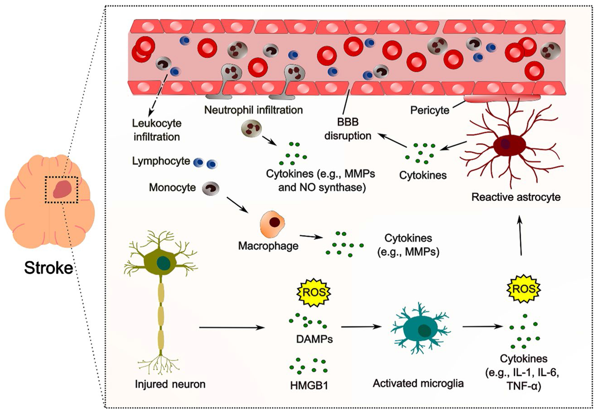
Recent findings: Neuroinflammation is a crucial process following acute ischemic stroke (AIS) and hemorrhagic stroke (HS). In AIS, neuroinflammation is initiated within minutes of the ischemia onset and continues for several days. In HS, neuroinflammation is initiated by blood byproducts in the subarachnoid space and/or brain parenchyma. In both cases, neuroinflammation is characterized by the activation of resident immune cells, such as microglia and astrocytes, and infiltration of peripheral immune cells, leading to the release of pro-inflammatory cytokines, chemokines, and reactive oxygen species. These inflammatory mediators contribute to blood-brain barrier disruption, neuronal damage, and cerebral edema, promoting neuronal apoptosis and impairing neuroplasticity, ultimately exacerbating the neurologic deficit. However, neuroinflammation can also have beneficial effects by clearing cellular debris and promoting tissue repair. The role of neuroinflammation in AIS and ICH is complex and multifaceted, and further research is necessary to develop effective therapies that target this process. Intracerebral hemorrhage (ICH) will be the HS subtype addressed in this review. Neuroinflammation is a significant contributor to brain tissue damage following AIS and HS. Understanding the mechanisms and cellular players involved in neuroinflammation is essential for developing effective therapies to reduce secondary injury and improve stroke outcomes. Recent findings have provided new insights into the pathophysiology of neuroinflammation, highlighting the potential for targeting specific cytokines, chemokines, and glial cells as therapeutic strategies.
Keywords: Astrocytes; Brain damage; Chemokines; Cytokines; Edema; Glial cells; Hemorrhagic stroke; Immune cells; Inflammatory response; Intracerebral hemorrhage; Ischemic stroke; Microglia; Neuroinflammation; Pathophysiology; Secondary injury; Therapeutic targets.
© 2023. The Author(s), under exclusive licence to Springer Science+Business Media, LLC, part of Springer Nature.
Figures
References
-
- Kleindorfer DO, Towfighi A, Chaturvedi S, Cockroft KM, Gutierrez J, Lombardi-Hill D, et al. 2021 Guideline for the prevention of stroke in patients with stroke and transient ischemic attack: a guideline from the American Heart Association/American Stroke Association. Stroke. 2021;52(7):e364–467. 10.1161/STR.0000000000000375. - DOI - PubMed
-
- Jung S, Gilgen M, Slotboom J, El-Koussy M, Zubler C, Kiefer C, et al. Factors that determine penumbral tissue loss in acute ischaemic stroke. Brain. 2013;136(Pt 12):3554–60. 10.1093/brain/awt246. - DOI - PubMed
-
This article examines the factors that contribute to the loss of penumbral tissue in cases of acute ischemic stroke. The study identifies variables such as time to treatment, collateral circulation, and infarct core size as critical determinants of penumbral tissue loss.
-
- Schaeffer S, Iadecola C. Revisiting the neurovascular unit. Nat Neurosci. 2021;24(9):1198–209. 10.1038/s41593-021-00904-7. - DOI - PMC - PubMed
-
This article revisits the concept of the neurovascular unit, emphasizing its crucial role in maintaining brain function and highlighting its relevance in neurological disorders. The review provides insights into the molecular and cellular mechanisms underlying neurovascular unit dysfunction and highlights its potential as a therapeutic target for various neurological conditions.
Publication types
MeSH terms
Substances
Grants and funding
LinkOut - more resources
Full Text Sources
Medical
Research Materials
