Review
. 2023 Apr;10(2):023522.
doi: 10.1117/1.NPh.10.2.023522. Epub 2023 Jun 30.

Applications of near-infrared spectroscopy in neurocritical care

Affiliations
Review

Applications of near-infrared spectroscopy in neurocritical care

Rachel Thomas et al. Neurophotonics. 2023 Apr.

Abstract

Significance: Acute brain injuries are commonly encountered in the intensive care unit. Alterations in cerebrovascular physiology triggered by the initial insult can lead to neurological worsening, further brain injury, and poor outcomes. Robust methods for assessing cerebrovascular physiology continuously at the bedside are limited.

Aim: In this review, we aim to assess the potential of near-infrared spectroscopy (NIRS) as a bedside tool to monitor cerebrovascular physiology in critically ill patients with acute brain injury as well as those who are at high risk for developing brain injury.

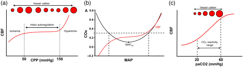
Approach: We first review basic principles of cerebral blood flow regulation and how these are altered after brain injury. We then discuss the potential role for NIRS in different acute brain injuries. We pay specific attention to the potential for NIRS to (1) identify new brain injuries and clinical worsening, (2) non-invasively measure intracranial pressure (ICP) and cerebral autoregulation, and (3) identify optimal blood pressure (BP) targets that may improve patient outcomes.

Results: A growing body of work supports the use of NIRS in the care of brain injured patients. NIRS is routinely used during cardiac surgeries to identify acute neurologic events, and there is some evidence that treatment algorithms using cerebral oximetry may result in improved outcomes. In acute brain injury, NIRS can be used to measure autoregulation to identify an "optimum" BP where autoregulation status is best preserved. Finally, NIRS has been utilized to identify oximetry thresholds that correlate with poor outcome as well as identify new focal intracranial hemorrhages.

Conclusions: NIRS is emerging as a tool that can non-invasively measure brain function in critically ill patients. Future work will be aimed at technical refinements to improve diagnostic accuracy, as well as larger scale clinical trials that can establish a definitive impact on patient outcomes.

Keywords: cerebral autoregulation; near-infrared spectroscopy; stroke; traumatic brain injury.


Figures

Fig. 1
Fig. 1
Major concepts in cerebral autoregulation and vascular reactivity. (a) Graph depicting relationship of CPP to CBF with intact cerebral autoregulation. Within the zone of intact autoregulation (dashed lines), changes in vessel diameter maintain consistent CBF. When CPP drops below the zone of autoregulation, ischemia ensues. Conversely, elevated pressures above the autoregulation zone lead to hyperemia and edema. (b) Optimal MAP (MAPopt) is calculated by comparing the cerebral TOI as measured by NIRS across the patient’s range of BP. The autoregulatory curve is superimposed in red, and the gray box denotes the patient’s MAP range that corresponds to those that fall within preserved autoregulation status (COx0.3). MAPopt corresponds to the nadir of the COx curve. (c) Cerebrovascular reactivity to CO2 is depicted with lower paCO2 causing vasoconstriction.
Fig. 2
Fig. 2
Examples of using NIRS derived autoregulation indices to identify MAPopt. (a) Data from a pediatric cardiac arrest patient. Top graph shows the time course of fluctuations in MAP and StO2 over a 24-h period. Second from top, temporal fluctuations in COx are shown. Periods of time with poor autoregulation (i.e., COx>0.3) are shaded gray. Second from bottom, method for deriving MAPopt (MAP where COx is minimized) and both ULA and LLA (BP where COx > 0.3) is shown. Bottom graph shows temporal trends in MAPopt superimposed on top of the patient’s actual BP. Areas shaded dark gray represent periods of time when actual MAP was significantly below MAPopt (defined as periods where MAP<MAPopt5  mmHg). (b) Similar data from adult patients with non-traumatic subarachnoid hemorrhage. Black trace represents patient’s actual MAP, red trace represents trends in MAPopt calculated from COx. Shaded red areas represent the range of intact autoregulation. The top curve shows a patient with BP trends where MAP largely stayed within ULA and LLA, whereas bottom curve shows a patient where BP often exceeded the ULA. [(a)- taken from Ref.  and (b)- taken from Ref. 31].

References

    1. Deschamps A., et al. , “Cerebral oximetry monitoring to maintain normal cerebral oxygen saturation during high-risk cardiac surgery: a randomized controlled feasibility trial,” Anesthesiology 124, 826–836 (2016).ANESAV10.1097/ALN.0000000000001029 - DOI - PubMed
    1. Yokose N., et al. , “Bedside monitoring of cerebral blood oxygenation and hemodynamics after aneurysmal subarachnoid hemorrhage by quantitative time-resolved near-infrared spectroscopy,” World Neurosurg 73, 508–513 (2010).10.1016/j.wneu.2010.02.061 - DOI - PubMed
    1. Budohoski K. P., et al. , “Cerebral autoregulation after subarachnoid hemorrhage: comparison of three methods,” J. Cereb. Blood Flow Metab. 33, 449–456 (2013).10.1038/jcbfm.2012.189 - DOI - PMC - PubMed
    1. Hametner C., et al. , “Noninvasive cerebral oximetry during endovascular therapy for acute ischemic stroke: an observational study,” J. Cereb. Blood Flow Metab. 35, 1722–1728 (2015).10.1038/jcbfm.2015.181 - DOI - PMC - PubMed
    1. Ritzenthaler T., et al. , “Cerebral near-infrared spectroscopy: a potential approach for thrombectomy monitoring,” Stroke 48, 3390–3392 (2017).SJCCA710.1161/STROKEAHA.117.019176 - DOI - PubMed