

Case Reports
doi: 10.1016/j.case.2023.01.003.
eCollection 2023 Jun.
Congenital Absence of the Left Atrial Appendage: Role of Multimodality Imaging
Affiliations
- PMID: 37396472
- PMCID: PMC10307590
- DOI: 10.1016/j.case.2023.01.003
Item in Clipboard
Case Reports
Congenital Absence of the Left Atrial Appendage: Role of Multimodality Imaging
CASE (Phila).
.
No abstract available
Figures
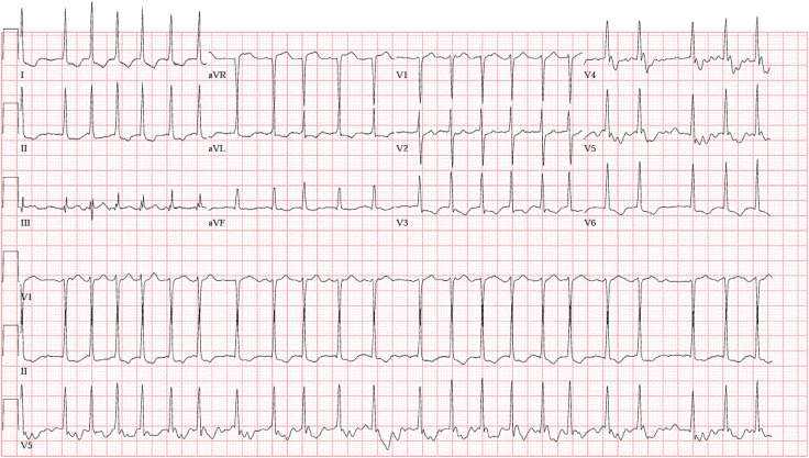
Twelve-lead electrocardiogram showed AF with rapid ventricular response at a heart rate of 137 bpm with ST depression and T-wave inversion in the lateral leads.

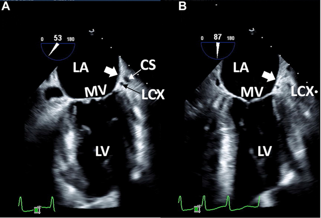
Case 1: two-dimensional TEE, midesophageal long-axis view, systolic view highlighting the mitral commissure (A; 53°) and conventional 2-chamber (B; 87°) view indicating the usual location of the LAA (thick white arrow). The CS and LCx are well visualized in the atrioventricular groove indicating the usual anatomic location of the LAA. CS, Coronary sinus; LCx, left circumflex coronary artery; LV, left ventricle; MV, mitral valve.
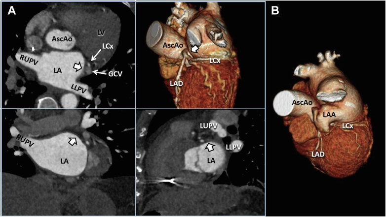
Case 1: CCT imaging demonstrates 3D cross-correlation of case 1 (A; quad panel) compared with a representative normal 3D volume-rendered example (B; right single panel). The multiplanar reformat quad display allows comprehensive demonstration of the abLAA (thick white arrow) in axial (upper left), coronal (lower left), and sagittal (lower right) orientations. AscAo, Ascending thoracic aorta; GCV, great cardiac vein; LAD, left anterior descending coronary artery; LCx, left circumflex coronary artery; LLPV, left lower pulmonary vein; LUPV, left upper pulmonary vein; LV, left ventricle; RUPV, right upper pulmonary vein.
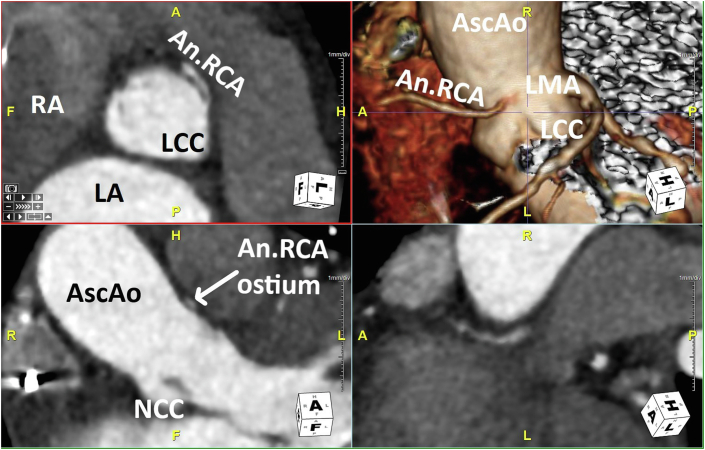
Case 1: CCT multiplanar reformat quad display demonstrates the anomalous RCA origin through a slit-like ostium (arises from the LCC) and interarterial course from multiple perspectives. An.RCA, Anomalous right coronary artery; AscAo, ascending thoracic aorta; LCC, Left coronary cusp; LMA, left main coronary artery; NCC, noncoronary cusp; RA, right atrium.
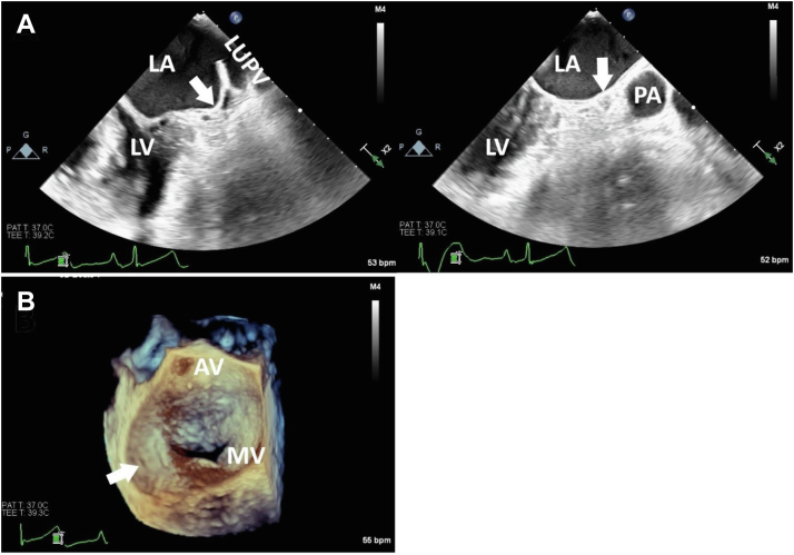
Case 2: two-dimensional TEE, midesophageal, long-axis systolic views at 45° (A; top left) and 90° (A; top right) demonstrates the abLAA (arrows). The TEE 3D display, surgeon’s view, demonstrates the en face view of the mitral valve from the perspective of the LA with the abLAA (arrow). AV, Aortic valve; MV, mitral valve; LUPV, left upper pulmonary vein; LV, left ventricle; PA, main pulmonary artery.
References
-
- Pourafkari L., Sadeghpour A., Baghbani-Oskouei A., Savadi-Oskouei S., Pouraliakbar H., Fazelifar A.F., et al. Absent left atrial appendage: case report and review of the literature. Cardiovasc Pathol. 2020;45:107178. - PubMed
-
- Guo L.J., Ding M.Y., Sun D.D., Zhao H.Z., Pan S.Q., Zhu F. Congenital absence of left atrial appendage combined with type A Wolff-Parkinson-White syndrome diagnosed by multimodal imaging. J Clin Ultrasound. 2022;50:28–30. - PubMed
-
- Pashun R.A., Gannon M.P., Tomassetti C., Rahmani N., Saba S.G. Congenital absence of the left atrial appendage. J Cardiovasc Comput Tomogr. 2020;14:e115–e117. - PubMed
Publication types
LinkOut - more resources
Full Text Sources
