

The pathological potential of ependymal cells in mild traumatic brain injury
- PMID: 37396927
- PMCID: PMC10312375
- DOI: 10.3389/fncel.2023.1216420
The pathological potential of ependymal cells in mild traumatic brain injury
Abstract
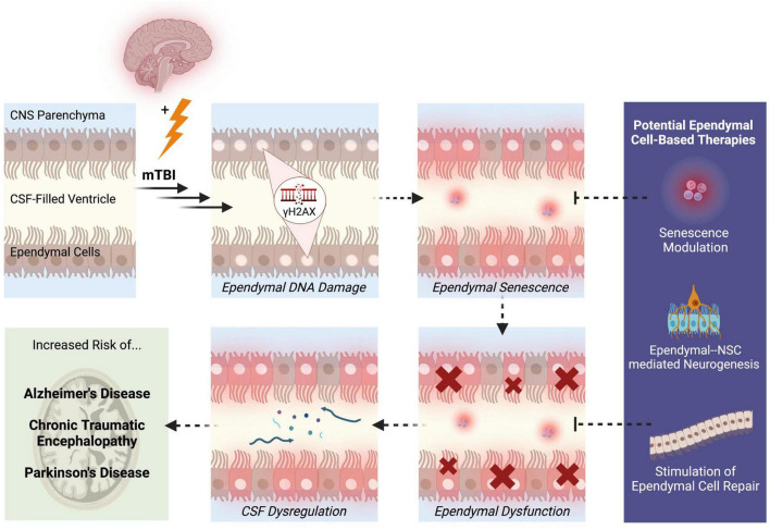
Mild traumatic brain injury (mTBI) is a common neurological condition affecting millions of individuals worldwide. Although the pathology of mTBI is not fully understood, ependymal cells present a promising approach for studying the pathogenesis of mTBI. Previous studies have revealed that DNA damage in the form of γH2AX accumulates in ependymal cells following mTBI, with evidence of widespread cellular senescence in the brain. Ependymal ciliary dysfunction has also been observed, leading to altered cerebrospinal fluid homeostasis. Even though ependymal cells have not been extensively studied in the context of mTBI, these observations reflect the pathological potential of ependymal cells that may underlie the neuropathological and clinical presentations of mTBI. This mini review explores the molecular and structural alterations that have been reported in ependymal cells following mTBI, as well as the potential pathological mechanisms mediated by ependymal cells that may contribute to overall dysfunction of the brain post-mTBI. Specifically, we address the topics of DNA damage-induced cellular senescence, dysregulation of cerebrospinal fluid homeostasis, and the consequences of impaired ependymal cell barriers. Moreover, we highlight potential ependymal cell-based therapies for the treatment of mTBI, with a focus on neurogenesis, ependymal cell repair, and modulation of senescence signaling pathways. Further insight and research in this field will help to establish the role of ependymal cells in the pathogenesis of mTBI and may lead to improved treatments that leverage ependymal cells to target the origins of mTBI pathology.
Keywords: DNA damage; brain barriers; cellular senescence; cerebrospinal fluid; ependymal cells; mild traumatic brain injury; neural stem cells.
Copyright © 2023 Nelles and Hazrati.
Conflict of interest statement
The authors declare that the research was conducted in the absence of any commercial or financial relationships that could be construed as a potential conflict of interest.
Figures
Similar articles
-
DNA repair deficiency and senescence in concussed professional athletes involved in contact sports.Acta Neuropathol Commun. 2019 Nov 14;7(1):182. doi: 10.1186/s40478-019-0822-3. Acta Neuropathol Commun. 2019. PMID: 31727161 Free PMC article.
-
Ependymal cells and neurodegenerative disease: outcomes of compromised ependymal barrier function.Brain Commun. 2022 Nov 4;4(6):fcac288. doi: 10.1093/braincomms/fcac288. eCollection 2022. Brain Commun. 2022. PMID: 36415662 Free PMC article. Review.
-
Early onset senescence and cognitive impairment in a murine model of repeated mTBI.Acta Neuropathol Commun. 2021 May 8;9(1):82. doi: 10.1186/s40478-021-01190-x. Acta Neuropathol Commun. 2021. PMID: 33964983 Free PMC article.
-
Cellular Senescence in Traumatic Brain Injury: Evidence and Perspectives.Front Aging Neurosci. 2021 Sep 28;13:742632. doi: 10.3389/fnagi.2021.742632. eCollection 2021. Front Aging Neurosci. 2021. PMID: 34650425 Free PMC article. Review.
-
Iron Metabolism Disorders for Cognitive Dysfunction After Mild Traumatic Brain Injury.Front Neurosci. 2021 Mar 16;15:587197. doi: 10.3389/fnins.2021.587197. eCollection 2021. Front Neurosci. 2021. PMID: 33796002 Free PMC article. Review.
Cited by
-
Ependymal cells: roles in central nervous system infections and therapeutic application.J Neuroinflammation. 2024 Oct 9;21(1):255. doi: 10.1186/s12974-024-03240-2. J Neuroinflammation. 2024. PMID: 39385253 Free PMC article. Review.
-
Multiciliated ependymal cells: an update on biology and pathology in the adult brain.Acta Neuropathol. 2024 Sep 10;148(1):39. doi: 10.1007/s00401-024-02784-0. Acta Neuropathol. 2024. PMID: 39254862 Review.
References
Publication types
LinkOut - more resources
Full Text Sources