

Management of Impacted Maxillary Canine with Immediate Implant and Sticky Bone Auto Tooth Graft
- PMID: 37396960
- PMCID: PMC10313465
- DOI: 10.1155/2023/2761700
Management of Impacted Maxillary Canine with Immediate Implant and Sticky Bone Auto Tooth Graft
Abstract
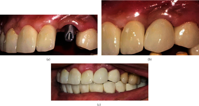
The management of the upper impacted canines includes a range of options, including orthodontic options in their various forms, up to extraction and replacing the tooth with a dental implant. Auto tooth graft (ATG) has achieved good clinical efficacy and was recently used as a grafting material for its bone induction and conduction properties. The use of platelet-rich fibrin (PRF) is highly effective in regenerative dentistry, and its use with bone grafts has improved tissue healing. This case report shows for the first time managing impacted canine with extraction and converting it into ATG and mixing it with injectable PRF to obtain sticky bone ATG and insertion of an immediate implant in a female patient who complains about a missing upper left canine. The results show the good bone formation and satisfactory clinical aspects.
Copyright © 2023 Wajeha Albatal et al.
Conflict of interest statement
The authors declare that they have no conflicts of interest.
Figures


Similar articles
-
Clinical Considerations and Management of a Labially Impacted Maxillary Canine With Immediate Implant Placement and Alveolar Bone Reconstruction: A Case Report.Cureus. 2024 Oct 15;16(10):e71529. doi: 10.7759/cureus.71529. eCollection 2024 Oct. Cureus. 2024. PMID: 39544558 Free PMC article.
-
Bone augmentation with sticky bone and platelet-rich fibrin by ridge-split technique and nasal floor engagement for immediate loading of dental implant after extracting impacted canine.Natl J Maxillofac Surg. 2019 Jan-Jun;10(1):98-101. doi: 10.4103/njms.NJMS_37_18. Natl J Maxillofac Surg. 2019. PMID: 31205397 Free PMC article.
-
Effect of Platelet-rich Fibrin and Free Gingival Graft in the Treatment of Soft Tissue Defect preceding Implant Placement.J Contemp Dent Pract. 2018 Jul 1;19(7):895-899. J Contemp Dent Pract. 2018. PMID: 30066697
-
Current knowledge and perspectives for the use of platelet-rich plasma (PRP) and platelet-rich fibrin (PRF) in oral and maxillofacial surgery part 2: Bone graft, implant and reconstructive surgery.Curr Pharm Biotechnol. 2012 Jun;13(7):1231-56. doi: 10.2174/138920112800624472. Curr Pharm Biotechnol. 2012. PMID: 21740370 Review.
-
The use of platelet-rich fibrin to enhance the outcomes of implant therapy: A systematic review.Clin Oral Implants Res. 2018 Oct;29 Suppl 18(Suppl Suppl 18):6-19. doi: 10.1111/clr.13275. Clin Oral Implants Res. 2018. PMID: 30306698 Free PMC article.
Cited by
-
Clinical Considerations and Management of a Labially Impacted Maxillary Canine With Immediate Implant Placement and Alveolar Bone Reconstruction: A Case Report.Cureus. 2024 Oct 15;16(10):e71529. doi: 10.7759/cureus.71529. eCollection 2024 Oct. Cureus. 2024. PMID: 39544558 Free PMC article.
References
-
- Cardaropoli D., Debernardi C., Cardaropoli G. Immediate placement of implant into impacted maxillary canine extraction socket. The International Journal of Periodontics and Restorative Dentistry . 2007;27(1):71–77. - PubMed
-
- Yadav A., Vinayak V., Grover H., Bhardwaj A. Extraction of impacted maxillary canine with simultaneous implant placement. Journal of Dental Sciences and Oral Rehabilitation . 2012;1:41–43.
-
- Cohen E. Atlas of Cosmetic and Reconstructive Periodontal Surgery . Vol. 3. Fairfield County, Connecticut, USA: People’s Medical Publishing House, Shelton, Connecticut,; 2007. p. p. 373.
Publication types
LinkOut - more resources
Full Text Sources
