

Case Reports
doi: 10.1080/23320885.2023.2228887.
eCollection 2023.
Mixed lesion of traumatic pseudoaneurysm and pyogenic granuloma on a digit
Affiliations
- PMID: 37397126
- PMCID: PMC10312018
- DOI: 10.1080/23320885.2023.2228887
Item in Clipboard
Case Reports
Mixed lesion of traumatic pseudoaneurysm and pyogenic granuloma on a digit
Case Reports Plast Surg Hand Surg.
.
Abstract
Traumatic aneurysms occurring in the digit are extremely rare. We report a case of a traumatic pseudoaneurysm arising from a terminal branch of the finger artery and presenting as a mixed lesion with pyogenic granuloma that was exposed to the outside of the body and treated surgically.
Keywords: False aneurysm; digital artery; hand surgery; microvascular surgery; ultrasound.
© 2023 The Author(s). Published by Informa UK Limited, trading as Taylor & Francis Group.
Conflict of interest statement
No potential conflict of interest was reported by the author(s).
Figures

Appearance of the right middle finger at the time of initial examination. A smooth erythematous nodule 7 mm in size was observed on the ulnar lateral nail fold. The nail is shifted to the radial side. The subcutaneous area under the nodule was indurated, swollen, and tender. (left) Dorsal side of the affected finger. (right) Palmar side of the affected finger.

Ultrasound findings. B-mode images revealed a well-defined hypoechoic region seen protruding from the subcutis to the outside of the body. Color doppler images showed pulsatile vascular inflow from deep within the lesion (arrowhead).
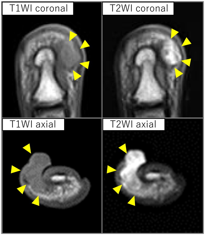
Magnetic resonance imaging findings. An iso-intensity in T1 and high-intensity in T2 weighted images lesion was observed protruding subcutaneously to the body surface on the ulnar aspect of the distal phalanx (arrowhead).

Intraoperative findings. (a) Intraoperative Photograph. A terminal branch of the digital artery flowed into the deep part of the lesion (arrowhead). (b) Photograph of the resected lesion. The lesion was hourglass-shaped, separated by the skin.

Histopathological findings. Hematoxylin-eosin stained images at 40x magnification. Scale bar = 500 µm. (a) Pathological findings for the lesion. The three-layered structure of the arterial wall was disrupted, consistent with the finding of a pseudoaneurysm. (b) Pathological findings of one region of the lesion. Capillary dilation and edematous stromal hyperplasia were observed, which were characteristic of pyogenic granuloma.

Appearance 6 months after surgery. No recurrence of lesions was noted, and finger morphology and nail shift improved.
References
Publication types
LinkOut - more resources
Full Text Sources