[Preprint]. 2025 Feb 20:2023.05.31.542897.
doi: 10.1101/2023.05.31.542897.

Single-cell type analysis of wing premotor circuits in the ventral nerve cord of Drosophila melanogaster

Affiliations

Single-cell type analysis of wing premotor circuits in the ventral nerve cord of Drosophila melanogaster

Erica Ehrhardt et al. bioRxiv. .

Abstract

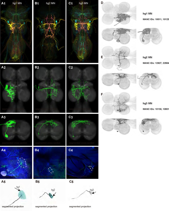
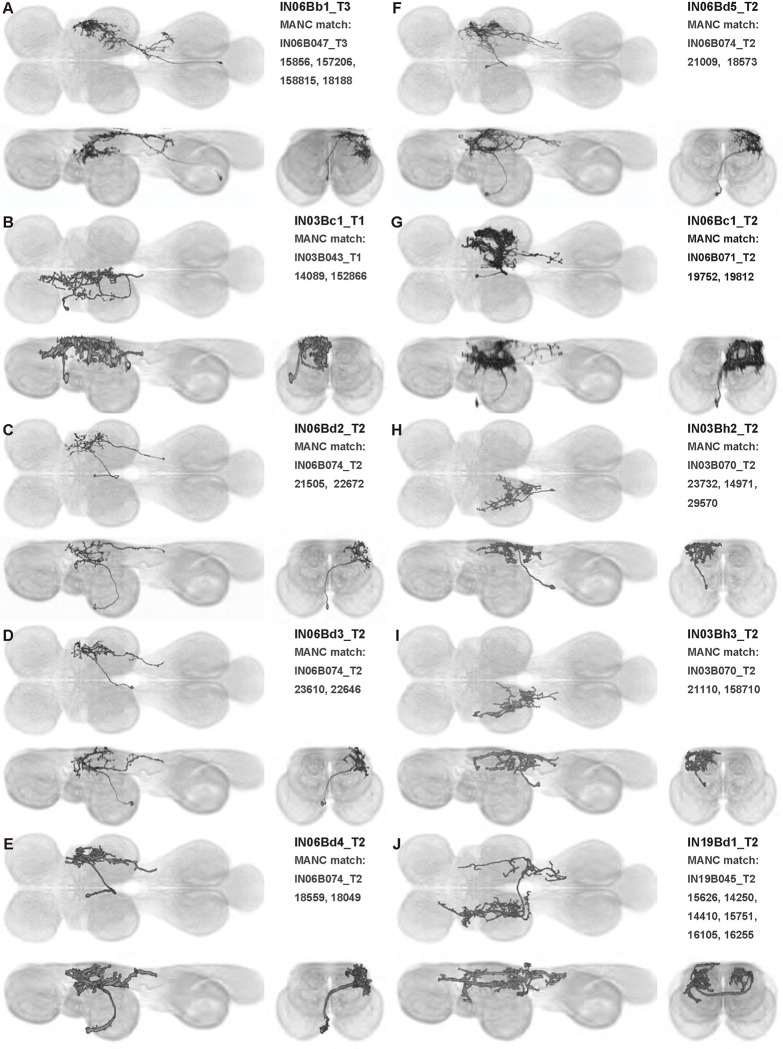
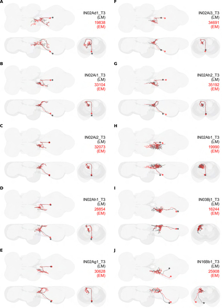
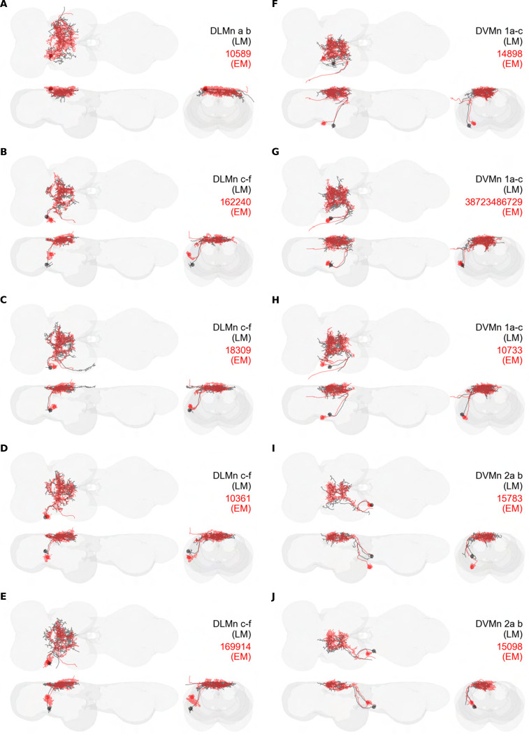
To perform most behaviors, animals must send commands from higher-order processing centers in the brain to premotor circuits that reside in ganglia distinct from the brain, such as the mammalian spinal cord or insect ventral nerve cord. How these circuits are functionally organized to generate the great diversity of animal behavior remains unclear. An important first step in unraveling the organization of premotor circuits is to identify their constituent cell types and create tools to monitor and manipulate these with high specificity to assess their functions. This is possible in the tractable ventral nerve cord of the fly. To generate such a toolkit, we used a combinatorial genetic technique (split-GAL4) to create 195 sparse transgenic driver lines targeting 196 individual cell types in the ventral nerve cord. These included wing and haltere motoneurons, modulatory neurons, and interneurons. Using a combination of behavioral, developmental, and anatomical analyses, we systematically characterized the cell types targeted in our collection. In addition, we identified correspondences between the cells in this collection and a recent connectomic data set of the ventral nerve cord. Taken together, the resources and results presented here form a powerful toolkit for future investigations of neuronal circuits and connectivity of premotor circuits while linking them to behavioral outputs.

Keywords: Drosophila; Split-GAL4; courtship song; flight; motoneuron; ventral nerve cord; wing.


Figures

Figure 1:
Figure 1:. Isolating neurons in the ventral nerve cord (VNC).
(A) The fly central nervous system (gray) with the ventral nerve cord (VNC) highlighted in black. Illustrations of wing motoneuron and haltere sensory afferent shown in blue and purple, respectively. (B) Top-down (dorsal) view of VNC showing example neuron types: wing motoneuron (blue), descending neuron (black), and haltere-to-wing neuropil interneuron (orange). Boundaries between the pro-, meso-, and metathoracic neuromeres—i.e. T1, T2, and T3—are also shown (gray dotted lines and labels). (C) Schematic of VNC neuropils. Abbreviations used: T1 (prothoracic segment), T2 (mesothoracic segment), T3 (metathoracic segment), VAC (ventral association center), mVAC (medial ventral association center), and AMNp (accessory mesothoracic neuropil). (D) Example usage of the split-GAL4 technique for narrowing driver line expression profile (white) in the VNC (orange). R59G07 (D1) and R50G08 (D2) are used to drive half of the GAL4 transcription factor in SS54506 (D3), resulting in a sparse expression pattern. (E) Multiple interneurons segmented from the expression pattern of SS54506 using multicolor flip-out (MCFO).
Figure 2:
Figure 2:. Morphology of DLM power muscle motoneurons.
(A1) Color MIP of full expression pattern of a split line targeting DLMns, SS44039, crossed with pJFRC51–3xUAS-Syt::smGFP-HA in su(Hw)attP1; pJFRC225–5xUAS-IVS-myr::smGFP-FLAG in VK00005, aligned to the JRC 2018 VNC Unisex template. Scale bar in A1 represents 50 μm. (A2) Segmented images of DLM wing motoneurons in upright VNCs. VNCs were aligned to the JRC 2018 Unisex template. (A3) Transverse views of the segmented neurons shown in A2. (A4) Images of the muscles and their motoneuron innervation in thoraxes stained with phalloidin conjugated with Alexa 633. Phalloidin staining is shown in blue, GFP in green, and nc82 (Bruchpilot, to label the nervous system) in gray. (A5) Segmented muscle images. (B-F) Multicolor flipout (MCFO) was used to separate the power motoneurons, isolating individual cells where possible. Males were used for all motoneuron images.
Figure 3:
Figure 3:. Morphology of DVM power muscle motoneurons.
(A1) Color MIP of full expression pattern of a split line targeting DVMns, SS31950, crossed with pJFRC51–3xUAS-Syt::smGFP-HA in su(Hw)attP1; pJFRC225–5xUAS-IVS-myr::smGFP-FLAG in VK00005, aligned to the JRC 2018 VNC Unisex template. Scale bar in A1 represents 50 μm. (A2) Segmented images of DVM wing motoneurons in upright VNCs. VNCs were aligned to the JFC 2018 Unisex template. (A3) Transverse views of the segmented neurons shown in A2. (A4) Images of the muscles and their motoneuron innervation in thoraxes stained with phalloidin conjugated with Alexa 633. Phalloidin staining is shown in blue, GFP in green, and nc82 (Bruchpilot, to label the nervous system) in gray. (A5) Segmented muscle images. (B-H) Multicolor flipout (MCFO) was used to separate the power motoneurons, isolating individual cells where possible. Males were used for all motoneuron images.
Figure 4:
Figure 4:. Morphology of tergopleural wing steering muscle motoneurons targeted by our sparse split lines.
(A-C1) Color MIPs of full expression patterns of the split lines (respectively SS51528, SS41052, SS47120), crossed with pJFRC51–3xUAS-Syt::smGFP-HA in su(Hw)attP1; pJFRC225–5xUAS-IVS-myr::smGFP-FLAG in VK00005, aligned to the JRC 2018 VNC Unisex template. Scale bar in A1 represents 50 μm. (A-C2) Segmented images of tergopleural wing motoneurons in upright VNCs. Multicolor flipout (MCFO) was used to separate left and right neurons. VNCs were aligned to the JFC 2018 Unisex template. (A-C3) Transverse views of the segmented neurons shown in A-C2. (A-C4) Images of the muscles and their motoneuron innervation in thoraxes stained with phalloidin conjugated with Alexa 633. Phalloidin staining is shown in blue, GFP in green, and nc82 (Bruchpilot, to label the nervous system) in gray. (A-C5) Segmented muscle images. (D-F) segmented images of steering motoneurons including side views. Males were used for all motoneuron images except tpN, as our tpN line lacked expression in males.
Figure 5:
Figure 5:. Morphology of other indirect wing steering muscle motoneurons targeted by our sparse split lines.
(A-B1) Color MIPs of full expression patterns of the split lines (SS47204, SS47125), crossed with pJFRC51–3xUAS-Syt::smGFP-HA in su(Hw)attP1; pJFRC225–5xUAS-IVS-myr::smGFP-FLAG in VK00005, aligned to the JRC 2018 VNC Unisex template. Scale bar in A1 represents 50 μm. (A-B2) Segmented images of other indirect wing motoneurons in upright VNCs. Multicolor flipout (MCFO) was used to separate left and right neurons. VNCs were aligned to the JFC 2018 Unisex template. (A-B3) Transverse views of the segmented neurons shown in A-B2. (A-B4) Images of the muscles and their motoneuron innervation in thoraxes stained with phalloidin conjugated with Alexa 633. Phalloidin staining is shown in blue, GFP in green, and nc82 (Bruchpilot, to label the nervous system) in gray. (A-B5) Segmented muscle images. (C-D) segmented images of steering motoneurons including side views. Males were used for all motoneuron images.
Figure 6:
Figure 6:. Morphology of basalar wing steering muscle motoneurons targeted by our sparse split lines.
(A-B1) Color MIPs of full expression patterns of the split lines (SS40980, SS45772, SS45779), crossed with pJFRC51–3xUAS-Syt::smGFP-HA in su(Hw)attP1; pJFRC225–5xUAS-IVS-myr::smGFP-FLAG in VK00005, aligned to the JRC 2018 VNC Unisex template. Scale bar in A1 represents 50 μm. (A-B2) Segmented images of basalar wing motoneurons in upright VNCs. Multicolor flipout (MCFO) was used to separate left and right neurons. VNCs were aligned to the JFC 2018 Unisex template. (A-B3) Transverse views of the segmented neurons shown in A-B2. (A-B4) Images of the muscles and their motoneuron innervation in thoraxes stained with phalloidin conjugated with Alexa 633. Phalloidin staining is shown in blue, GFP in green, and nc82 (Bruchpilot, to label the nervous system) in gray. (A-B5) Segmented muscle images. (C-D) segmented images of steering motoneurons including side views. Males were used for all motoneuron images.
Figure 7:
Figure 7:. Morphology of first axillary wing steering muscle motoneurons targeted by our sparse split lines.
(A-B1) Color MIPs of full expression patterns of the split lines (SS41039, SS45782), crossed with pJFRC51–3xUAS-Syt::smGFP-HA in su(Hw)attP1; pJFRC225–5xUAS-IVS-myr::smGFP-FLAG in VK00005, aligned to the JRC 2018 VNC Unisex template. Scale bar in A1 represents 50 μm. (A-B2) Segmented images of first axillary wing motoneurons in upright VNCs. Multicolor flipout (MCFO) was used to separate left and right neurons. VNCs were aligned to the JFC 2018 Unisex template. (A-B3) Transverse views of the segmented neurons shown in A-B2. (A-B4) Images of the muscles and their motoneuron innervation in thoraxes stained with phalloidin conjugated with Alexa 633. Phalloidin staining is shown in blue, GFP in green, and nc82 (Bruchpilot, to label the nervous system) in gray. (A-B5) Segmented muscle images. (C-D) segmented images of steering motoneurons including side views. Males were used for all motoneuron images.
Figure 8:
Figure 8:. Morphology of third axillary wing steering muscle motoneurons targeted by our sparse split lines.
(A-B1) Color MIPs of full expression patterns of the split lines (SS41027, SS45779, SS41027), crossed with pJFRC51–3xUAS-Syt::smGFP-HA in su(Hw)attP1; pJFRC225–5xUAS-IVS-myr::smGFP-FLAG in VK00005, aligned to the JRC 2018 VNC Unisex template. Scale bar in A1 represents 50 μm. (A-B2) Segmented images of third axillary wing motoneurons in upright VNCs. Multicolor flipout (MCFO) was used to separate left and right neurons. VNCs were aligned to the JFC 2018 Unisex template. (A-B3) Transverse views of the segmented neurons shown in A-B2. (A-B4) Images of the muscles and their motoneuron innervation in thoraxes stained with phalloidin conjugated with Alexa 633. Phalloidin staining is shown in blue, GFP in green, and nc82 (Bruchpilot, to label the nervous system) in gray. (A-B5) Segmented muscle images. (C-D) segmented images of steering motoneurons including side views. Males were used for all motoneuron images.
Figure 9:
Figure 9:. Morphology of fourth axillary wing steering muscle motoneurons targeted by our sparse split lines.
(A-C1) Color MIPs of full expression patterns of the split lines (SS32023,SS37253, SS49039), crossed with pJFRC51–3xUAS-Syt::smGFP-HA in su(Hw)attP1; pJFRC225–5xUAS-IVS-myr::smGFP-FLAG in VK00005, aligned to the JRC 2018 VNC Unisex template. Scale bar in A1 represents 50 μm. (A-C2) Segmented images of fourth axillary wing motoneurons in upright VNCs. Multicolor flipout (MCFO) was used to separate left and right neurons. VNCs were aligned to the JFC 2018 Unisex template. (A-C3) Transverse views of the segmented neurons shown in A-C2. (A-C4) Images of the muscles and their motoneuron innervation in thoraxes stained with phalloidin conjugated with Alexa 633. Phalloidin staining is shown in blue, GFP in green, and nc82 (Bruchpilot, to label the nervous system) in gray. (A-C5) Segmented muscle images. (D-F) segmented images of steering motoneurons including side views. Males were used for all motoneuron image.
Figure 10:
Figure 10:. Optogenetic stimulation of wing motor neurons evokes changes to flight kinematics.
(A) Schematic of tethered flight measurement apparatus. Tethered flies are positioned in front of a display screen that presents both open- and closed-loop visual stimuli. The fly is illuminated near-infrared LEDs and filmed by three cameras recording at 100 fps. An additional red LED (617 nm) provides optogenetic stimulation in 100 ms pulses. (B) Stills from two of the three cameras recording the tethered fly in flight. Top panel (side view) illustrates the forward (fwd) and backward (back) deviation angles; bottom panel (bottom view) illustrates the stroke amplitude. (C) Averaged wing kinematics for two flies undergoing 50 repetitions of a closed loop trial with 100 ms optogenetic pulse. Dark lines and envelopes show the mean and 95% confidence interval, respectively. Traces in blue correspond to a i2-GAL4>UAS-CsChrimson fly; traces in gray show an example genetic control, SS01062>UAS-CsChrimson, where SS01062 is an empty split Gal4 line. (D-G) Statistics across flies for the four kinematic variables shown in (C): stroke amplitude (D), wingbeat frequency (E), forward deviation angle (F), and backward deviation angle (G). Open circles show per-fly measurements; bars and horizontal lines show interquartile range and population median, respectively. The number of flies per genotype is shown between E and G. Significance is determined via Wilcoxon rank sum test with Bonferroni correction (***, p<0.001; **, p<0.01; *, p<0.05).
Figure 11:
Figure 11:. Chronic silencing of wing motoneurons results in courtship song deficits.
(A) Image from a typical courtship assay, showing a male (above) extending its left wing to sing to a female (below). (B) Example trace of song recording from a control group fly (SS01055>UAS-Kir2.1). Bouts of sine and pulse song are labelled on the left and right of the trace, respectively. (C) Example slow (top) and fast (bottom) pulse modes for a single fly. Thick black lines show the mean pulse shape; thin gray lines show individual pulses. (D) Courtship song statistics for motoneuron driver lines crossed to UAS-Kir2.1. Rows show different song parameters: total fraction of the trial spent singing (top), fraction of song spent singing pulse mode song (middle), and fraction of song spent singing sine mode song (bottom). Open circles show per-fly measurements; bars and horizontal lines show interquartile range and population median, respectively. The number of flies per genotype is given in D, with the sample sizes for control and experimental groups on the top and bottom, respectively. (E)€ Analysis of pulse type. The top and middle and rows show waveforms for slow and fast pulse modes, respectively, for each genotype in C (blue), overlaid onto control (dark gray). Thick line shows the grand mean across flies; envelope gives 95% confidence interval for mean from bootstrap. Bottom row shows the fraction of pulses that are classified as slow for each genotype. Significance assigned using the Wilcoxon rank sum test and Fisher’s exact test (***, p<0.001; **, p<0.01; *, p<0.05).
Figure 12:
Figure 12:. Morphology of haltere motoneurons targeted by our sparse split lines.
(A-B1) Color MIPs of full expression patterns of the split lines (SS51523, SS41075), crossed with pJFRC51–3xUAS-Syt::smGFP-HA in su(Hw)attP1; pJFRC225–5xUAS-IVS-myr::smGFP-FLAG in VK00005, aligned to the JRC 2018 VNC Unisex template. Scale bar in A1 represents 50 μm. (A-B2) Images of the muscles and their motoneuron innervation in thoraxes stained with phalloidin conjugated with Alexa 633. Phalloidin staining is shown in blue, GFP in green, and nc82 (Bruchpilot, to label the nervous system) in gray. (A-B3) Segmented muscle images. (C-D) segmented images of haltere motoneurons.
Figure 13:
Figure 13:. Morphology of haltere motoneurons targeted by our broad split lines.
(A-B1) Color MIPs of full expression patterns of the split lines (SS37231, SS47195), crossed with pJFRC51–3xUAS-Syt::smGFP-HA in su(Hw)attP1; pJFRC225–5xUAS-IVS-myr::smGFP-FLAG in VK00005, aligned to the JRC 2018 VNC Unisex template. Scale bar in A1 represents 50 μm. (A-B2) Images of the muscles and their motoneuron innervation in thoraxes stained with phalloidin conjugated with Alexa 633. Phalloidin staining is shown in blue, GFP in green, and nc82 (Bruchpilot, to label the nervous system) in gray. (A-B3) Segmented muscle images. (C-E) segmented images of haltere motoneurons.
Figure 14:
Figure 14:. Morphology of VUMs in our split lines.
(A1-E1) Color MIPs of the full expression pattern of each split (SS40867, SS40868, SS42385, SS45766, SS51508) in the VNC of males crossed with pJFRC51–3xUAS-Syt::smGFP-HA in su(Hw)attP1; pJFRC225–5xUAS-IVS-myr::smGFP-FLAG in VK00005, aligned to the JRC 2018 VNC Unisex template. Scale bar in A1 represents 50 μm. Wing and haltere neuropils are indicated by dashed white outlines. (A-E2) Medial views of the muscles and their motoneuron innervation in thoraxes stained with phalloidin conjugated with Alexa 633. Phalloidin staining is shown in blue, GFP in green, and nc82 (Bruchpilot, to label the nervous system) in gray. (A-E3) Segmented medial muscle images. (A-E4) Lateral views of the muscles and their motoneuron innervation. (A-E5) Segmented lateral muscle images. Males were used for all images.
Figure 15:
Figure 15:. Morphology of individual VUMs and a VPM in our splits, and a table of expression in our T2VUM splits.
Segmented multicolor flipout images aligned to the JRC 2018 VNC Unisex template. The driver lines were SS40867, SS46645, SS42385, SS40867, SS51508, SS48268 respectively.
Figure 16:
Figure 16:. Morphology of identified intrasegmental interneurons.
MCFO was used to separate interneurons. Each neuron is shown from three views—horizontal (top left), sagittal (bottom left), and transverse (bottom right)—along with neuron name and hemilineage identity (top right). Images taken from preparations of male flies, segmented to isolate individual neurons, and aligned to the JRC 2018 Unisex VNC template. The driver lines used are: SS44314, SS43546, SS36094, SS36094, SS44314, SS31472, SS49042, SS33409, SS49042, SS25511.
Figure 17:
Figure 17:. Morphology of identified intersegmental interneurons.
MCFO was used to separate interneurons. Each neuron is shown from three views—horizontal (top left), sagittal (bottom left), and transverse (bottom right)—along with neuron name and hemilineage identity (top right). Images taken from preparations of male flies, segmented to isolate individual neurons, and aligned to the JRC 2018 Unisex VNC template. The driver lines used are: SS54495, SS48619, SS60603, SS31899, SS32400, SS54480, SS49807, SS54495, VT014604 (no single cell images were produced from the split GAL4 driver lines targeting this cell, SS31309, SS30330, SS54480, SS54474 or SS54495, so a generation 1 GAL4 image was used), SS25553.
Figure 18:
Figure 18:. Comparison of interneuron single-cell light microscopy images and MANC electron microscopy matches.
Panels (A-I) each correspond to a single cell type, showing the skeletonized light microscopy (LM) image (black) and the skeleton of the best match obtained from electron microscopy data (red; MANC connectome). The labels in each panel give the neuron name according to our nomenclature (black) and the body ID for the cell in MANC (red).
Figure 19:
Figure 19:. Sexually dimorphic anatomy in split lines targeting two hemilineages.
A shows the male expression pattern of a split targeting 3B t1, crossed with pJFRC51–3xUAS-Syt::smGFP-HA in su(Hw)attP1; pJFRC225–5xUAS-IVS-myr::smGFP-FLAG in VK00005, not aligned B-E show the male expression patterns of 17A t2 in four different split lines, crossed with pJFRC51–3xUAS-Syt::smGFP-HA in su(Hw)attP1; pJFRC225–5xUAS-IVS-myr::smGFP-FLAG in VK00005. F-J show the female expression patterns of these same lines, crossed with UAS-CsChrimson. All images are aligned to the JRC 2018 VNC template.
Figure 1—figure supplements 1–5:
Figure 1—figure supplements 1–5:. VNC expression patterns of split-GAL4 driver lines stabilized in this project.
Each split line was crossed with pJFRC51–3xUAS-Syt::smGFP-HA in su(Hw)attP1; pJFRC225–5xUAS-IVS-myr::smGFP-FLAG in VK00005. This figure shows the male expression pattern, except in lines which had no male expression, such as SS51528. Each VNC stack was aligned to the JRC 2018 VNC Unisex template. These maximum intensity projections are color coded to indicate the depth of each pixel. Blue represents the ventral layers of the VNC while red represents the dorsal layers. Panels are arranged by split number, except that splits targeting leg neurons which were serendipitously produced in this project are arranged in the last 18 panels.
Figure 3—figure supplement 1:
Figure 3—figure supplement 1:. Power MN groups based on morphology in the VNC, illustrated using segmented MCFO images aligned to the unisex JRC2018 template.
(A) DLMna/b. (B) DLMnc-f. (C) The MNs innervating DVM 1 and 2. (D) The MNs innervating DVM 3. (E) Schematic depicting the muscle fibers innervated by the MNs in panels A-D in the medial (left) and lateral (right) thorax.
Figure 9—figure supplement 1:
Figure 9—figure supplement 1:. Wing control MN groups based on morphology in the VNC, illustrated using segmented MCFO images aligned to the unisex JRC2018 template.
(A) Unilateral steering MNs with arborization mainly in the wing neuropil: b1 MN, iii3 MN, iii4 MN. (B) Unilateral steering MNs with arborization mainly in the wing neuropil: tpN MN, tp1 MN, tp2 MN, ps1 MN, hg2 MN, hg3 MN. (C) Steering MNs with approximately equal arborization in the wing neuropil and intermediate tectulum: b3 MN, i1 MN, i2 MN, hg1 MN. (D) Steering MNs with little or no arborization in the wing neuropil: tt MN, b2 MN, iii1 MN.
Figure 11—figure supplement 1:
Figure 11—figure supplement 1:. Additional analyses of the effects of wing motoneuron silencing on courtship song.
(A) Courtship song statistics for motoneuron driver lines crossed to UAS-Kir2.1. Rows show inter pulse interval (top) and sine song carrier frequency (bottom) for each motoneuron driver line tested (columns). Open circles show per-fly measurements; bars and horizontal lines show interquartile range and population median, respectively. The number of flies per genotype is given in each figure axis. (B) Transition matrices giving median probabilities across flies for transitioning between the three song modes: pulse, sine, and null. Rows correspond to data from genetic control flies (top) and motoneuron-silenced flies (bottom). Orange asterisks indicate significant differences between genetic control and silenced groups. Significance assigned using the Wilcoxon rank sum test (***, p<0.001; **, p<0.01; *, p<0.05).
Figure 13—figure supplement 1:
Figure 13—figure supplement 1:. Haltere MN groups based on morphology in the VNC, illustrated using segmented MCFO images aligned to the unisex JRC2018 template.
(A) Haltere MNs with unilateral, intersegmental arborization: hb1/2 MN. (B) Haltere MNs with bilateral arborization mainly in the haltere neuropil: hDVMn, hi1 MN, hi2 MN.
Figure 16—figure supplement 1:
Figure 16—figure supplement 1:. Morphology of identified intrasegmental interneurons.
MCFO was used to separate interneurons. Each neuron is shown from three views—horizontal (top left), sagittal (bottom left), and transverse (bottom right)—along with neuron name and hemilineage identity (top right). Images taken from preparations of male flies, segmented to isolate individual neurons, and aligned to the JRC 2018 Unisex VNC template. The driver lines used are: SS42439, SS42442, SS42442, SS42442, SS42438, SS42439, SS48215, SS48215, SS48268, SS47192.
Figure 16—figure supplement 2:
Figure 16—figure supplement 2:. Morphology of identified intrasegmental interneurons.
MCFO was used to separate interneurons. Each neuron is shown from three views—horizontal (top left), sagittal (bottom left), and transverse (bottom right)—along with neuron name and hemilineage identity (top right). Images taken from preparations of male flies, segmented to isolate individual neurons, and aligned to the JRC 2018 Unisex VNC template. The driver lines used are: 22E12 (no single-cell image appeared in the MCFO of the split GAL4 line targeting this cell, SS53421, so a generation 1 GAL4 line image was used), SS45830, SS45843, SS45843, SS45830, SS45843, SS25482, SS31309, SS46725 26A08 (no single-cell image appeared in the MCFO of the split GAL4 line targeting this cell, SS48272, so a generation 1 GAL4 line image was used).
Figure 16—figure supplement 3:
Figure 16—figure supplement 3:. Morphology of identified intrasegmental interneurons.
MCFO was used to separate interneurons. Each neuron is shown from three views—horizontal (top left), sagittal (bottom left), and transverse (bottom right)—along with neuron name and hemilineage identity (top right). Images taken from preparations of male flies, segmented to isolate individual neurons, and aligned to the JRC 2018 Unisex VNC template. The driver lines used are: SS48272, SS48272, SS49799, SS49766, SS42438, SS40783, SS42442, SS46675, SS46675, SS40783.
Figure 16—figure supplement 4:
Figure 16—figure supplement 4:. Morphology of identified intrasegmental interneurons.
MCFO was used to separate interneurons. Each neuron is shown from three views—horizontal (top left), sagittal (bottom left), and transverse (bottom right)—along with neuron name and hemilineage identity (top right). Images taken from preparations of male flies, segmented to isolate individual neurons, and aligned to the JRC 2018 Unisex VNC template. The driver lines used are: SS42438, SS42447, SS49779, SS51830, SS37306, SS52389, SS52389, SS52389, SS52389, SS52389.
Figure 16—figure supplement 5:
Figure 16—figure supplement 5:. Morphology of identified intrasegmental interneurons.
MCFO was used to separate interneurons. Each neuron is shown from three views—horizontal (top left), sagittal (bottom left), and transverse (bottom right)—along with neuron name and hemilineage identity (top right). Images taken from preparations of male flies, segmented to isolate individual neurons, and aligned to the JRC 2018 Unisex VNC template. The driver lines used are: SS44002, SS44003, SS48268, SS47192, SS37233, SS20796, SS20796, SS49041, SS40778, SS54506.
Figure 16—figure supplement 6:
Figure 16—figure supplement 6:. Morphology of identified intrasegmental interneurons.
MCFO was used to separate interneurons. Each neuron is shown from three views—horizontal (top left), sagittal (bottom left), and transverse (bottom right)—along with neuron name and hemilineage identity (top right). Images taken from preparations of male flies, segmented to isolate individual neurons, and aligned to the JRC 2018 Unisex VNC template. The driver lines used are: SS44314, SS54506, SS54506, SS54506, SS25502, SS54506, SS54506, 50G08 (no single-cell images were produced from the split GAL4 line targeting this cell, SS54506, so a generation 1 GAL4 image was used), SS49125, SS31274.
Figure 16—figure supplement 7:
Figure 16—figure supplement 7:. Morphology of identified intrasegmental interneurons.
MCFO was used to separate interneurons. Each neuron is shown from three views—horizontal (top left), sagittal (bottom left), and transverse (bottom right)—along with neuron name and hemilineage identity (top right). Images taken from preparations of male flies, segmented to isolate individual neurons, and aligned to the JRC 2018 Unisex VNC template. The driver lines used are: SS25478, SS45611.
Figure 16—figure supplement 8:
Figure 16—figure supplement 8:. Local-segmental VNC interneuron categories, illustrated using segmented MCFO images aligned to the unisex JRC2018 template.
(A) Local-segmental interneurons that mainly innervate the neck, AMN or intermediate tectulum. (B) Local-segmental interneurons with unilateral arborization mainly in the wing neuropil. (C) Local-segmental interneurons with bilateral arborization mainly in the wing neuropil. (D) Local-segmental interneurons with arborization mainly in the haltere neuropil.
Figure 17—figure supplement 1:
Figure 17—figure supplement 1:. Morphology of identified intersegmental interneurons.
MCFO was used to separate interneurons. Each neuron is shown from three views—horizontal (top left), sagittal (bottom left), and transverse (bottom right)—along with neuron name and hemilineage identity (top right). Images taken from preparations of male flies, segmented to isolate individual neurons, and aligned to the JRC 2018 Unisex VNC template. The driver lines used are: SS25553, SS42079, SS42079, SS42079, SS49809, SS33489, SS42475, SS49784, SS49784, SS49784.
Figure 17—figure supplement 2:
Figure 17—figure supplement 2:. Morphology of identified intersegmental interneurons.
MCFO was used to separate interneurons. Each neuron is shown from three views—horizontal (top left), sagittal (bottom left), and transverse (bottom right)—along with neuron name and hemilineage identity (top right). Images taken from preparations of male flies, segmented to isolate individual neurons, and aligned to the JRC 2018 Unisex VNC template. The driver lines used are: SS28361, SS46735, SS42464, SS42464, SS31289, SS31290, SS31290, SS49800, SS49778.
Figure 17—figure supplement 3:
Figure 17—figure supplement 3:. Morphology of identified intersegmental interneurons.
MCFO was used to separate interneurons. Each neuron is shown from three views—horizontal (top left), sagittal (bottom left), and transverse (bottom right)—along with neuron name and hemilineage identity (top right). Images taken from preparations of male flies, segmented to isolate individual neurons, and aligned to the JRC 2018 Unisex VNC template. The driver lines used are: SS49807, SS29871, SS49779, 22E12 (no single cell images were produced from the split GAL4 driver lines targeting this cell, SS40456, SS42447 or SS53421, so a generation 1 GAL4 image was used), 22E12 (no single cell images were produced from the split GAL4 driver line targeting this cell, SS53421, so a generation 1 GAL4 image was used,) 22E12 (no single cell images were produced from the split GAL4 driver lines targeting this cell, SS53421, SS42447 or SS40456, so a generation 1 GAL4 image was used), SS40456, SS49799, SS49800, SS49800, SS40764.
Figure 17—figure supplement 4:
Figure 17—figure supplement 4:. Morphology of identified intersegmental interneurons.
MCFO was used to separate interneurons. Each neuron is shown from three views—horizontal (top left), sagittal (bottom left), and transverse (bottom right)—along with neuron name and hemilineage identity (top right). Images taken from preparations of male flies, segmented to isolate individual neurons, and aligned to the JRC 2018 Unisex VNC template. The driver lines used are: SS40764, SS45385, SS45385, SS42446, SS42446, SS42446, SS48204, SS42446, SS48204, SS25553.
Figure 17—figure supplement 5:
Figure 17—figure supplement 5:. Morphology of identified intersegmental interneurons.
MCFO was used to separate interneurons. Each neuron is shown from three views—horizontal (top left), sagittal (bottom left), and transverse (bottom right)—along with neuron name and hemilineage identity (top right). Images taken from preparations of male flies, segmented to isolate individual neurons, and aligned to the JRC 2018 Unisex VNC template. The driver lines used are: SS42499, SS42438, SS46295, SS49779, SS51830, SS49802, SS44005, SS40969, SS40969, SS29600.
Figure 17—figure supplement 6:
Figure 17—figure supplement 6:. Morphology of identified intersegmental interneurons.
MCFO was used to separate interneurons. Each neuron is shown from three views—horizontal (top left), sagittal (bottom left), and transverse (bottom right)—along with neuron name and hemilineage identity (top right). Images taken from preparations of male flies, segmented to isolate individual neurons, and aligned to the JRC 2018 Unisex VNC template. The driver lines used are: SS51531, SS48709, SS51531, SS46260, SS46735, SS49777, 59F02 (no single-cell image appeared in the split GAL4 line targeting this cell, SS49777, so a generation 1 GAL4 image was used), SS42499, SS47214, SS47215.
Figure 17—figure supplement 7:
Figure 17—figure supplement 7:. Morphology of identified intersegmental interneurons.
MCFO was used to separate interneurons. Each neuron is shown from three views—horizontal (top left), sagittal (bottom left), and transverse (bottom right)—along with neuron name and hemilineage identity (top right). Images taken from preparations of male flies, segmented to isolate individual neurons, and aligned to the JRC 2018 Unisex VNC template. The driver lines used are: SS44002, SS47215, SS49853, SS47215, SS49853, SS29535, SS29535. SS44276, SS31246, SS30816.
Figure 17—figure supplement 8:
Figure 17—figure supplement 8:. Morphology of identified intersegmental interneurons.
MCFO was used to separate interneurons. Each neuron is shown from three views—horizontal (top left), sagittal (bottom left), and transverse (bottom right)—along with neuron name and hemilineage identity (top right). Images taken from preparations of male flies, segmented to isolate individual neurons, and aligned to the JRC 2018 Unisex VNC template. The driver lines used are: SS42050, SS49806, SS49806, SS47215, SS29602.
Figure 17—figure supplement 9:
Figure 17—figure supplement 9:. Intersegmental VNC interneuron categories, illustrated using segmented MCFO images aligned to the unisex JRC2018 template.
(A) Intersegmental interneurons with their main arborization in the neck neuropil. (B) Intersegmental interneurons with mainly arborization in the AMN. (C) Intersegmental interneurons with arborization mainly in the tectulum. (D) Intersegmental interneurons with unilateral arborization mainly in the wing neuropil. (E) Intersegmental interneurons with bilateral arborization mainly in the wing neuropil. (F) Intersegmental interneurons with unilateral arborization mainly in the haltere neuropil. (G) Intersegmental interneurons with bilateral arborization mainly in the haltere neuropil.
Figure 18—figure supplement 1–16:
Figure 18—figure supplement 1–16:. Comparison of interneuron single-cell light microscopy images and MANC electron microscopy matches.
Figure 18—figure supplement 1–16:
Figure 18—figure supplement 1–16:. Comparison of interneuron single-cell light microscopy images and MANC electron microscopy matches.
Figure 18—figure supplement 1–16:
Figure 18—figure supplement 1–16:. Comparison of interneuron single-cell light microscopy images and MANC electron microscopy matches.
Figure 18—figure supplement 1–16:
Figure 18—figure supplement 1–16:. Comparison of interneuron single-cell light microscopy images and MANC electron microscopy matches.
Figure 18—figure supplement 1–16:
Figure 18—figure supplement 1–16:. Comparison of interneuron single-cell light microscopy images and MANC electron microscopy matches.
Figure 18—figure supplement 1–16:
Figure 18—figure supplement 1–16:. Comparison of interneuron single-cell light microscopy images and MANC electron microscopy matches.
Figure 18—figure supplement 1–16:
Figure 18—figure supplement 1–16:. Comparison of interneuron single-cell light microscopy images and MANC electron microscopy matches.
Figure 18—figure supplement 1–16:
Figure 18—figure supplement 1–16:. Comparison of interneuron single-cell light microscopy images and MANC electron microscopy matches.
Figure 18—figure supplement 1–16:
Figure 18—figure supplement 1–16:. Comparison of interneuron single-cell light microscopy images and MANC electron microscopy matches.
Figure 18—figure supplement 1–16:
Figure 18—figure supplement 1–16:. Comparison of interneuron single-cell light microscopy images and MANC electron microscopy matches.
Figure 18—figure supplement 1–16:
Figure 18—figure supplement 1–16:. Comparison of interneuron single-cell light microscopy images and MANC electron microscopy matches.
Figure 18—figure supplement 1–16:
Figure 18—figure supplement 1–16:. Comparison of interneuron single-cell light microscopy images and MANC electron microscopy matches.
Figure 18—figure supplement 1–16:
Figure 18—figure supplement 1–16:. Comparison of interneuron single-cell light microscopy images and MANC electron microscopy matches.
Figure 18—figure supplement 1–16:
Figure 18—figure supplement 1–16:. Comparison of interneuron single-cell light microscopy images and MANC electron microscopy matches.
Figure 18—figure supplement 1–16:
Figure 18—figure supplement 1–16:. Comparison of interneuron single-cell light microscopy images and MANC electron microscopy matches.
Figure 18—figure supplement 1–16:
Figure 18—figure supplement 1–16:. Comparison of interneuron single-cell light microscopy images and MANC electron microscopy matches.
Figure 18—figure supplement 17–20:
Figure 18—figure supplement 17–20:. Comparison of motoneuron single-cell light microscopy images and MANC electron microscopy matches.
Figure 18—figure supplement 17–20:
Figure 18—figure supplement 17–20:. Comparison of motoneuron single-cell light microscopy images and MANC electron microscopy matches.
Figure 18—figure supplement 17–20:
Figure 18—figure supplement 17–20:. Comparison of motoneuron single-cell light microscopy images and MANC electron microscopy matches.
Figure 18—figure supplement 17–20:
Figure 18—figure supplement 17–20:. Comparison of motoneuron single-cell light microscopy images and MANC electron microscopy matches.
Figure 18—figure supplement 21:
Figure 18—figure supplement 21:. Comparison of VUM single-cell light microscopy images and MANC electron microscopy matches.

Similar articles

Cited by

References

    1. AGEE H. R. & ORONA E. 1988. Studies of the Neural Basis of Evasive Flight Behavior in Response to Acoustic Stimulation in Heliothis zea (Lepidoptera: Noctuidae): Organization of the Tympanic Nerves. Annals of the Entomological Society of America, 81, 977–985.
    1. ANDO N., WANG H., SHIRAI K., KIGUCHI K. & KANZAKI R. 2011. Central projections of the wing afferents in the hawkmoth, Agrius convolvuli. J Insect Physiol, 57, 1518–36. - PubMed
    1. ARBER S. 2012. Motor Circuits in Action: Specification, Connectivity, and Function. Neuron, 74, 975–989. - PubMed
    1. ARTHUR B. J., SUNAYAMA-MORITA T., COEN P., MURTHY M. & STERN D. L. 2013. Multi-channel acoustic recording and automated analysis of Drosophila courtship songs. BMC biology, 11, 11–11. - PMC - PubMed
    1. ASO Y., HATTORI D., YU Y., JOHNSTON R. M., IYER N. A., NGO T.-T. B., DIONNE H., ABBOTT L. F., AXEL R., TANIMOTO H. & RUBIN G. M. 2014. The neuronal architecture of the mushroom body provides a logic for associative learning. eLife, 3, e04577–e04577. - PMC - PubMed

Publication types