[Preprint]. 2023 Jun 13:2023.06.12.544338.
doi: 10.1101/2023.06.12.544338.

Zfp697 is an RNA-binding protein that regulates skeletal muscle inflammation and regeneration

Affiliations

Zfp697 is an RNA-binding protein that regulates skeletal muscle inflammation and regeneration

Jorge C Correia et al. bioRxiv. .

Update in

  • Zfp697 is an RNA-binding protein that regulates skeletal muscle inflammation and remodeling.
    Correia JC, Jannig PR, Gosztyla ML, Cervenka I, Ducommun S, Præstholm SM, Dias JM, Dumont KD, Liu Z, Liang Q, Edsgärd D, Emanuelsson O, Gregorevic P, Westerblad H, Venckunas T, Brazaitis M, Kamandulis S, Lanner JT, Teixeira AI, Yeo GW, Ruas JL. Correia JC, et al. Proc Natl Acad Sci U S A. 2024 Aug 20;121(34):e2319724121. doi: 10.1073/pnas.2319724121. Epub 2024 Aug 14. Proc Natl Acad Sci U S A. 2024. PMID: 39141348 Free PMC article.

Abstract

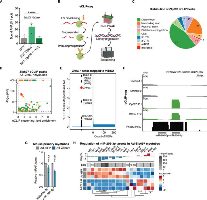
Muscular atrophy is a mortality risk factor that happens with disuse, chronic disease, and aging. Recovery from atrophy requires changes in several cell types including muscle fibers, and satellite and immune cells. Here we show that Zfp697/ZNF697 is a damage-induced regulator of muscle regeneration, during which its expression is transiently elevated. Conversely, sustained Zfp697 expression in mouse muscle leads to a gene expression signature of chemokine secretion, immune cell recruitment, and extracellular matrix remodeling. Myofiber-specific Zfp697 ablation hinders the inflammatory and regenerative response to muscle injury, compromising functional recovery. We uncover Zfp697 as an essential interferon gamma mediator in muscle cells, interacting primarily with ncRNAs such as the pro-regenerative miR-206. In sum, we identify Zfp697 as an integrator of cell-cell communication necessary for tissue regeneration.


Conflict of interest statement

Competing Interests: G.W.Y. is an SAB member of Jumpcode Genomics and a co-founder, member of the Board of Directors, on the SAB, equity holder, and paid consultant for Locanabio and Eclipse BioInnovations. G.W.Y. is a distinguished visiting professor at the National University of Singapore. G.W.Y.’s interests have been reviewed and approved by the University of California, San Diego in accordance with its conflict-of-interest policies.

Figures

Fig. 1.
Fig. 1.. Zfp697 expression increases during muscle recovery from atrophy.
(A) Overview of the mouse hindlimb unloading and reloading protocol. (B) Change in mouse gastrocnemius mass (normalized by tibia length) during hindlimb unloading and reloading (n = 6–18). One-way ANOVA with Tukey’s multiple comparisons test. (C) RNA-seq of mouse gastrocnemius muscle after 10 days of hindlimb unloading, 10 days of unloading followed by 1 day of reloading, and controls (n = 2–4 per condition). Heatmap highlights genes differentially expressed between unloading compared with control mice, and reloading compared with unloading. (D) Gene set enrichment analysis (GSEA) for hallmark pathways in mouse gastrocnemius after hindlimb unloading and reloading. FDR, false discovery rate. (E-F) MA plots of gene expression levels in mouse gastrocnemius after 10 days of unloading compared with control, and 1 day of reloading compared with 10 days of unloading. Red dots indicate genes with significantly increased expression, while blue dots indicate genes with significantly decreased expression (padj < 0.05). Known and putative transcriptional factors are highlighted. TF, transcription factor; padj, adjusted p-value. (G) Genome browser tracks of the mouse Zfp697 gene highlighting RNA-seq data for control, unloaded and reloaded gastrocnemius, and PhastCons60 vertebrate conservation score. (H) Schematic representation of the Zfp697 protein and its predicted glutamine-rich and zinc finger structural domains. The numbers shown refer to amino acid positions in the mouse Zfp697 protein. (I) Zfp697 gene expression in mouse gastrocnemius during hindlimb unloading and reloading (n = 6–8). One-way ANOVA with Tukey’s multiple comparisons test. Data represent mean values and error bars represent SEM.
Fig. 2.
Fig. 2.. Zfp697 is necessary for IFNγ signal transduction in myotubes.
(A) Zfp697 gene expression in mouse primary myotubes with Zfp697 gain- or loss-of-function. For gain-of-function experiments, fully differentiated myotubes were transduced with adenovirus expressing GFP alone (Ad-GFP) or together with Zfp697 (Ad-Zfp697; n = 4 independent experiments). For loss-of-function, cells were transduced with adenovirus expressing Zfp697-specific (Ad-shZfp697) or scrambled control shRNAs (Ad-shControl; n = 3 independent experiments). Two-tailed Student’s paired t test versus respective control. (B) Gene expression analysis of chemokines in mouse primary myotubes with Zfp697 gain- or loss-of-function. Two-tailed Student’s paired t test versus respective control. (C) Gene set enrichment analysis for hallmark pathways in mouse primary myotubes overexpressing Zfp697. FDR, false discovery rate. (D) MA plot showing mean gene expression versus log2 fold-change in mouse primary myotubes overexpressing Zfp697. Differentially expressed genes are shown in blue (padj < 0.05). Interferon gamma response genes are highlighted in red. padj, adjusted p-value. (E-F) Gene expression analysis of Zfp697, chemokines and interferon gamma (IFNγ) response genes in mouse primary myotubes after Zfp697 loss-of-function and treated with IFNγ or control (20 ng/mL for 8 hours; n = 3 independent experiments). Two-way ANOVA with Tukey’s multiple comparisons test. Data represent mean values and error bars represent SEM.
Fig. 3.
Fig. 3.. Zfp697 is an RNA-binding protein that interacts with ncRNAs and miRNAs.
(A) In vitro RNA-binding assay performed with GST alone (control), full-length Zfp697, and Zfp697 N-terminus (aa 1–192) lacking all the zinc finger motifs. Results are displayed as percentage of input recovered (n = 3–4). One-way ANOVA with Fisher’s LSD. (B) Overview of the enhanced UV crosslinking followed by immunoprecipitation (eCLIP) sequencing protocol. (C-D) Volcano plot (bottom left panel) and distribution (top right panel) of significantly enriched Zfp697 eCLIP peaks in mouse primary myotubes overexpressing Zfp697. Cutoffs of eCLIP IP/SMInput log2 fold enrichment and -log10 padj are denoted by dashed lines (padj < 0.001, fold enrichment > 8, IDR < 0.01). Color coding represents location of Zfp697 binding sites across genic regions. padj, adjusted p-value; IDR, Irreproducible Discovery Rate. (E) Proportion of significantly enriched binding sites mapped to miRNA for Zfp697 (red) compared to other RNA binding proteins (RBPs) with eCLIP data in ENCODE 3. (F) Genome browser tracks showing Zfp697 binding site at miR-206–3p. PhastCons60 represents vertebrate conservation score. SMInput, size-matched input; RPM, reads per million. (G) Expression of miR-206–3p and miR-206–5p in mouse primary myotubes overexpressing Zfp697 (n = 3 independent experiments). Two-tailed Student’s paired t test versus Ad-GFP. (H) Expression heatmap and log2 fold-change of miR-206–3p target genes in mouse primary myotubes overexpressing Zfp697 determined by RNA-seq. padj, adjusted p-value; ns, not significant.
Fig. 4.
Fig. 4.. Skeletal muscle-specific Zfp697 knockout blunts recovery from injury.
(A) Schematic representation of the strategy adopted to generate skeletal-muscle specific Zfp697 knockout mice (Zfp697 mKO) and flox controls. Red arrows indicate loxP sites location. (B) Experimental approach for hindlimb unloading and reloading of Zfp697 mKO and flox littermates. (C) Zfp697 gene expression in the gastrocnemius (left panel) and soleus (right panel) of Zfp697 mKO and flox littermates following hindlimb unloading and reloading. Two-tailed Student’s t test compared with time-matched flox controls. (D) Muscle mass change after 10 days of hindlimb unloading comparing Zfp697 mKO and flox littermates with pre-unloading genotype-matched controls (n = 6). Two-tailed Student’s t test. (E) Muscle mass change after 1 or 3 days of hindlimb reloading comparing Zfp697 mKO and flox littermates with genotype-matched 10 days of unloaded mice (n = 6). Two-way ANOVA with Šídák’s multiple comparisons test. (F) Change in body-weight-normalized grip strength following a single bout of strenuous downhill running in Zfp697-mKO mice and flox littermates (n = 10). Repeated measures two-way ANOVA with Šídák’s multiple comparisons test. (G) Gait analysis of Zfp697 mKO mice and floxed littermates (flox, n = 5; mKO, n = 4) at baseline and following chemically induced muscle injury. Cardiotoxin was intramuscularly injected in the gastrocnemius and tibialis anterior muscles of one leg (right) and control solution in the same site of contralateral leg (left). Repeated measures two-way ANOVA. Data represent mean values and error bars represent SEM.
Fig. 5.
Fig. 5.. Zfp697 mKOs fail to activate gene expression necessary for regeneration.
(A) Principal component analysis for RNA-seq performed in the gastrocnemius muscle of control Zfp697 flox and mKO, after 10 days of hindlimb unloading and 3 days of reloading (n = 3 per condition and genotype). (B) Heatmap of differentially expressed genes for the interaction effect between conditions and genotypes. (C) Gene set enrichment analysis for hallmark pathways accounting for the interaction effect between conditions (control, unloading and reloading) and genotypes (mKO and flox). FDR, false discovery rate. (D) Heatmap of genes belonging to inflammatory and interferon gamma response pathways. (E) Fraction of muscle-resident cell populations identified by digital cytometry (CIBERSORTx) applied to bulk muscle RNA-seq data from control, hindlimb unloaded and reloaded Zfp697 flox and mKO mice (n = 3 per condition and genotype). Two-way ANOVA with Šídák’s multiple comparisons test. *p<0.05 between indicated cell types. (F) Immunostaining for proliferating cells using KI67 in gastrocnemius muscles of Zfp697 flox and mKO and respective quantification (n = 4 per condition and genotype). Two-way ANOVA with Fisher’s LSD. P values represent comparison with time-matched flox controls. Scale bar = 500 µm.

References

    1. Chow L. S., Gerszten R. E., Taylor J. M., Pedersen B. K., van Praag H., Trappe S., Febbraio M. A., Galis Z. S., Gao Y., Haus J. M., Lanza I. R., Lavie C. J., Lee C.-H., Lucia A., Moro C., Pandey A., Robbins J. M., Stanford K. I., Thackray A. E., Villeda S., Watt M. J., Xia A., Zierath J. R., Goodpaster B. H., Snyder M. P., Exerkines in health, resilience and disease. Nat Rev Endocrinol. 18, 273–289 (2022). - PMC - PubMed
    1. Furrer R., Hawley J. A., Handschin C., The molecular athlete: exercise physiology from mechanisms to medals. Physiological Reviews (2023), doi:10.1152/physrev.00017.2022. - DOI - PMC - PubMed
    1. Hindi S. M., Millay D. P., All for One and One for All: Regenerating Skeletal Muscle. Cold Spring Harb Perspect Biol. 14, a040824 (2022). - PMC - PubMed
    1. Joe A. W. B., Yi L., Natarajan A., Le Grand F., So L., Wang J., Rudnicki M. A., Rossi F. M. V., Muscle injury activates resident fibro/adipogenic progenitors that facilitate myogenesis. Nat Cell Biol. 12, 153–163 (2010). - PMC - PubMed
    1. Bernard C., Zavoriti A., Pucelle Q., Chazaud B., Gondin J., Role of macrophages during skeletal muscle regeneration and hypertrophy-Implications for immunomodulatory strategies. Physiol Rep. 10, e15480 (2022). - PMC - PubMed

Publication types