[Preprint]. 2023 Jun 15:2023.06.15.544806.
doi: 10.1101/2023.06.15.544806.

Guanine-containing ssDNA and RNA induce dimeric and tetrameric SAMHD1 in cryo-EM and binding studies

Affiliations

Guanine-containing ssDNA and RNA induce dimeric and tetrameric SAMHD1 in cryo-EM and binding studies

Benjamin Orris et al. bioRxiv. .

Update in

Abstract

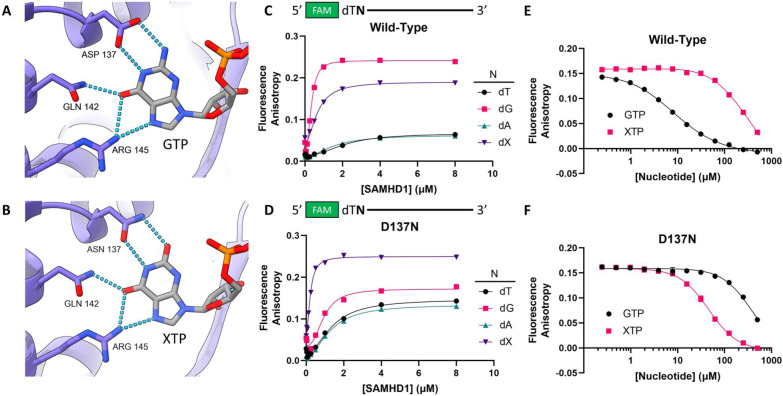
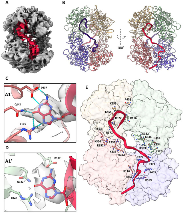
The dNTPase activity of tetrameric SAM and HD domain containing deoxynucleoside triphosphate triphosphohydrolase 1 (SAMHD1) plays a critical role in cellular dNTP regulation. SAMHD1 also associates with stalled DNA replication forks, DNA repair foci, ssRNA, and telomeres. The above functions require nucleic acid binding by SAMHD1, which may be modulated by its oligomeric state. Here we establish that the guanine-specific A1 activator site of each SAMHD1 monomer is used to target the enzyme to guanine nucleotides within single-stranded (ss) DNA and RNA. Remarkably, nucleic acid strands containing a single guanine base induce dimeric SAMHD1, while two or more guanines with ~20 nucleotide spacing induce a tetrameric form. A cryo-EM structure of ssRNA-bound tetrameric SAMHD1 shows how ssRNA strands bridge two SAMHD1 dimers and stabilize the structure. This ssRNA-bound tetramer is inactive with respect to dNTPase and RNase activity.


Conflict of interest statement

Conflict of interest statement M.W.S, Y.X., K.W.H, S.H., B.B., and D.S. are employees of Pfizer Inc. and may hold shares in the company.

Figures

Figure 1.
Figure 1.. A1 site confers binding specificity for guanine and xanthine bases in ssDNA.
(A) Guanine-specific hydrogen bonding network formed in the A1 site of wild-type SAMHD1 (PDB: 6TXC). (B) Xanthine-specific hydrogen bonding network formed in the A1 site of SAMHD1 D137N (PDB: 6TXA). (C) Binding of wild-type SAMHD1 to a series of 5’FAM-labeled 40mer ssDNA oligonucleotides (50 nM) consisting of a dT38 homopolymer with nucleotides (N) dT, dG, dA, or dX (deoxyxanthosine) in position two. Error bars indicate standard error of three independent replicate measurements at each SAMHD1 concentration. (D) Binding of SAMHD1 D137N to the same series of oligonucleotides as in panel C. Error bars indicate standard error of three independent replicate measurements at each SAMHD1 concentration. Error bars indicate standard error of three independent replicate measurements at each nucleotide concentration. (E) Displacement of the dG-containing 40mer (0.5 μM) from wild-type SAMHD1 (1 μM) with GTP (black) or XTP (xanthosine triphosphate, pink). (F) Displacement of the dX-containing 40mer (0.5 μM) from SAMHD1 D137N (1 μM) with GTP (black) or XTP (pink). Error bars indicate standard error of three independent replicate measurements at each nucleotide concentration.
Figure 2.
Figure 2.. dG positional and oligonucleotide length effects on binding.
(A) Binding isotherms of SAMHD1 to a series of 5′FAM-labeled ssDNA 40mers containing a single dG nucleotide in varying positions in a dT homopolymer backbone (50 nM each). (B) Plot of K0.5 (left) and maximum anisotropy (Amax, right) for the ssDNA binding isotherms in (A) (black) and the ssRNA binding isotherms in Supplemental Figure S4B (pink). (C) Binding of SAMHD1 to a series of 5′FAM-labeled ssDNA oligonucleotides consisting of a single dG base at the 5′ end followed by dT homopolymer of varying lengths (n = 9 to 29). (D) K0.5 (left) and maximum anisotropy (Amax, right) observed for the different homopolymer lengths used in panel (C). All Error bars for both K0.5 and Amax indicate standard errors as determined by least-squares regression fit to the Hill equation (eq 2).
Figure 3.
Figure 3.. Single G-bases near the 5’ end of ssDNA and ssRNA induce SAMHD1 dimerization.
All crosslinking experiments were carried out as follows: SAMHD1 (1 μM) was incubated under each condition for 10 minutes, then crosslinked with 50 mM glutaraldehyde. All gels included dedicated marker lanes for SAMHD1 alone, SAMHD1 and 50 μM GTP, and SAMHD1 and 100 μM dGTPαS to confirm monomer (M), dimer (D) and tetramer (T) species. (A) Oligomeric states of SAMHD1 induced by 1 μM 5′FAM-dTdNdT38 (where N = dT, dG or dA). (B) Binding of SAMHD1 to 5′FAM-dTdGdT38 (1 μM) indicates a stoichiometry of two SAMHD1 monomers per DNA strand. (C) Oligomeric states induced by 1 μM 5′FAM- UNU38 (where N = U, G or A). (D) Binding of SAMHD1 to 5′FAM-UGU38 (1 μM) indicates a stoichiometry of one SAMHD1 monomer per RNA strand. (E) Oligomeric states induced by 5’FAM-labeled dT homopolymer ssDNA 40mers (1 μM) as a function of the position of the dG base within the sequence. (F) Oligomeric states induced by dT homopolymers of increasing lengths with a single dG base in position 1 ([DNA] = 1 μM).
Figure 4.
Figure 4.. Two or more G bases in ssDNA and ssRNA with ~20 nt spacing induce SAMHD1 tetramerization.
GAXL crosslinking experiments were carried out as in Fig. 3. (A) Oligomeric states of SAMHD1 induced by ssDNA32. Lanes from left to right reflect serial two-fold dilutions in the range 10 to 0.3 μM. (B) Oligomeric states of SAMHD1 induced by ssRNA32. Lanes from left to right reflect serial two-fold dilutions of each NA in the range 10 to 0.31 μM. (C) Stoichiometric binding of SAMHD1 to ssDNA32 (1 μM). (D) Stoichiometric binding of SAMHD1 to ssRNA32. (E) Oligomeric states induced by a 40mer ssDNA with a single 5′ G base fixed in position 1 and a second G base positioned at the indicated spacings (n) (5′FAM-dGdTndGdTx). (F) Oligomeric states induced by a 40mer ssRNA with a single 5′ G base fixed in position 1 and a second G base positioned at the indicated spacings (n) (5′FAM-GUnGUx).
Figure 5.
Figure 5.. CryoEM analysis of ssRNA32 complexes with SAMHD1.
(A) Overview of particles observed in cryo-EM study and proposed mechanism of T* formation. Abbreviations: D (dimer), T*op, T*cl (open and closed conformational states of the ssRNA bound tetramer). For low resolution species, (D and D·D) SAMHD1 dimers were rigidly docked into the density using the fitmap command in ChimeraX. For both conformations of T*, density corresponding to RNA is colored violet. (B) Structure of the canonical dNTP-saturated tetramer induced by dGTPαS (T, PDB: 7UJN). (C) Dimer-dimer interface interactions in the T complex with dGTPαS. A charged hydrogen bonding network involving the interfacial residues D361, H364, and R372 is observed. (D) Structure of the ssRNA32-bound tetramer (T*cl). The RNA was deleted from this depiction of T*cl to facilitate comparisons between T and T*cl. (E) Dimer-dimer interface interactions in the T*cl. complex with ssRNA32. A significant displacement of the two interfacial helices disrupts the hydrogen bonding of residues D361, H364, and R372.
Figure 6.
Figure 6.. Molecular basis for ssRNA32 binding (T*cl).
(A) Smoothed density map of T*cl shown at low contour to highlight ssRNA connectivity between allosteric sites. Density corresponding to ssRNA32 is highlighted in pink. (B) Fragments of ssRNA32 can be posed in the tube density through molecular dynamics flexible fitting using ISOLDE positioning G8 and G25 in the A1 sites on each face of the tetramer. (C) A guanosine nucleotide modeled in the A1 site of chains A (purple) & B (pink), where it forms a hydrogen bond network with D137, Q142, and R145. (D) A guanosine nucleotide modeled in the A1 sites of chains C (green) and D (yellow) (denoted A1′). The guanosine binding pose is rotated as compared to panel (C) and does not form hydrogen bonds with D137, Q142, and R145. This difference in poses between A1 and A1′ sites may arise from the different 5’-3’ orientation of the RNA strand at the A1′ sites. (E) Cationic residues located near the dimer-dimer interface of T*. Residues K116, K332, R333, K336, R352, K354, R371, R372, K377, R451, K455, K523, K559 are positioned to interact with the flexible sugar-phosphate backbone of ssRNA32.
Figure 7.
Figure 7.. SAMHD1 interacts with long mRNA molecules as an ensemble of dimers and tetramers.
Transmission electron micrographs (TEM) shown at 150,000X magnification using a 1% uranyl formate negative stain. (A) TEM image of in vitro transcribed 2 kb RNA (10 ng/μL) alone (top) and in the presence of 100 nM SAMHD1 (bottom). The mixed dimeric (D) and tetrameric (T*) states of SAMHD1 observed in the images were confirmed by the GAXL method (gel lane on the right). (B) SAMHD1 (100 nM) bound to ssRNA32 (100 nM). The largely tetrameric (T*) state of SAMHD1 observed in the images was confirmed by the GAXL method (gel lane on the right). (C) Proposed model for SAMHD1 binding to long RNA involves G binding to A1 sites and the formation of enzyme dimers. Where the spacing of guanine residues is correct, dimeric units can coalesce to form tetramer.

References

    1. Morris E. R. et al. Crystal structures of SAMHD1 inhibitor complexes reveal the mechanism of water-mediated dNTP hydrolysis. Nat. Commun. 11, 3165 (2020). - PMC - PubMed
    1. Zhang Z. et al. Involvement of SAMHD1 in dNTP homeostasis and the maintenance of genomic integrity and oncotherapy (Review). Int. J. Oncol. 56, 879–888 (2020). - PubMed
    1. Hansen E. C., Seamon K. J., Cravens S. L. & Stivers J. T. GTP activator and dNTP substrates of HIV-1 restriction factor SAMHD1 generate a long-lived activated state. Proc. Natl. Acad. Sci. U. S. A. 111, E1843–51 (2014). - PMC - PubMed
    1. Ji X. et al. Mechanism of allosteric activation of SAMHD1 by dGTP. Nat. Struct. Mol. Biol. 20, 1304–1309 (2013). - PMC - PubMed
    1. Seamon K. J., Bumpus N. N. & Stivers J. T. Single-Stranded Nucleic Acids Bind to the Tetramer Interface of SAMHD1 and Prevent Formation of the Catalytic Homotetramer. Biochemistry 55, 6087–6099 (2016). - PMC - PubMed

Publication types