[Preprint]. 2023 Jun 1:2023.05.30.542928.
doi: 10.1101/2023.05.30.542928.

MFGE8 inhibits insulin signaling through PTP1B

Affiliations

MFGE8 inhibits insulin signaling through PTP1B

Ritwik Datta et al. bioRxiv. .

Update in

Abstract

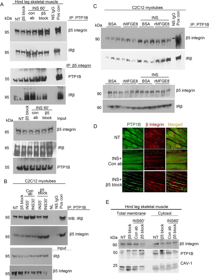
The role of integrins in regulating insulin signaling is incompletely understood. We have previously shown that binding of the integrin ligand milk fat globule epidermal growth factor like 8 (MFGE8) to the αvβ5 integrin promotes termination of insulin receptor signaling in mice. Upon ligation of MFGE8, β5 complexes with the insulin receptor beta (IRβ) in skeletal muscle resulting in dephosphorylation of IRβ and reduction of insulin-stimulated glucose uptake. Here we investigate the mechanism by which the interaction between β5 and IRβ impacts IRβ phosphorylation status. We show that β5 blockade inhibits and MFGE8 promotes PTP1B binding to and dephosphorylation of IRβ resulting in reduced or increased insulin-stimulated myotube glucose uptake respectively. The β5-PTP1B complex is recruited by MFGE8 to IRβ leading to termination of canonical insulin signaling. β5 blockade enhances insulin-stimulated glucose uptake in wild type but not Ptp1b KO mice indicating that PTP1B functions downstream of MFGE8 in modulating insulin receptor signaling. Furthermore, in a human cohort, we report serum MFGE8 levels correlate with indices of insulin resistance. These data provide mechanistic insights into the role of MFGE8 and β5 in regulating insulin signaling.


Conflict of interest statement

Conflict of interest None

Figures

Figure 1:
Figure 1:. β5 blockade reduces insulin-stimulated PTP1B phosphatase activity
(A,B) PTP1B phosphatase activity in the presence of β5 blocking or isotype control antibody in (A) skeletal muscle lysates harvested from WT mice 15 and 60 minutes after IP insulin (1U/kg) administration and (B) in Hela cells 30 minutes after EGF or insulin (100 nm) treatment. N=4 male mice in each group for panel A. Data merged from 2 independent experiments. N=3 independent experiments for panel B. Data are expressed as mean ± SEM; *p < 0.05, **p < 0.01, and analyzed by One-way ANOVA followed by Bonferroni’s post-test.
Figure 2:
Figure 2:. β5 complexes with PTP1B and IRβ
(A) Co-immunoprecipitation studies showing interaction of PTP1B with β5 and IRβ in hind leg skeletal muscle lysates from mice in presence of β5 blocking or isotype control antibody 60 minutes after IP insulin (1U/Kg) treatment. Blots represent 2 independent experiments with N=4 male mice for insulin-treated groups and 2 mice without insulin treatment. (B,C) Co-immunoprecipitation studies showing interaction of PTP1B with β5 and IRβ in C2C12 myotubes treated with (B) β5 blocking (5μg/mL) or isotype control antibody or (C) MFGE8 (10 μg/mL) or BSA control in the presence and absence of insulin for 30 minutes. N=4 male mice in total. Panel B represents 2 independent experiments. For panel C, cell culture, treatment and protein isolation performed on 3 different days. After immunoprecipitation, protein samples were run on the same gel for western blot. (D) Immunostaining of PTP1B (green) and β5 integrin (red) in skeletal muscle cross sections from mice treated with insulin for 60 minutes in presence of β5 blocking or isotype control antibody. Images are representative from 2 independent experiments. (E) Cell fractionation of insulin-treated skeletal muscle tissue samples from mice pretreated with β5 blocking or isotype control antibody and insulin for 60 minutes, followed by Western blot for PTP1B and β5 in the cytoplasmic and membrane fraction. Western blotting for CAVEOLIN-1 (CAV-1) confirmed enrichment of the membrane fractions. N=1 per group per experiment. Western blots are representative of 3 independent experiments.
Figure 3:
Figure 3:. β5 blockade activates canonical insulin signaling
(A) Representative western blot showing phosphorylated (serine 473 residue, S473) and total AKT levels in C2C12 myotubes in the presence of β5 blocking or isotype control antibody treated with insulin for 30 minutes. N=3 independent experiments. (B) Densitometric analysis of western blots from panel A. (C-D) Representative western blot showing phosphorylated AKT and total AKT levels in C2C12 myotubes treated with rMFGE8 (10μg/mL) and insulin for 20 minutes. N=6 independent samples from 2 independent experiments. (D) Densitometric analysis of western blots from panel C. (E) Representative western blot of phosphorylated and total AKT levels in skeletal muscle lysates from mice treated with insulin for 5, 15 and 30 minutes in presence of β5 blocking or isotype control antibody. N=1 male mouse per group per experiment with 3 independent experiment. (F) Densitometric analysis of western blots from panel E. (G) Effect of GLUT-1 inhibitor (1μM) or control peptide on 2NBDG uptake in C2C12 myotubes treated with β5 blocking or isotype control antibody after insulin stimulation. N=3 independent experiments. Data expressed as relative fold change to untreated cells (NT). (H) Effect of β5 blockade (5μg/mL) or rMFGE8 treatment (10 μg/mL) on 2NBDG uptake in 3T3 fibroblasts in the presence or absence of insulin. N=3 independent experiments. (I) Western blot showing GLUT-1 and GLUT-4 levels in cytosolic and membrane fractions isolated from C2C12 myotubes treated with insulin for 30 minutes in presence of β5 blocking or isotype control antibody. Na+K+ATPase and GAPDH were used as loading controls for membrane and cytosolic fractions respectively. The western blot represents 3 independent experiments. Data are expressed as mean ± SEM; *p < 0.05, **p < 0.01, ***p < 0.001, and analyzed by Oneway ANOVA followed by Bonferroni’s post-test.
Figure 4:
Figure 4:. β5 blockade impacts insulin signaling through PTP1B
(A) 2NBDG uptake assay in WT and Ptp1b KO myotubes in presence of β5 blocking or isotype control antibody and insulin. N=4 independent experiments. (B) Glucose tolerance test in 7 to 8-week-old male WT and Ptp1b KO mice after IP injection of β5 blocking or isotype control antibody. N=6 male mice per group from 2 independent experiments are presented. Data are expressed as mean ± SEM. Data in panel (A) were analyzed by One-way ANOVA followed by Bonferroni’s post-test. **p < 0.05. Data in panel (B) were analyzed by 2-way ANOVA followed by Tukey’s post-test. **p<0.01, *p < 0.05, when comparing WT+Con ab versus WT+β5 blockade groups; #p<0.05 when comparing WT+Con ab versus Ptp1bKO+Con ab groups.
Figure 5:
Figure 5:. Model depicting how MFGE8 impacts insulin signaling through PTP1B.
MFGE8 binding of the β5 integrin on the outer cell membrane leads to recruitment of the β5-PTP1B complexes to IRβ. PTP1B subsequently dephosphorylates IRβ leading to reduced translocation of GLUT4 to the cell surface and reduced glucose uptake in response to insulin.
Figure 6:
Figure 6:. Physiological regulation of PTP1B/β5 interaction
Co-immunoprecipitation studies showing interaction of PTP1B with β5 and IRβ in hind-leg skeletal muscle lysates from mice fasted for 16h or mice refed for 1h after a 16h fast. N=4 mice in each group.

References

    1. Khalifeh-Soltani A., Ha A., Podolsky M. J., McCarthy D. A., McKleroy W., Azary S., Sakuma S., Tharp K. M., Wu N., Yokosaki Y., Hart D., Stahl A., and Atabai K. (2016) alpha8beta1 integrin regulates nutrient absorption through an Mfge8-PTEN dependent mechanism. Elife 5 - PMC - PubMed
    1. Khalifeh-Soltani A., McKleroy W., Sakuma S., Cheung Y. Y., Tharp K., Qiu Y., Turner S. M., Chawla A., Stahl A., and Atabai K. (2014) Mfge8 promotes obesity by mediating the uptake of dietary fats and serum fatty acids. Nature medicine 20, 175–183 - PMC - PubMed
    1. Datta R., Gholampour M. A., Yang C. D., Volk R., Lin S., Podolsky M. J., Arnold T., Rieder F., Zaro B. W., Verzi M., Lehner R., Abumrad N., Lizama C. O., and Atabai K. (2023) MFGE8 links absorption of dietary fatty acids with catabolism of enterocyte lipid stores through HNF4gamma-dependent transcription of CES enzymes. Cell Rep 42, 112249. - PMC - PubMed
    1. Khalifeh-Soltani A., Gupta D., Ha A., Iqbal J., Hussain M., Podolsky M. J., and Atabai K. (2016) Mfge8 regulates enterocyte lipid storage by promoting enterocyte triglyceride hydrolase activity. JCI Insight 1, e87418. - PMC - PubMed
    1. Datta R., Lizama C. O., Soltani A. K., McKleroy W., Podolsky M. J., Yang C. D., Huynh T. L., Cautivo K. M., Wang B., Koliwad S. K., Abumrad N. A., and Atabai K. (2021) Autoregulation of insulin receptor signaling through MFGE8 and the alphavbeta5 integrin. Proceedings of the National Academy of Sciences of the United States of America 118 - PMC - PubMed

Publication types