[Preprint]. 2023 Jun 16:rs.3.rs-2997323.
doi: 10.21203/rs.3.rs-2997323/v1.

Transmembrane Stem Factor Nanodiscs Enhanced Revascularization in a Hind Limb Ischemia Model in Diabetic, Hyperlipidemic Rabbits

Affiliations

Transmembrane Stem Factor Nanodiscs Enhanced Revascularization in a Hind Limb Ischemia Model in Diabetic, Hyperlipidemic Rabbits

Eri Takematsu et al. Res Sq. .

Update in

Abstract

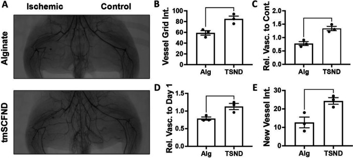
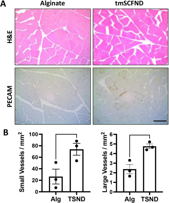
Therapies to revascularize ischemic tissue have long been a goal for the treatment of vascular disease and other disorders. Therapies using stem cell factor (SCF), also known as a c-Kit ligand, had great promise for treating ischemia for myocardial infarct and stroke, however clinical development for SCF was stopped due to toxic side effects including mast cell activation in patients. We recently developed a novel therapy using a transmembrane form of SCF (tmSCF) delivered in lipid nanodiscs. In previous studies, we demonstrated tmSCF nanodiscs were able to induce revascularization of ischemia limbs in mice and did not activate mast cells. To advance this therapeutic towards clinical application, we tested this therapy in an advanced model of hindlimb ischemia in rabbits with hyperlipidemia and diabetes. This model has therapeutic resistance to angiogenic therapies and maintains long term deficits in recovery from ischemic injury. We treated rabbits with local treatment with tmSCF nanodiscs or control solution delivered locally from an alginate gel delivered into the ischemic limb of the rabbits. After eight weeks, we found significantly higher vascularity in the tmSCF nanodisc-treated group in comparison to alginate treated control as quantified through angiography. Histological analysis also showed a significantly higher number of small and large blood vessels in the ischemic muscles of the tmSCF nanodisc treated group. Importantly, we did not observe inflammation or mast cell activation in the rabbits. Overall, this study supports the therapeutic potential of tmSCF nanodiscs for treating peripheral ischemia.

Keywords: Peripheral Ischemia; Peripheral Vascular Disease; Revascularization; Therapeutic Angiogenesis; diabetes; hyperlipidemia; nanodisc; rabbit ischemia model; transmembrane stem cell factor.


Conflict of interest statement

Competing interests The authors (ET and ABB) have patented the technology presented in this manuscript. The other authors (MM, GH, JG, PF, LM, GC, ADS, and RS) on the study do not have competing interests.

Figures

Figure 1
Figure 1. Transmembrane SCF nanodiscs enhance revascularization in the thigh of diabetic, hyperlipidemic rabbits with hindlimb ischemia.
(A) Angiograms of the thighs of the rabbits at week 10. The control limb is shown on the right and the limb with the ligated femoral artery is on the left. (B) Quantification of vessel grid intersections counted in the ischemic thigh at the model endpoint. The vessels are counted as number of intersections with an overlayed grid. (C) Relative vascularity of the ischemic thigh ratioed to the contralateral control thigh at the model endpoint. (D) Relative vascularity of the ischemic thigh ratioed to the thigh at day 1 prior to ligation. (E) Quantification of new vessels counted in the ischemic thigh at the model endpoint. *p < 0.05 vs. alginate; **p < 0.01 vs. alginate. (n=3).
Figure 2
Figure 2. tmSCFND enhances revascularization in the calf and foot of diabetic, hyperlipidemic rabbits with hindlimb ischemia.
(A) Angiograms of the lower limb of the rabbits at week 10. (B) Quantification of vessels counted in the ischemic calf and foot at the model endpoint. The vessels are counted as number of intersections with an overlayed grid. (C) Relative vascularity of the ischemic limb ratioed to the contralateral control limb at the model endpoint. (D) Relative vascularity of the ischemic calf and foot ratioed to the calf and foot at day 1 prior to ligation. *p < 0.05 versus alginate; **p < 0.05 versus alginate (n=3).
Figure 3
Figure 3. Histological analysis of vascularization of calf treated by tmSCF nanodiscs.
(A) Upper: H&E staining analysis of biopsies from the ischemic hind limbs of rabbits. Bottom: PECAM staining analysis of biopsies from the ischemic hind limbs of rabbits. Scale bar = 100 μm. (B) Left: Quantification of small vessels counted in the ischemic calf at week 10. Right: Quantification of arterioles vessels counted in the ischemic calf at week 10. *p < 0.05 versus alginate group (n = 3).

References

    1. Liang J, Wu Y-L, Chen B-J, Zhang W, Tanaka Y, Sugiyama H. The C-Kit Receptor-Mediated Signal Transduction and Tumor-Related Diseases. International Journal of Biological Sciences. 2013;9:435–443. doi: 10.7150/ijbs.6087 - DOI - PMC - PubMed
    1. Futrega K, King M, Lott WB, Doran MR. Treating the whole not the hole: necessary coupling of technologies for diabetic foot ulcer treatment. Trends in Molecular Medicine. 2014;20:137–142. doi: 10.1016/j.molmed.2013.12.004 - DOI - PubMed
    1. Rajendran P, Rengarajan T, Thangavel J, Nishigaki Y, Sakthisekaran D, Sethi G, Nishigaki I. The vascular endothelium and human diseases. International journal of biological sciences. 2013;9:1057–1069. doi: 10.7150/ijbs.7502 - DOI - PMC - PubMed
    1. Avogaro A, Albiero M, Menegazzo L, de Kreutzenberg S, Fadini GP. Endothelial Dysfunction in Diabetes. The role of reparatory mechanisms. 2011;34:S285–S290. doi: 10.2337/dc11-s239 %J Diabetes Care - DOI - PMC - PubMed
    1. Thiruvoipati T, Kielhorn CE, Armstrong EJ. Peripheral artery disease in patients with diabetes: Epidemiology, mechanisms, and outcomes. World journal of diabetes. 2015;6:961–969. doi: 10.4239/wjd.v6.i7.961 - DOI - PMC - PubMed

Publication types

LinkOut - more resources