[Preprint]. 2024 Mar 25:2023.06.03.542675.
doi: 10.1101/2023.06.03.542675.

Structural characterization of ligand binding and pH-specific enzymatic activity of mouse Acidic Mammalian Chitinase

Affiliations

Structural characterization of ligand binding and pH-specific enzymatic activity of mouse Acidic Mammalian Chitinase

Roberto Efraín Díaz et al. bioRxiv. .

Update in

Abstract

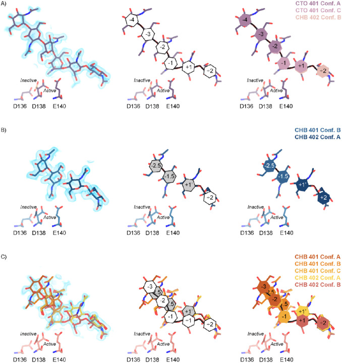
Chitin is an abundant biopolymer and pathogen-associated molecular pattern that stimulates a host innate immune response. Mammals express chitin-binding and chitin-degrading proteins to remove chitin from the body. One of these proteins, Acidic Mammalian Chitinase (AMCase), is an enzyme known for its ability to function under acidic conditions in the stomach but is also active in tissues with more neutral pHs, such as the lung. Here, we used a combination of biochemical, structural, and computational modeling approaches to examine how the mouse homolog (mAMCase) can act in both acidic and neutral environments. We measured kinetic properties of mAMCase activity across a broad pH range, quantifying its unusual dual activity optima at pH 2 and 7. We also solved high resolution crystal structures of mAMCase in complex with oligomeric GlcNAcn, the building block of chitin, where we identified extensive conformational ligand heterogeneity. Leveraging these data, we conducted molecular dynamics simulations that suggest how a key catalytic residue could be protonated via distinct mechanisms in each of the two environmental pH ranges. These results integrate structural, biochemical, and computational approaches to deliver a more complete understanding of the catalytic mechanism governing mAMCase activity at different pH. Engineering proteins with tunable pH optima may provide new opportunities to develop improved enzyme variants, including AMCase, for therapeutic purposes in chitin degradation.


Conflict of interest statement

Competing interests S.J.V.D. and R.M.L. are listed as inventors on a patent for the use of chitinases to treat fibrotic lung disease. S.J.V.D, R.M.L., and J.S.F. are listed as inventors on a patent for mutant chitinases with enhanced expression and activity.

Figures

Figure 1 |
Figure 1 |. Kinetic properties of mAMCase catalytic domain at various pH.
A) Chemical depiction of the conserved two-step mechanism where the glycosidic oxygen is protonated by an acidic residue and a nucleophile adds into the anomeric carbon leading to elimination of the hydrolyzed product. B) The rate of 4MU-chitobioside catalysis (1/sec) by mAMCase catalytic domain is plotted as a function of 4MU-chitobioside concentration (μM). Each data point represents n = 4 with error bars representing the standard deviation. Michaelis-Menten equation without substrate inhibition was used to estimate the kcat and KM from the initial rate of reaction at various substrate concentrations. C) The rate of substrate turnover (1/sec) by mAMCase catalytic domain is plotted as a function of pH. Error bars represent the 95% confidence interval. D) The Michaelis-Menten constant of mAMCase catalytic domain is plotted as a function of pH. Error bars represent the 95% confidence interval. E) The catalytic efficiency (kcat/KM) of mAMCase catalytic domain is plotted as a function of pH. F) Hypothetical catalytic activity modeled explained by a low pH mechanism (red), and high pH mechanism (blue) and their corresponding total activity (dashed line).
Figure 2 |
Figure 2 |. Schematic representation of sugar-binding subsites in mAMCase.
A) PDB ID: 8GCA, chain A. Stick representation of all GlcNAc2 sugar-binding events observed in n sugar-binding subsites with 2mFo-DFc map shown as a 1.2 Å contour (blue), the subsite nomenclature, and a schematic of alternative conformation ligand modeling. B) PDB ID: 8FRA, chain D. Stick representation of all GlcNAcn binding events observed in n+0.5 sugar-binding subsites with 2mFo-DFc map shown as a 1.2 Å contour (blue), the subsite nomenclature, and a schematic of alternative conformation ligand modeling. C) PDB ID: 8FR9, chain B. Stick representation of all GlcNAcn binding events observed in n and n+0.5 sugar-binding subsites with 2mFo-DFc map shown as a 1.2 Å contour (blue), the subsite nomenclature, and a schematic of alternative conformation ligand modeling.
Figure 3 |
Figure 3 |. Asp138 orientation correlates with ligand subsite occupancy.
A) PDB ID: 8FR9, chain B. Schematic of the alternative conformation ligand modeling. B) Linear correlation between sugar-binding subsite occupancy and Asp138 active conformation occupancy.
Figure 4 |
Figure 4 |. pKa of GH18 chitinases in the D2 inactive and active conformation.
A) PDB ID: 8GCA, chain A. Distribution of pKa across Asp136, Asp138, Glu140 of mAMCase structures in either Asp138 inactive or Asp138 active conformation. B) PDB ID: 3FXY, 3RM4, 3RM8, 3RME (inactive conformation); 2YBU, 3FY1 (active conformation). Distribution of pKa across Asp136, Asp138, Glu140 of hAMCase structures in either Asp138 inactive or Asp138 active conformation. C) PDB ID: 3ALF, 3AQU, 3FXY, 3RM4, 3RM8, 3RME (inactive conformation); 2UY2, 2UY3, 2YBU, 4HME, 4MNJ, 4R5E, 4TXE (active conformation). Distribution of pKa across the catalytic triad D1xD2xE of GH18 chitinases in either D2 inactive or active conformation.
Figure 5 |
Figure 5 |. Distribution of distances observed every 10 ps of each simulation and their respective time courses.
A) Asp138 χ1 angles over a 10 ns simulation. B) Representative minimum distance snapshots of structure during pH 6.5 inactive simulation (left), and pH 2.0 active simulation (right). C) Distribution of Asp138 χ1 angles over a 10 ns simulation.
Figure 6 |
Figure 6 |. Proposed model for ligand translocation towards the active site and ligand release post-catalysis.
A) PDB ID: 8GCA, chain A with no ligand (step 1); with GlcNAc4 generated by phenix.elbow using PubChem ID: 10985690 (step 2); with GlcNAc6 generated by phenix.elbow using PubChem ID: 6918014 (step 3–4, 8); with oxazolinium intermediate generated by phenix.elbow using PubChem ID: 25260046 (steps 5.1–5.2); with GlcNAc2 and GlcNAc4 generated by phenix.elbow using PubChem ID: 439544 and 10985690, respectively (steps 6–7). Chemical representation of GH18 catalytic cycle with corresponding molecular models of each step. Catalytic residues Asp136, Asp138, Glu140, and ligands are shown as sticks. Protons are shown as gray spheres. B) PDB ID: 8GCA, chain A. Animated movie of the mAMcase catalytic cycle at pH 2.0 (separate file) and C) at pH 6.5 (separate file). Catalytic residues Asp136, Asp138, Glu140, and ligands are shown as sticks. Protons are shown as gray spheres.

References

    1. Cabib E. & Bowers B. Timing and function of chitin synthesis in yeast. J. Bacteriol. 124, 1586–1593 (1975). - PMC - PubMed
    1. Zhu K. Y., Merzendorfer H., Zhang W., Zhang J. & Muthukrishnan S. Biosynthesis, turnover, and functions of chitin in insects. Annu. Rev. Entomol. 61, 177–196 (2016). - PubMed
    1. Tang W. J., Fernandez J., Sohn J. J. & Amemiya C. T. Chitin is endogenously produced in vertebrates. Curr. Biol. 25, 897–900 (2015). - PMC - PubMed
    1. Elieh Ali Komi D., Sharma L. & Dela Cruz C. S. Chitin and Its Effects on Inflammatory and Immune Responses. Clin. Rev. Allergy Immunol. 54, 213–223 (2018). - PMC - PubMed
    1. Van Dyken S. J. et al. Spontaneous Chitin Accumulation in Airways and Age-Related Fibrotic Lung Disease. Cell 169, 497–509.e13 (2017). - PMC - PubMed

Publication types