

Primary Sclerosing Encapsulating Peritonitis (PSEP) With Meckel's Diverticulum: A Rare Case Report
- PMID: 37398790
- PMCID: PMC10311126
- DOI: 10.7759/cureus.39756
Primary Sclerosing Encapsulating Peritonitis (PSEP) With Meckel's Diverticulum: A Rare Case Report
Abstract
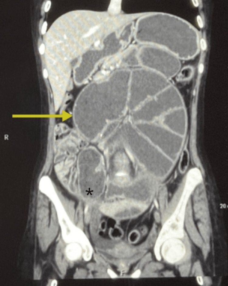
Sclerosing encapsulating peritonitis (SEP) is a rare disease. Preoperative diagnosis of SEP can be made with imaging, such as computed tomography (CT). SEP is characterized by a partial or complete encasement of the small intestine by a layer of a thick grayish-white fibro collagenous membrane similar to an abdominal cocoon. The most common symptoms of SEP are abdominal pain, nausea, and vomiting. This rare disease often leads to acute or sub-acute intestinal obstruction. We discuss, in this report, how we managed a case of primary sclerosing encapsulating peritonitis with Meckel's diverticulum at our institution.
Keywords: abdominal cocoon; abdominal pain; ascites; intestinal obstruction; meckel´s diverticulum; sclerosing encapsulating peritonitis; small intestine.
Copyright © 2023, Chaudhary et al.
Conflict of interest statement
The authors have declared that no competing interests exist.
Figures


References
-
- Imaging features of encapsulating peritoneal sclerosis in continuous ambulatory peritoneal dialysis patients. Ti JP, Al-Aradi A, Conlon PJ, Lee MJ, Morrin MM. AJR Am J Roentgenol. 2010;195:0–4. - PubMed
-
- Peritonitis chronica fibrosa incapsulata. Owtschinnikow PJ. Arch fu¨r Klin Chir. 1907;83:623–634.
-
- Unusual small intestinal obstruction in adolescent girls: the abdominal cocoon. Foo KT, Ng KC, Rauff A, Foong WC, Sinniah R. Br J Surg. 1978;65:427–430. - PubMed
-
- Length of time on peritoneal dialysis and encapsulating peritoneal sclerosis — position paper for ISPD: 2017 update. Brown EA, Bargman J, van Biesen W, et al. Perit Dial Int. 2017;37:362–374. - PubMed
Publication types
LinkOut - more resources
Full Text Sources