. 2023 Sep 1;80(9):891-902.
doi: 10.1001/jamaneurol.2023.1988.

Mapping Lesion-Related Epilepsy to a Human Brain Network

Affiliations

Mapping Lesion-Related Epilepsy to a Human Brain Network

Frederic L W V J Schaper et al. JAMA Neurol. .

Abstract

Importance: It remains unclear why lesions in some locations cause epilepsy while others do not. Identifying the brain regions or networks associated with epilepsy by mapping these lesions could inform prognosis and guide interventions.

Objective: To assess whether lesion locations associated with epilepsy map to specific brain regions and networks.

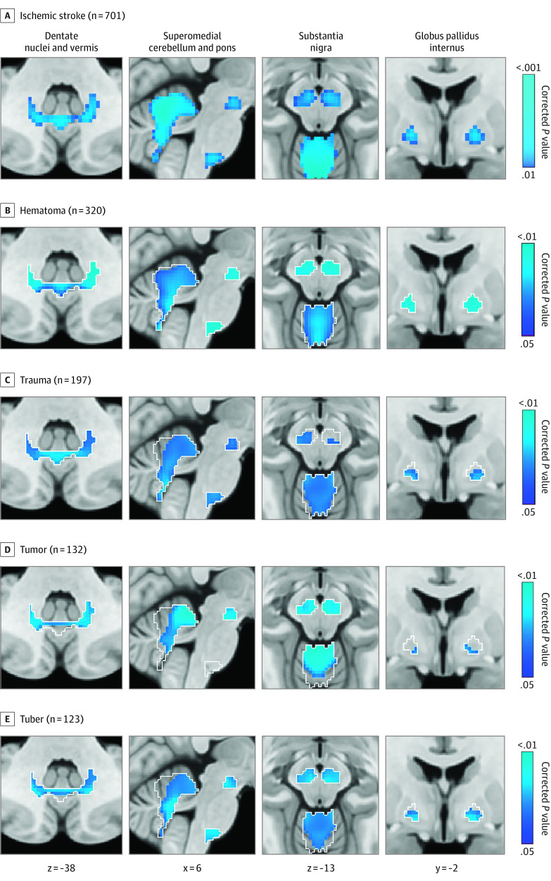
Design, setting, and participants: This case-control study used lesion location and lesion network mapping to identify the brain regions and networks associated with epilepsy in a discovery data set of patients with poststroke epilepsy and control patients with stroke. Patients with stroke lesions and epilepsy (n = 76) or no epilepsy (n = 625) were included. Generalizability to other lesion types was assessed using 4 independent cohorts as validation data sets. The total numbers of patients across all datasets (both discovery and validation datasets) were 347 with epilepsy and 1126 without. Therapeutic relevance was assessed using deep brain stimulation sites that improve seizure control. Data were analyzed from September 2018 through December 2022. All shared patient data were analyzed and included; no patients were excluded.

Main outcomes and measures: Epilepsy or no epilepsy.

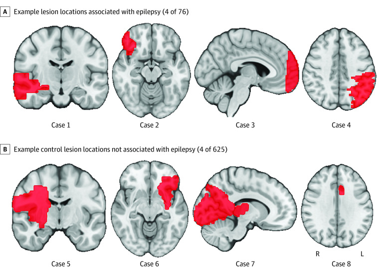
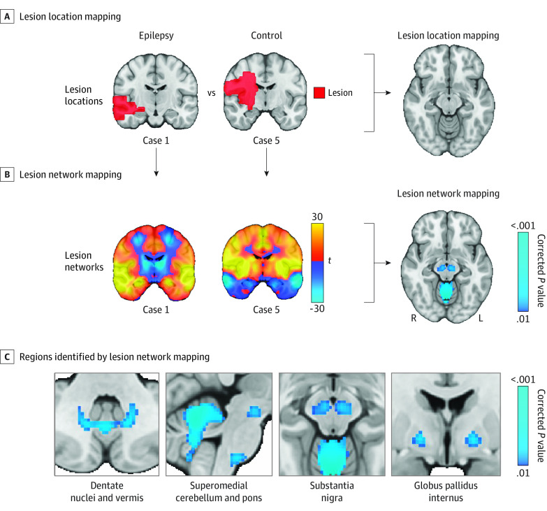
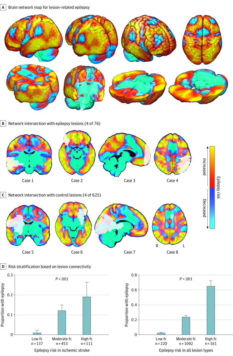
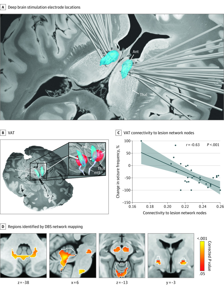
Results: Lesion locations from 76 patients with poststroke epilepsy (39 [51%] male; mean [SD] age, 61.0 [14.6] years; mean [SD] follow-up, 6.7 [2.0] years) and 625 control patients with stroke (366 [59%] male; mean [SD] age, 62.0 [14.1] years; follow-up range, 3-12 months) were included in the discovery data set. Lesions associated with epilepsy occurred in multiple heterogenous locations spanning different lobes and vascular territories. However, these same lesion locations were part of a specific brain network defined by functional connectivity to the basal ganglia and cerebellum. Findings were validated in 4 independent cohorts including 772 patients with brain lesions (271 [35%] with epilepsy; 515 [67%] male; median [IQR] age, 60 [50-70] years; follow-up range, 3-35 years). Lesion connectivity to this brain network was associated with increased risk of epilepsy after stroke (odds ratio [OR], 2.82; 95% CI, 2.02-4.10; P < .001) and across different lesion types (OR, 2.85; 95% CI, 2.23-3.69; P < .001). Deep brain stimulation site connectivity to this same network was associated with improved seizure control (r, 0.63; P < .001) in 30 patients with drug-resistant epilepsy (21 [70%] male; median [IQR] age, 39 [32-46] years; median [IQR] follow-up, 24 [16-30] months).

Conclusions and relevance: The findings in this study indicate that lesion-related epilepsy mapped to a human brain network, which could help identify patients at risk of epilepsy after a brain lesion and guide brain stimulation therapies.


Conflict of interest statement

Conflict of Interest Disclosures: Dr Schaper reported grants from the American Epilepsy Society, the National Institute of Neurological Disorders and Stroke, the Royal Netherlands Academy of Arts and Sciences, the Dr Jan Meerwaldt Stichting, and Stichting De Drie Lichten during the conduct of the study. Dr Horn reported personal fees from Boston Scientific outside the submitted work. Dr Siddiqi reported consultant fees from Magnus Medical Scientific, Acacia Mental Health, and Kaizen Brain Center; speaker fees from Brainsway; and investigator-initiated funding from Neuronetics during the conduct of the study and owns patents on using brain connectivity to guide brain stimulation. Dr Rheims reported consulting or speaker fees from UCB Pharma, Eisai, Angelini, Jazz Pharmaceuticals, Zogenix, GW Pharmaceuticals, Idiorsia, Livanova, and Arvelle Therapeutics. Dr Guénot reported personal fees from Dixi Medical outside the submitted work. Dr Colon has received speaker honoraria from Medtronic. Dr Bubrick reported personal fees from uniQure outside the submitted work. Dr Peters reported personal fees from Neurelis outside the submitted work. Dr Wu reported US patent 7 512 435 with royalties paid from General Electric, Siemens, Imaging Biometrics, and Olea Medical and US patent for 11 436 732 issued for automatic segmentation of acute ischemic stroke lesions in computed tomography data. Dr Joutsa reported grants from Finnish Medical Foundation, Instrumentarium Research Foundation, and Turku University Hospital (ERVA funds) during the conduct of the study; grants from Finnish Foundation for Alcohol Studies, Sigrid Juselius Foundation, and University of Turku (private donation) outside the submitted work; speaker honoraria from Lundbeck and Novartis; and conference travel support from AbbVie and Abbott. Dr Fox reported grants from National Institute of Neurological Disorders and Stroke during the conduct of the study and personal fees from Magnus Medical, Solterix; nonfinancial support from Boston Scientific; and grants from National Institute of Mental Health, National Institute on Aging, Ellison-Baszucki Family Foundation, Kaye Family Research Endowment, and Manley family outside the submitted work; in addition, Dr Fox had a patent for use of brain connectivity imaging to guide brain stimulation issued with no royalties and a patent for lesion network mapping pending with no royalties. No other disclosures were reported.

Figures

Figure 1.
Figure 1.. Lesion Locations
Brain slices are shown in radiological orientation.
Figure 2.
Figure 2.. Lesion Location and Network Mapping
A, Lesion location mapping methods were not able to identify associations between damage to a specific brain region and epilepsy. B, Lesion network mapping was then performed, which computes the functional connectivity between each lesion location (red) and all other brain voxels, using the resting-state functional connectivity data from 1000 healthy participants (ie, the human connectome). C, Lesion network mapping identified regions in the basal ganglia and cerebellum (ie, lesion network nodes) that were more negatively connected to lesion locations associated with epilepsy vs control lesions; 2-sided P values are shown after familywise error rate correction for multiple testing. Brain slices are shown in radiological orientation.
Figure 3.
Figure 3.. Generalizability to Other Lesion Types
Lesion network nodes in the basal ganglia and cerebellum derived from ischemic stroke data (A) were used as an a priori search space (white outlines) to test for similar findings in four validation data sets with different lesion etiologies (B). Negative functional connectivity to voxels in the basal ganglia and cerebellum was significantly associated with epilepsy in hematomas, traumas, tumors, and tubers. One-sided P values are shown after false discovery rate correction for multiple testing. Brain slices are shown in radiological orientation.
Figure 4.
Figure 4.. Relevance for Estimating Epilepsy Risk
A, Functional connectivity (fc) with the lesion network nodes in the basal ganglia and cerebellum (Figure 2C) defines a distributed brain network map of areas at increased risk or decreased risk of epilepsy when lesioned. Regions of increased risk in this network include the temporal lobe, parietal lobe, areas around the central sulcus, and CA1 region of the hippocampus. Regions of decreased risk include the supplementary motor area, anterior cingulate, and subcortical regions. To illustrate this finding, we show the same lesion locations from Figure 1 (white outlines), now overlaid on our network map, including 5 representative lesions associated with epilepsy (B) and 5 lesions not associated with epilepsy (C). Note that the lesions associated with epilepsy intersect areas of high risk compared to lesions not associated with epilepsy. D, Patients were stratified into 3 risk groups based on intersection of their lesion location with this network, using leave-one–data set–out analysis. More patients in the high-fc group had epilepsy compared to patients in the low-fc group both for ischemic stroke and across all lesion types.
Figure 5.
Figure 5.. Relevance for Deep Brain Stimulation (DBS) in Epilepsy
A, DBS electrodes from 30 patients with drug-resistant epilepsy show slight variability in electrode location within the anterior thalamus. B, The stimulation site for each patient was identified by computing the volume of activated tissue based on individualized stimulation settings. C, Functional connectivity between patient-specific stimulation sites and the lesion network nodes in the basal ganglia and cerebellum was associated with better seizure outcome. D, Positive functional connectivity between patient-specific stimulation sites and multiple voxels within the lesion network nodes (white outlines) was significantly associated with therapeutic response after deep brain stimulation. One-sided P values are shown after false discovery rate correction for multiple testing. Brain slices are shown in radiological orientation. Ant indicates anterior nucleus of the thalamus; mtt, mammillothalamic tract; Thal, thalamus; VAT, volume of activated tissue.

Comment in

References

    1. Beghi E, Giussani G, Abd-Allah F, et al. ; GBD 2016 Epilepsy Collaborators . Global, regional, and national burden of epilepsy, 1990-2016: a systematic analysis for the Global Burden of Disease Study 2016. Lancet Neurol. 2019;18(4):357-375. doi:10.1016/S1474-4422(18)30454-X - DOI - PMC - PubMed
    1. Pitkänen A, Roivainen R, Lukasiuk K. Development of epilepsy after ischaemic stroke. Lancet Neurol. 2016;15(2):185-197. doi:10.1016/S1474-4422(15)00248-3 - DOI - PubMed
    1. Galovic M, Döhler N, Erdélyi-Canavese B, et al. . Prediction of late seizures after ischaemic stroke with a novel prognostic model (the SeLECT score): a multivariable prediction model development and validation study. Lancet Neurol. 2018;17(2):143-152. doi:10.1016/S1474-4422(17)30404-0 - DOI - PubMed
    1. Siddiqi SH, Kording KP, Parvizi J, Fox MD. Causal mapping of human brain function. Nat Rev Neurosci. 2022;23(6):361-375. doi:10.1038/s41583-022-00583-8 - DOI - PMC - PubMed
    1. Hariz M, Lees AJ, Blomstedt Y, Blomstedt P. Serendipity and observations in functional neurosurgery: from James Parkinson’s stroke to Hamani’s & Lozano’s flashbacks. Stereotact Funct Neurosurg. 2022;100(4):201-209. doi:10.1159/000525794 - DOI - PubMed

Publication types