. 2023 Sep;33(5):e13185.
doi: 10.1111/bpa.13185. Epub 2023 Jul 3.

Recurrent TRAK1::RAF1 Fusions in pediatric low-grade gliomas

Affiliations

Recurrent TRAK1::RAF1 Fusions in pediatric low-grade gliomas

Jamal K Benhamida et al. Brain Pathol. 2023 Sep.

Abstract

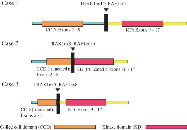
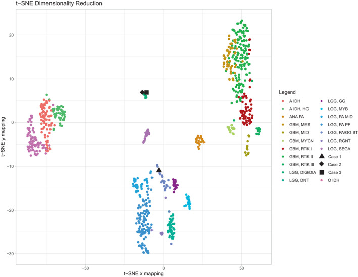
Fusions involving CRAF (RAF1) are infrequent oncogenic drivers in pediatric low-grade gliomas, rarely identified in tumors bearing features of pilocytic astrocytoma, and involving a limited number of known fusion partners. We describe recurrent TRAK1::RAF1 fusions, previously unreported in brain tumors, in three pediatric patients with low-grade glial-glioneuronal tumors. We present the associated clinical, histopathologic and molecular features. Patients were all female, aged 8 years, 15 months, and 10 months at diagnosis. All tumors were located in the cerebral hemispheres and predominantly cortical, with leptomeningeal involvement in 2/3 patients. Similar to previously described activating RAF1 fusions, the breakpoints in RAF1 all occurred 5' of the kinase domain, while the breakpoints in the 3' partner preserved the N-terminal kinesin-interacting domain and coiled-coil motifs of TRAK1. Two of the three cases demonstrated methylation profiles (v12.5) compatible with desmoplastic infantile ganglioglioma (DIG)/desmoplastic infantile astrocytoma (DIA) and have remained clinically stable and without disease progression/recurrence after resection. The remaining tumor was non-classifiable; with focal recurrence 14 months after initial resection; the patient remains symptom free and without further recurrence/progression (5 months post re-resection and 19 months from initial diagnosis). Our report expands the landscape of oncogenic RAF1 fusions in pediatric gliomas, which will help to further refine tumor classification and guide management of patients with these alterations.

Keywords: CRAF; RAF1; brain pathology; glioneuronal tumors; low-grade gliomas.


Conflict of interest statement

David Zagzag and Marc K. Rosenblum are members of the Editorial Board of Brain Pathology and were not involved in peer review of this submission.

Figures

FIGURE 1
FIGURE 1
MRI findings in TRAK1::RAF1 fusion positive gliomas: T1‐weighted, post‐contrast MRI demonstrates a hypointense, well‐circumscribed 1 cm ovoid lesion with heterogenous rim‐like enhancement, cortically‐based in the left inferotemporal lobe in Case 1 (A); after gross total resection, an area of focal nodular enhancement subjacent to the surgical site was noted on 14‐month surveillance scan (B). Tumors in Case 2 (C) and Case 3 (D) involved both brain and leptomeninges. T1‐weighted, post‐contrast MRI in Case 2 demonstrated a 3 cm solid and cystic enhancing mass with significant surrounding edema (C), while T1‐weighted, post‐contrast MRI in Case 3 demonstrated an infiltrative lesion in the R parieto‐occipital lobe with thick nodular leptomeningeal enhancement and ~4 cm non‐enhancing cystic‐solid component (D).
FIGURE 2
FIGURE 2
Histologic features of TRAK1::RAF1 fusion positive gliomas: The tumor in Case 1 demonstrated features compatible with a low grade glial‐glioneuronal tumor, comprised of bland astrocytic cells with ovoid nuclei and occasional perinuclear halos (A) . While generally well‐circumscribed, a slightly infiltrative border was noted (B). The tumors in Cases 2 and 3 demonstrated features compatible with diffuse leptomeningeal glioneuronal tumor, involving both leptomeninges and superficial cortex. In Case 2, tumor cells consisted of intermixed nests of gemistoytic astrocytes displaying plump, eosinophilic cytoplasm, and occasional ganglion‐like morphology (C). Tumors in Cases 2 and 3 demonstrated a desmoplastic, dense collagenous matrix with storiform spindled growth pattern (D [Case 2]; E [Case 3]), and in both cases a second population of more primitive‐appearing neurocyte‐like cells was present (F). Ki‐67 proliferative index was moderately elevated in Cases 1 (G) and 3 (H).
FIGURE 3
FIGURE 3
Features of recurrent TRAK1::RAF1 fusions in pediatric low grade glial‐glioneuronal tumors: Schematics showing the intrachromosomal rearrangement event on chromosome 3p leading to fusions TRAK1 (3p22.1) and RAF1 (3p25.2) in all 3 cases. Breakpoints in RAF1 (NM_002880) all occurred 5′ of the kinase domain (exons 7, 10 and 8 in Cases 1, 2, and 3 respectively). TRAK1 contains an N‐terminal kinesin‐interacting conserved region with three coiled‐coil domains, expected to facilitate homodimerization, and preserved by the TRAK1 breakpoints identified here (exon 15 [NM_001042646], exon 8 [NM_014965.4], and exon 5[NM_001042646]).
FIGURE 4
FIGURE 4
DNA methylation‐based classification of TRAK1::RAF1 fusion positive pediatric low grade glial‐glioneuronal tumors: T‐SNE dimensionality reduction was performed on the tumors from Case 1 (▲), Case 2 (♦), Case 3 (■), and a subset of CNS tumors from the DKFZ reference set (color coded according to methylation cluster assignation) with the Rtsne package version 0.15. Analyses were performed using the mnp.v11b6 package in R version 3.6.1; as previously described (PMID: 29539639).

References

    1. Ellison DW, Hawkins C, Jones DTW, Onar‐Thomas A, Pfister SM, Reifenberger G, et al. cIMPACT‐NOW update 4: diffuse gliomas characterized by MYB, MYBL1, or FGFR1 alterations or BRAF(V600E) mutation. Acta Neuropathol. 2019;137(4):683–7. - PubMed
    1. Ryall S, Tabori U, Hawkins C. Pediatric low‐grade glioma in the era of molecular diagnostics. Acta Neuropathol Commun. 2020;8(1):30. - PMC - PubMed
    1. Zhang J, Wu G, Miller CP, Tatevossian RG, Dalton JD, Tang B, et al. Whole‐genome sequencing identifies genetic alterations in pediatric low‐grade gliomas. Nat Genet. 2013;45(6):602–12. - PMC - PubMed
    1. Louis DN, Ohgaki H, Wiestler OD, Cavenee WK, Burger PC, Jouvet A, et al. The 2007 WHO classification of tumours of the central nervous system. Acta Neuropathol. 2007;114(2):97–109. - PMC - PubMed
    1. Lind KT, Chatwin HV, DeSisto J, Coleman P, Sanford B, Donson AM, et al. Novel RAF fusions in pediatric low‐grade gliomas demonstrate MAPK pathway activation. J Neuropathol Exp Neurol. 2021;80(12):1099–107. - PubMed

Publication types

Substances