. 2024 Jan 19;61(2):117-124.
doi: 10.1136/jmg-2023-109264.

SMARCA4 mutation causes human otosclerosis and a similar phenotype in mice

Affiliations

SMARCA4 mutation causes human otosclerosis and a similar phenotype in mice

Max Drabkin et al. J Med Genet. .

Abstract

Background: Otosclerosis is a common cause of adult-onset progressive hearing loss, affecting 0.3%-0.4% of the population. It results from dysregulation of bone homeostasis in the otic capsule, most commonly leading to fixation of the stapes bone, impairing sound conduction through the middle ear. Otosclerosis has a well-known genetic predisposition including familial cases with apparent autosomal dominant mode of inheritance. While linkage analysis and genome-wide association studies suggested an association with several genomic loci and with genes encoding structural proteins involved in bone formation or metabolism, the molecular genetic pathophysiology of human otosclerosis is yet mostly unknown.

Methods: Whole-exome sequencing, linkage analysis, generation of CRISPR mutant mice, hearing tests and micro-CT.

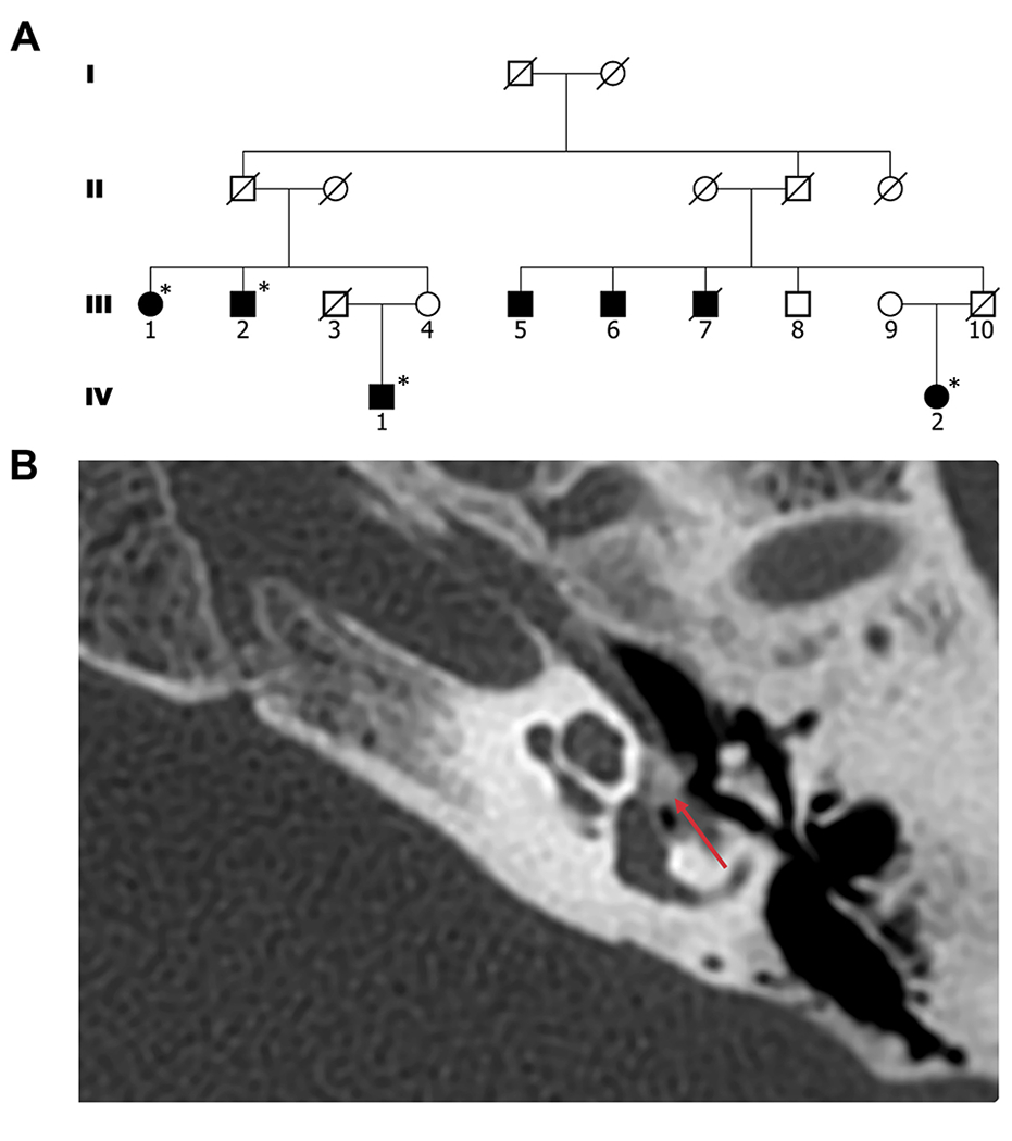
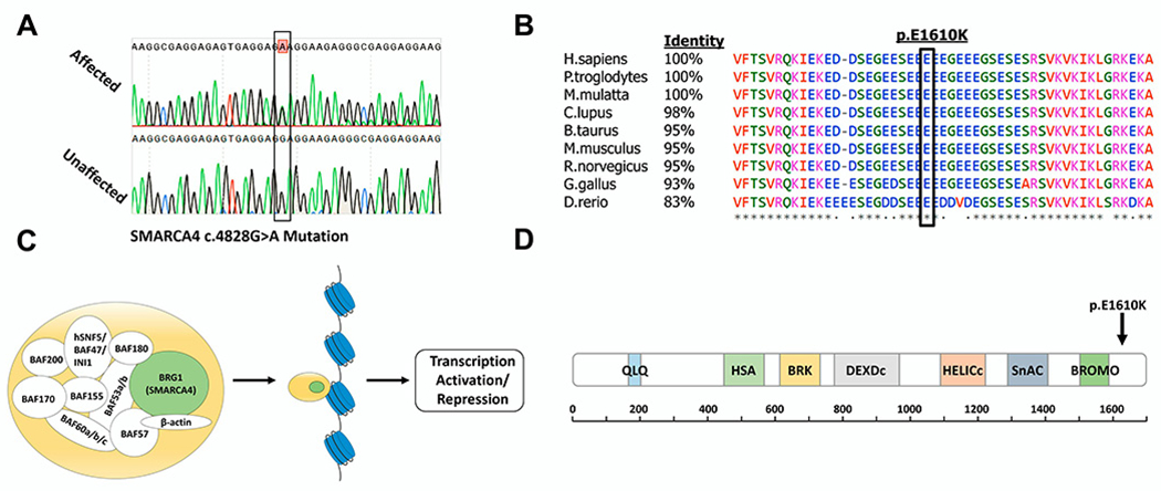
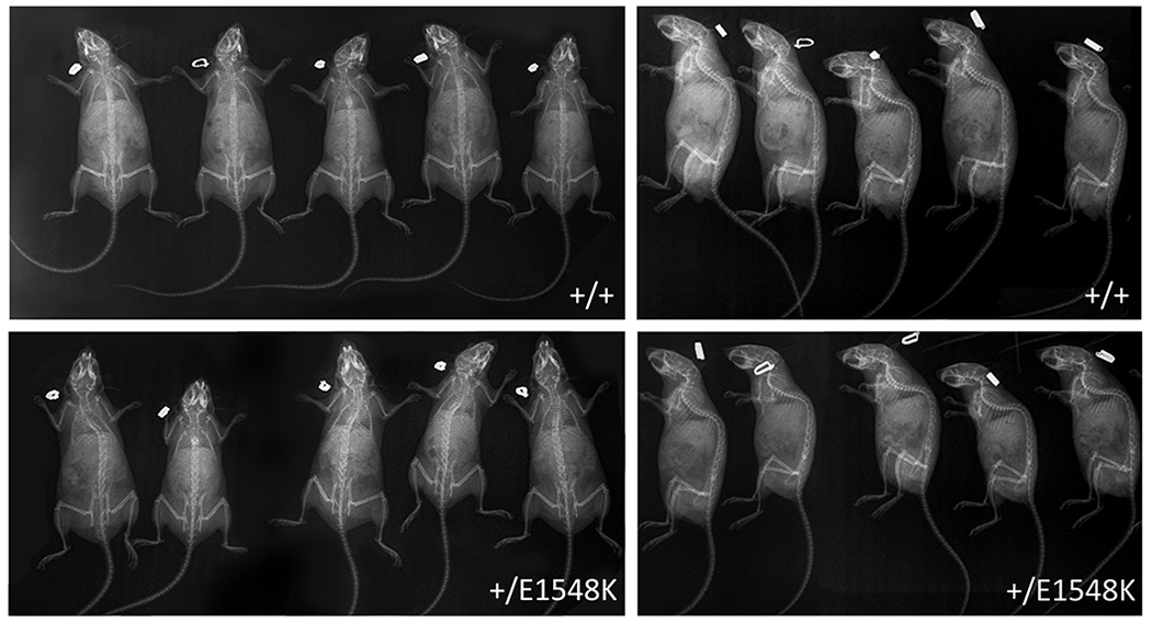
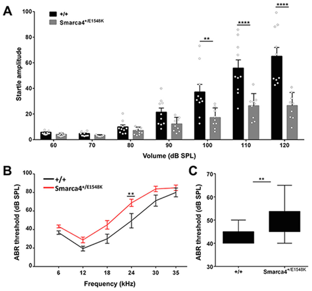
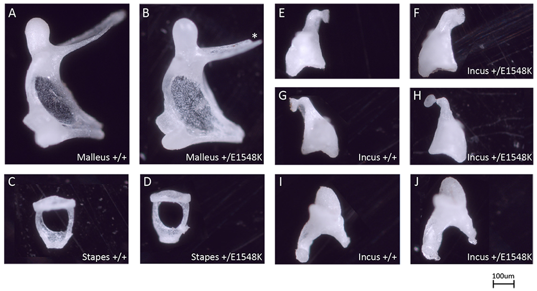
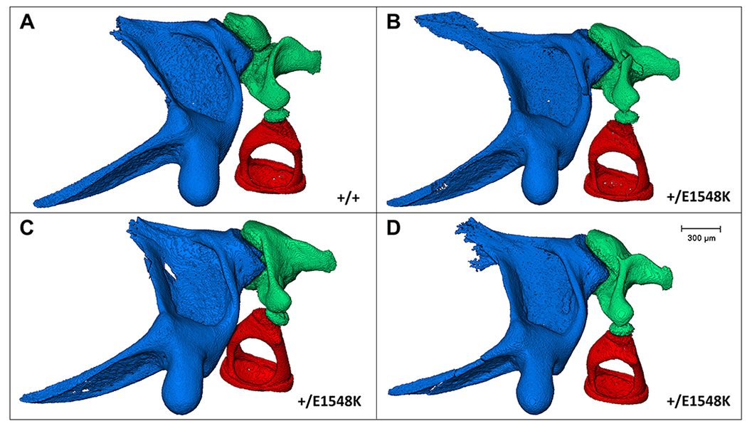
Results: Through genetic studies of kindred with seven individuals affected by apparent autosomal dominant otosclerosis, we identified a disease-causing variant in SMARCA4, encoding a key component of the PBAF chromatin remodelling complex. We generated CRISPR-Cas9 transgenic mice carrying the human mutation in the mouse SMARCA4 orthologue. Mutant Smarca4+/E1548K mice exhibited marked hearing impairment demonstrated through acoustic startle response and auditory brainstem response tests. Isolated ossicles of the auditory bullae of mutant mice exhibited a highly irregular structure of the incus bone, and their in situ micro-CT studies demonstrated the anomalous structure of the incus bone, causing disruption in the ossicular chain.

Conclusion: We demonstrate that otosclerosis can be caused by a variant in SMARCA4, with a similar phenotype of hearing impairment and abnormal bone formation in the auditory bullae in transgenic mice carrying the human mutation in the mouse SMARCA4 orthologue.

Keywords: genetics; human genetics; molecular biology; mutation, missense; phenotype.


Conflict of interest statement

Competing interests: None declared.

Figures

Figure 1.
Figure 1.. Pedigree and Cranial CT scan.
(A) The studied kindred: black-filled circles and squares denote affected individuals; * denotes affected individuals with confirmed otosclerosis diagnosed by surgery or computerized tomography; white circles and squares denote unaffected individuals. (B) Cranial CT scan of patient IV:2, showing the left temporal bone; red arrow indicates an otosclerosis focus.
Figure 2.
Figure 2.. Validation, conservation, and position of the SMARCA4 mutation.
(A) Sanger sequencing results of an affected individual heterozygous for the SMARCA4 mutation and an unaffected individual with the wild-type allele. (B) Multiple sequence alignment of SMARCA4 orthologues with percentage of protein sequence identity between the human protein and several mammalian proteins. Black box marks the position of the Glutamate residue mutated to Lysine. Symbols below sequences correspond to degree of conservation: asterisk indicates identical; period indicates weakly similar; blank indicates no similarity. (C) Illustration of the various subunits composing the PBAF complex[28], a member of the human SWI/SNF family of chromatin remodelers. The catalytic subunit of the complex, BRG, is encoded by SMARCA4 (highlighted in green). (D) Schematic representation of BRG1 showing all conserved domains of the protein, with approximate corresponding positions indicated below; information obtained from SMART protein domain annotation resource[46]. Arrow marks the site of the altered amino acid in residue 1610, adjacent to the Bromodomain. Domain names: QLQ, a Gln, Leu, Gln motif; HSA, helicase/SANT-associated; BRK, brahma and kismet; DEXDc, DEAD-like helicases superfamily; HELICc, helicase superfamily c-terminal domain; SnAC, Snf2-ATP coupling, chromatin remodeling complex; BROMO, bromodomain.
Figure 3.
Figure 3.. Mouse whole body x-ray images.
Anterior-posterior (left) and lateral (right) x-ray scans of wild-type and Smarca4+E1548K mice.
Figure 4.
Figure 4.. Hearing tests performed on wild type vs. Smarca4+/E1548K mice.
(A) Average startle amplitudes recorded in acoustic startle response tests performed on wild type (n=10) and Smarca4+/E1548K (n=9) mice between 60-120 dB SPL. Error bars indicate the SEM. **P<0.01, ****P<0.0001 (Two-way ANOVA with Holm-Sidak correction for multiple comparisons). (B) Average auditory brainstem response thresholds measured in response to pure tones at 6, 12, 18, 24, 30 and 35 kHz in wild type (n=9) and Smarca4+/E1548K (n=12) mice. Error bars indicate the SEM. *P<0.01 (Two-way ANOVA with Holm-Sidak correction for multiple comparisons). (C) Average auditory brainstem response thresholds measured in response to broadband (click) stimuli in wild type (n=9) and Smarca4+/E1548K (n=12) mice. **P<0.005 (Student’s t test).
Figure 5.
Figure 5.. Light microscopy images of isolated ossicles.
Individual ossicles isolated from wild type (A,C,E,G,I) and Smarca4+/E1548K (B,D,F,H,J) mice. Images are representative of 3 mice from each genotype. Asterisk marks technical defect.
Figure 6.
Figure 6.. Micro-CT images of mouse middle ear ossicles.
In-situ structure and interactions between the middle ear ossicles of wild type (A) and Smarca4+E1548K (B-D) mice following left auditory bullae micro-CT scanning, segmentation, and image processing. Blue, malleus; green, incus; red, stapes.

References

    1. Declau F, Van Spaendonck M, Timmermans JP, et al. Prevalence of otosclerosis in an unselected series of temporal bones. Otol Neurotol 2001;22:596–602. - PubMed
    1. Gordon MA. The genetics of otosclerosis: A review. Otol Neurotol 1989;10:426–38. - PubMed
    1. Quesnel AM, Ishai R, McKenna MJ. Otosclerosis: Temporal Bone Pathology. Otolaryngol Clin North Am 2018;51:291–303. - PubMed
    1. Chole RA, McKenna M. Pathophysiology of Otosclerosis. Otol Neurotol 2001;22:249–57. - PubMed
    1. Rudic M, Keogh I, Wagner R, et al. The pathophysiology of otosclerosis: Review of current research. Hear Res 2015;330:51–6. - PubMed

Publication types