. 2023 Jul 11;120(28):e2302143120.
doi: 10.1073/pnas.2302143120. Epub 2023 Jul 3.

MAPK/MAK/MRK overlapping kinase (MOK) controls microglial inflammatory/type-I IFN responses via Brd4 and is involved in ALS

Affiliations

MAPK/MAK/MRK overlapping kinase (MOK) controls microglial inflammatory/type-I IFN responses via Brd4 and is involved in ALS

Jesús A Pérez-Cabello et al. Proc Natl Acad Sci U S A. .

Abstract

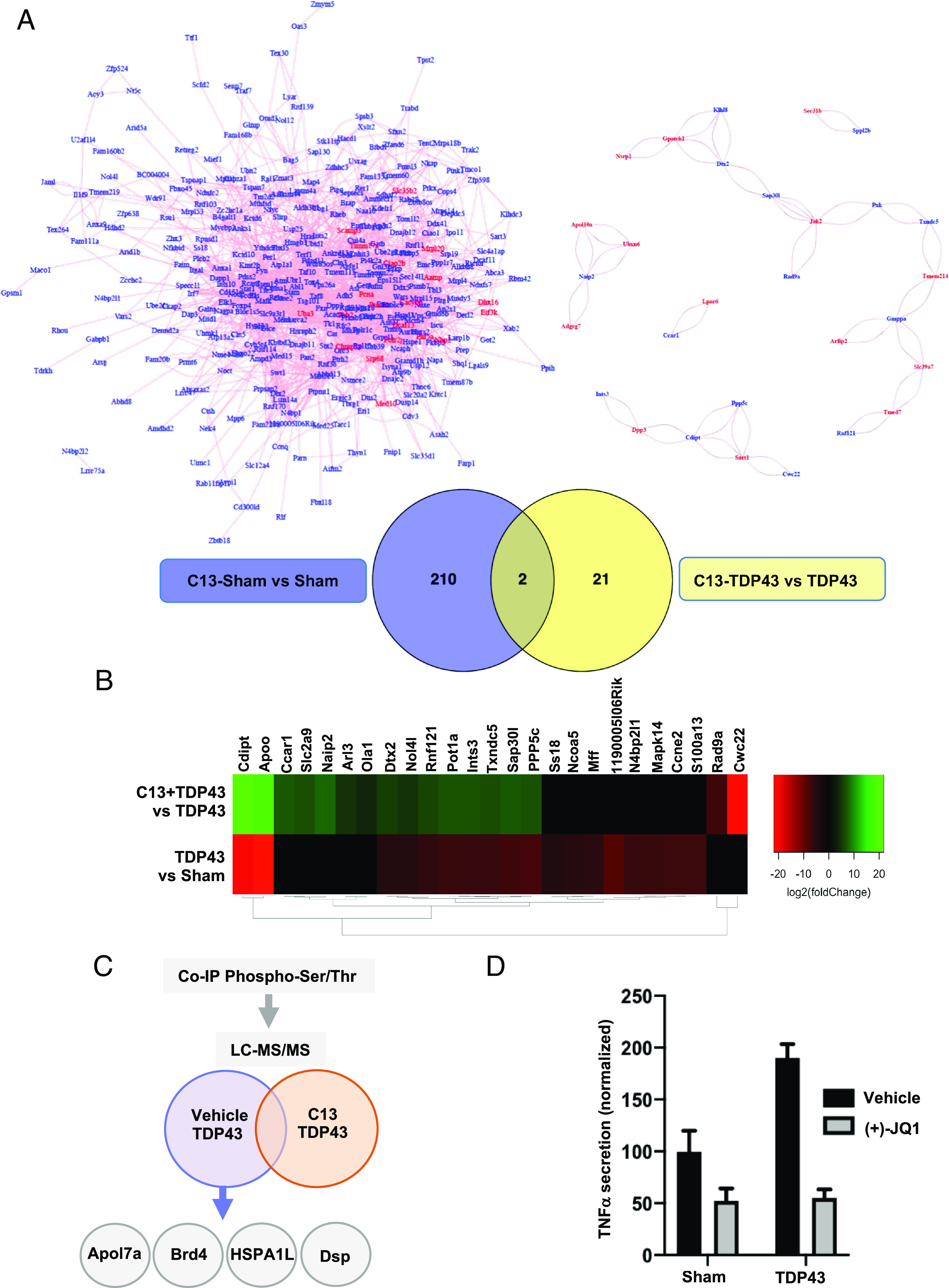
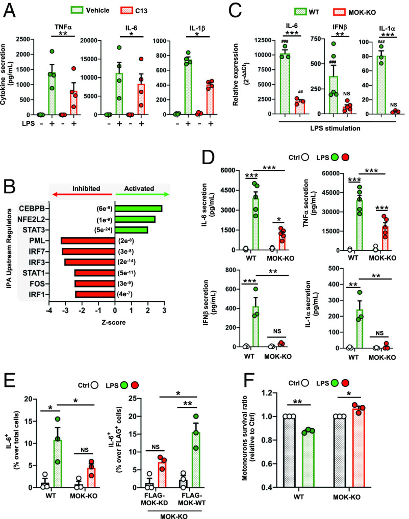
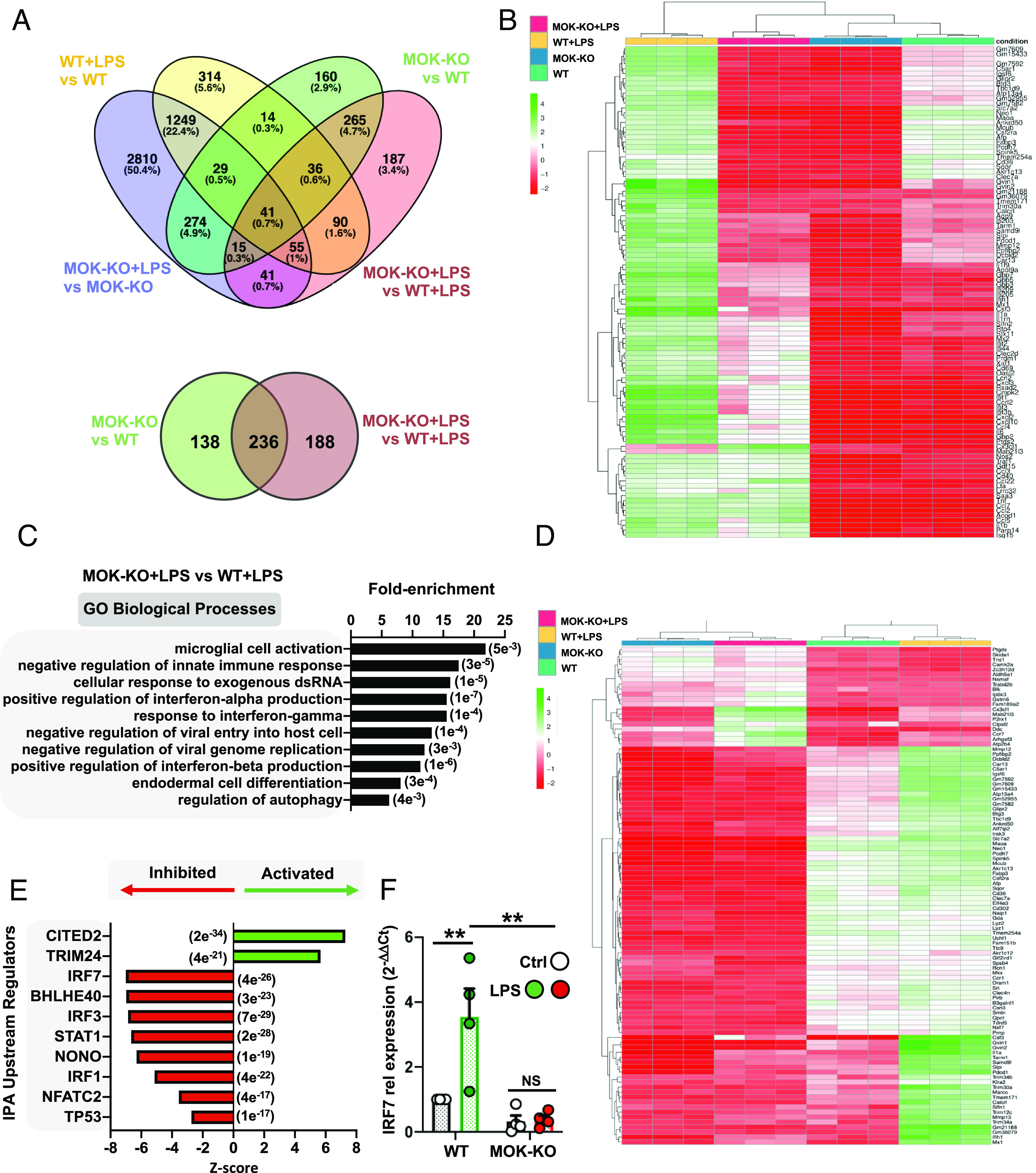
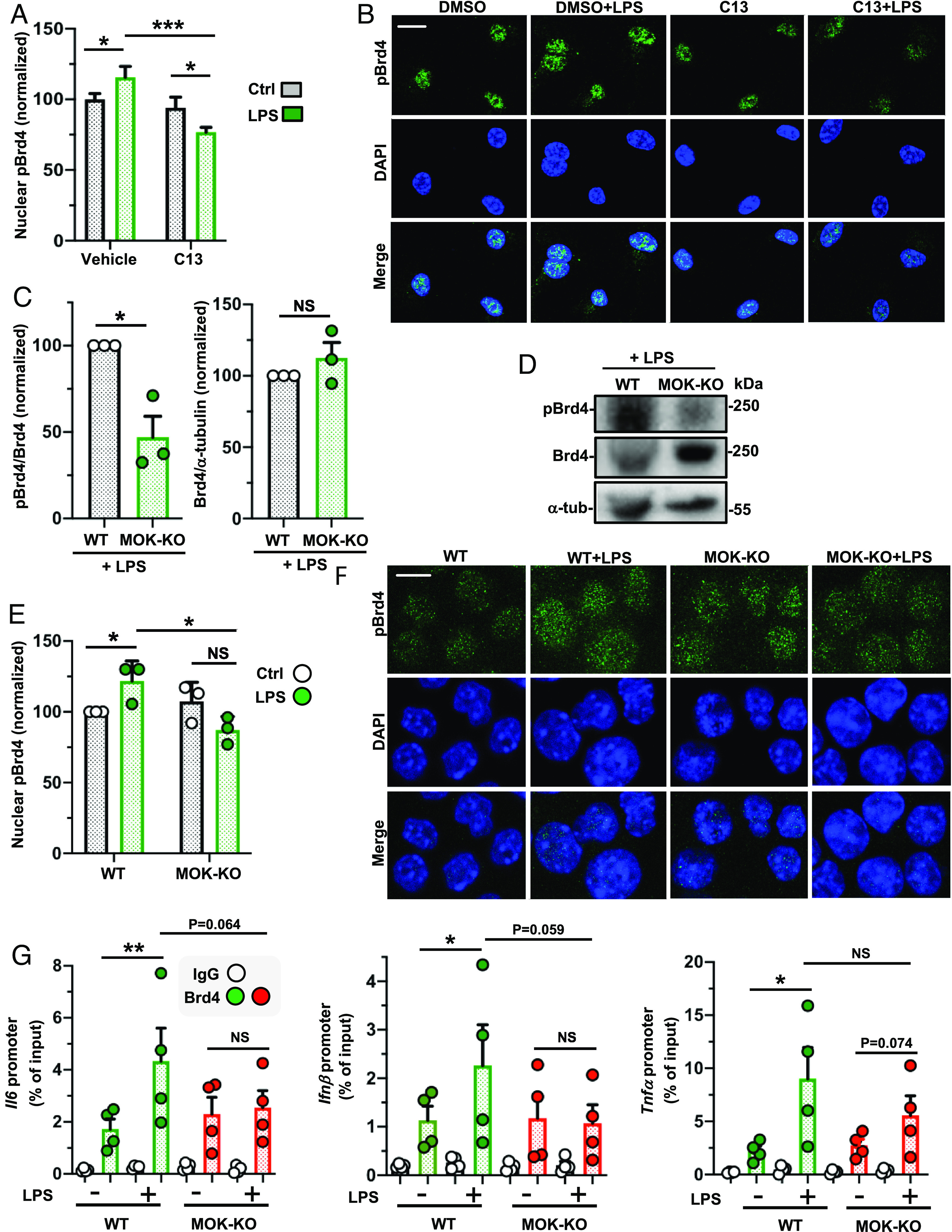
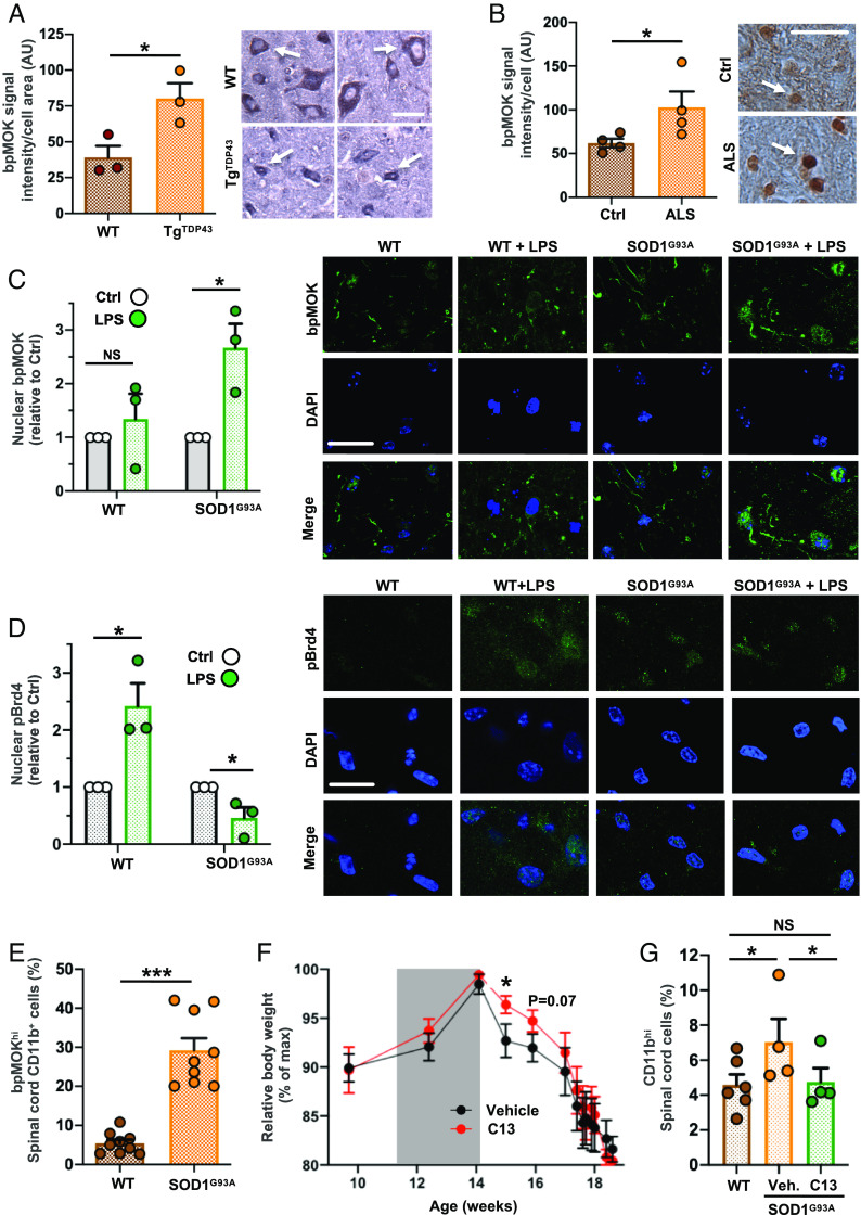
Amyotrophic lateral sclerosis (ALS) is a fatal and incurable neurodegenerative disease affecting motor neurons and characterized by microglia-mediated neurotoxic inflammation whose underlying mechanisms remain incompletely understood. In this work, we reveal that MAPK/MAK/MRK overlapping kinase (MOK), with an unknown physiological substrate, displays an immune function by controlling inflammatory and type-I interferon (IFN) responses in microglia which are detrimental to primary motor neurons. Moreover, we uncover the epigenetic reader bromodomain-containing protein 4 (Brd4) as an effector protein regulated by MOK, by promoting Ser492-phospho-Brd4 levels. We further demonstrate that MOK regulates Brd4 functions by supporting its binding to cytokine gene promoters, therefore enabling innate immune responses. Remarkably, we show that MOK levels are increased in the ALS spinal cord, particularly in microglial cells, and that administration of a chemical MOK inhibitor to ALS model mice can modulate Ser492-phospho-Brd4 levels, suppress microglial activation, and modify the disease course, indicating a pathophysiological role of MOK kinase in ALS and neuroinflammation.

Keywords: amyotrophic lateral sclerosis (ALS); glia; neurodegenerative disease; neuroinflammation; signaling kinase.


Conflict of interest statement

The authors declare no competing interest.

Figures

Fig. 1.
Fig. 1.
MOK-mediated mechanisms occur in microglial cells upon exposure to TDP-43 aggregates. (A, Bottom) Venn diagrams showing the number of DEGs from analyzed SLAM-Seq data obtained with SIM-A9 cells exposed to 5 µg/mL TDP-43 aggregates (TDP43) or sham aggregates (Sham) for 4 h, pretreated for 1 h with 10 µM C13 [or DMSO (dimethylsulfoxide) as vehicle]. Results are from three independent experiments (N = 3). Indicated are the number of DEGs for “C13 Sham vs. Sham” and “C13-TDP43 vs. TDP43” comparisons (PAdj. < 0.05 and PAdj. < 0.1, respectively). (A, Top) Predicted network analysis based on the two sets of identified DEGs by using GeneMANIA (19). Blue labels correspond to top DEGs and red labels, to inferred genes. (B) Heatmap representing the relative expression profiles (DEGs with PAdj. < 0.05) comparing “TDP43 vs. Sham” and “C13-TDP43 vs. TDP43” treatments. The differential regulation in gene expression changes between both comparisons indicates an effect of MOK inhibitor C13 in the transcriptional profile of microglial cells upon exposure to TDP-43 aggregates. (C) Schematic representation of LC–MS/MS analysis of eluates from antiphospho-Ser/Thr immunoprecipitation assays of lysates from primary microglial cells exposed to 5 µg/mL TDP-43 aggregates, pretreated for 1 h with 10 µM C13 (or DMSO). (D) Determination of TNFα by ELISA from primary microglial cells exposed to 5 µg/mL TDP-43 aggregates (TDP43) or sham aggregates (Sham) overnight, pretreated for 1 h with 10 µM (+)-JQ1 or DMSO (vehicle). Shown values were normalized to Sham control. Data are mean ± SD (N = 2).
Fig. 2.
Fig. 2.
MOK positively regulates microglial responses, including inflammatory and type-I IFN pathways. (A) Quantification of proinflammatory cytokines by ELISA in supernatants from primary microglial cells stimulated with 1 µg/mL LPS for 5 h (TNFα) or 16 h (IL-6, IL-1β) after pretreatment with 10 µM C13 (or DMSO) for 1 h (N = 4). One-way ANOVA followed by the Tukey post hoc test. (B) Top predicted upstream regulators by IPA from 158 DEGs (C13-treated vs. untreated, PAdj < 0.05) identified by RNA-Seq studies from primary microglial cells stimulated with 1 µg/mL LPS for 5 h. The P-values of overlap are shown in parentheses. (C) Assessment of gene expression levels for a set of key cytokines by qRT-PCR from WT and MOK-KO SIM-A9 cells stimulated with 1 µg/mL LPS for 5 h (N = 3). Data represent fold changes relative to unstimulated WT cells. #P < 0.05, ##P < 0.01, and ###P < 0.001 vs. unstimulated controls. One-way ANOVA followed by the Tukey post hoc test. (D) Quantification of TNFα, IL-6, IFNβ and IL-1α levels by ELISA in supernatants from WT and MOK-KO SIM-A9 cells stimulated with 1 µg/mL LPS for 5 h or overnight for IL-1α (N = 5 for IL-6, TNFα and N = 4 for IFNβ, IL-1α). One-way ANOVA followed by the Tukey post hoc test. (E) Flow cytometry quantification of IL-6+ cells in WT and MOK-KO SIM-A9 cells, previously stimulated with 1 µg/mL LPS for 5 h (Left). Rescue of phenotype in MOK-KO cells resulting from cell transfection with FLAG-MOK-WT or FLAG-MOK-KD constructs and stimulation with 1 µg/mL LPS for 5 h (Right) (N = 3). Student’s t test performed between shown groups, paired, one-tailed. (F) Fold changes of primary motor neurons survival (relative to each control) after exposure to conditioned media from LPS-stimulated WT or MOK-KO cells overnight (N = 3). Student’s t test between shown groups, unpaired, two-tailed. Data in A and CF are the mean ± SEM. from “N” independent experiments, and *P < 0.05, **P < 0.01, and ***P < 0.001. NS: not significant.
Fig. 3.
Fig. 3.
Transcriptomic analysis from RNA-Seq studies of MOK-KO cells and response to LPS stimulation. Data in AE are from 3 independent experiments (N = 3). (A) Venn diagrams depicting the number of total (Top) and protein-coding (Bottom) DEGs between MOK-KO and WT SIM-A9 cells under basal conditions and/or after 1 µg/mL LPS stimulation for 5 h (PAdj. < 0.05; log2 fold change >2 and <−2); (B) Heatmap showing unsupervised clustering analysis for the top 100 protein-coding genes based on relative expression levels in the four samples; (C) GO “biological process” term enrichment analysis of total DEGs for MOK-KO+LPS vs. WT+LPS. Shown are the top hits based on the P-value (indicated in parentheses) with at least three up-/down-regulated genes; (D) Heatmap depicting clustering analysis for 100 coding DEGs identified for the MOK-KO+LPS vs. WT+LPS comparison (consisting of the top 80% down-regulated and top 20% up-regulated, genes to maintain proportionality of all significant DEGs found); (E) Top 10 IPA-predicted upstream expression regulators from coding DEGs comparing MOK-KO vs. WT cells upon LPS stimulation, indicating positive (activated) or negative (inhibited) z-scores. The results are from all genes that were found to be differentially regulated in the RNA-Seq deseq2 analysis in any comparison with PAdj of <e-5. The P value of overlap is indicated in parentheses; (F) Assessment of IRF7 gene expression by qRT-PCR from WT and MOK-KO SIM-A9 cells stimulated with 1 µg/mL LPS for 5 h. Data represent fold changes relative to unstimulated WT cells (WT Ctrl) (N = 4). One-way ANOVA followed by the Tukey post hoc test. Data are the mean ± SEM. from N independent experiments, and *P < 0.05 and **P < 0.01.
Fig. 4.
Fig. 4.
MOK regulates phospho-Ser492-Brd4 levels under neuroinflammatory conditions. (A and B) Quantification of nuclear pBrd4 levels by confocal IF with anti-pBrd4 (green) and DAPI staining of primary microglial cells pretreated with 10 µM C13 (or DMSO) for 1 h and stimulated or not with 1 µg/mL LPS for 4 h. The data and images shown correspond to one experiment (N = 20 analyzed images per condition) and are representative of two independent experiments. (Scale bar: 10 µm.) (C and D) Quantification and representative image of western blot analyses with anti-pBrd4 and anti-Brd4 (normalized to Brd4 or a-tubulin signal, respectively) in lysates of WT and MOK-KO SIM-A9 cells stimulated with 1 µg/mL LPS for 1 h. Data are from three independent experiments (N = 3). Student’s t test, unpaired, one-tailed. (E and F) Quantification and representative images of confocal IF analyses of nuclear pBrd4 levels with anti-pBrd4 (green) and DAPI staining of WT and MOK-KO SIM-A9 cells stimulated or not with 1 µg/mL LPS for 1 h. The signal was enhanced by 50% in all four images in the first row for better visualization. Images are from one out of three independent experiments (N = 3). (Scale bar: 10 µm.) Student’s t test, unpaired, two-tailed. (G) ChIP-PCR assessment of Brd4 binding to specific cytokine promoters. Represented data are relative values (percentage of input) of PCR products for Il6, Ifnβ1, and Tnfα promoters after ChIP assay with either anti-Brd4 or IgG control antibodies with chromatin isolated from WT or MOK-KO cells treated or not with 1 µg/mL of LPS for 1 h (N = 4). Student’s t test between shown groups, ratio-paired, one-tailed. Data are the mean ± SEM. from N independent experiments, and *P < 0.05, **P < 0.01 and ***P < 0.001. NS: not significant.
Fig. 5.
Fig. 5.
MOK is altered in CNS cells in the context of ALS. Quantification of bpMOK levels by IHC analysis in spinal cord tissue samples (Left) and representative IHC images (Right) for spinal tissue samples from (A) TgTDP43 and WT mice (late stage; N = 3) and (B) sporadic ALS patients and age-matched control subjects (N = 4). Student’s t test, unpaired, one-tailed. (Scale bar: 25 µm.) The white arrow indicates one analyzed cell as an example. (C and D, Left) quantification of nuclear bpMOK (C) or pBrd4 (D) levels (green) and DAPI staining by confocal IF of spinal cord organotypic cultures, stimulated or not with 1 µg/mL LPS overnight, from WT and SOD1G93A mice (5 wk old, N = 3). Represented data are the fold change of nuclear bpMOK signal upon LPS stimulation normalized to the corresponding unstimulated controls. Student’s t test, unpaired, two-tailed. (Right) representative images of both IF assays corresponding to each condition. (Scale bar: 25 µm.) (E) Flow cytometry analysis of the bpMOKhi-expressing population in CD11b+ cells isolated from early-onset SOD1G93A or WT mouse spinal cords (14 wk old; N = 9). Student’s t test, unpaired, two-tailed. (F) Time-course body weight monitorization of SOD1G93A mice injected (i.p.) with C13 compound (20 µg per dose) or vehicle (N = 6) starting at 11 wk of age, every other day along 3 wk (gray window). Body weight values are normalized to each mouse’s maximum reached weight (% of maximum). One-way ANOVA followed by the Tukey post hoc test. (G) Flow cytometry analysis of the CD11bhi-expressing live cells isolated from the spinal cord from WT or C13- or vehicle-administered SOD1G93A mice (14 wk old; N = 6 for WT, N = 4 for C13/vehicle-treated SOD1G93A mice). The CD11bhi signal threshold corresponded to the top 5% (on average) for the WT mice. Student’s t test, paired, one-tailed. Data are the mean ± SEM. from N independent experiments, and *P < 0.05, **P < 0.01, and ***P < 0.001. NS: not significant.

References

    1. Caballero-Hernandez D., et al. , The ‘omics’ of amyotrophic lateral sclerosis. Trends Mol. Med. 22, 53–67 (2016). - PubMed
    1. Feldman E. L., et al. , Amyotrophic lateral sclerosis. Lancet 400, 1363–1380 (2022). - PMC - PubMed
    1. Ransohoff R. M., How neuroinflammation contributes to neurodegeneration. Science 353, 777–783 (2016). - PubMed
    1. Van Harten A. C. M., Phatnani H., Przedborski S., Non-cell-autonomous pathogenic mechanisms in amyotrophic lateral sclerosis. Trends Neurosci. 44, 658–668 (2021). - PMC - PubMed
    1. Clarke B. E., Patani R., The microglial component of amyotrophic lateral sclerosis. Brain 143, 3526–3539 (2020). - PMC - PubMed

Publication types

MeSH terms