. 2023 Jul 11;120(28):e2304256120.
doi: 10.1073/pnas.2304256120. Epub 2023 Jul 3.

The evolution of archaeal flagellar filaments

Affiliations

The evolution of archaeal flagellar filaments

Mark A B Kreutzberger et al. Proc Natl Acad Sci U S A. .

Abstract

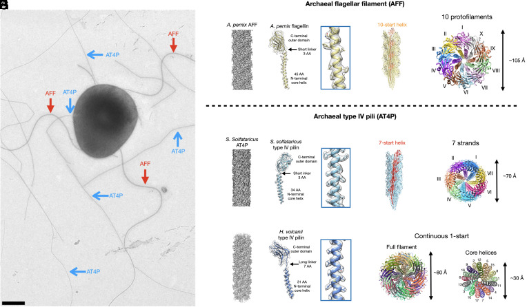
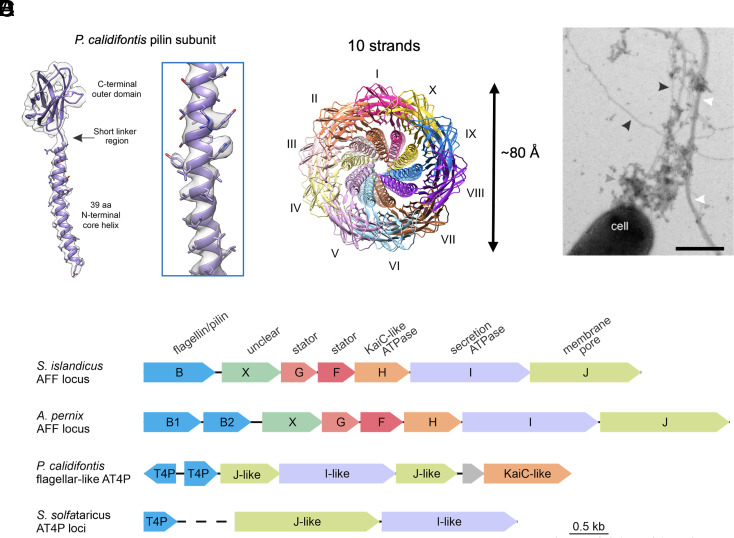
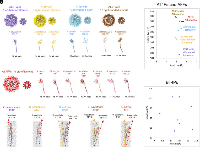
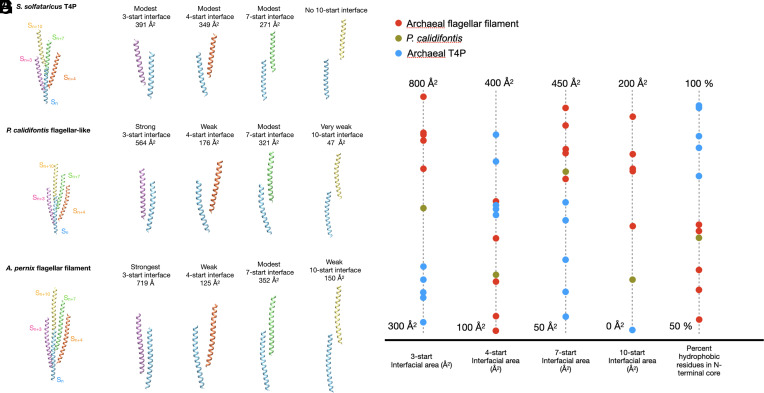
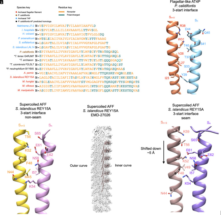
Flagellar motility has independently arisen three times during evolution: in bacteria, archaea, and eukaryotes. In prokaryotes, the supercoiled flagellar filaments are composed largely of a single protein, bacterial or archaeal flagellin, although these two proteins are not homologous, while in eukaryotes, the flagellum contains hundreds of proteins. Archaeal flagellin and archaeal type IV pilin are homologous, but how archaeal flagellar filaments (AFFs) and archaeal type IV pili (AT4Ps) diverged is not understood, in part, due to the paucity of structures for AFFs and AT4Ps. Despite having similar structures, AFFs supercoil, while AT4Ps do not, and supercoiling is essential for the function of AFFs. We used cryo-electron microscopy to determine the atomic structure of two additional AT4Ps and reanalyzed previous structures. We find that all AFFs have a prominent 10-strand packing, while AT4Ps show a striking structural diversity in their subunit packing. A clear distinction between all AFF and all AT4P structures involves the extension of the N-terminal α-helix with polar residues in the AFFs. Additionally, we characterize a flagellar-like AT4P from Pyrobaculum calidifontis with filament and subunit structure similar to that of AFFs which can be viewed as an evolutionary link, showing how the structural diversity of AT4Ps likely allowed for an AT4P to evolve into a supercoiling AFF.

Keywords: cryo-EM; helical polymers; motility.


Conflict of interest statement

The authors declare no competing interest.

Figures

Fig. 1.
Fig. 1.
Cryo-EM structures of an AFF and two archaeal type IV pili. (A) Electron micrograph of a Saccharolobus islandicus REY15A cell showing supercoiled AFF (red arrows) and nonsupercoiled archaeal type IV pili (AT4P, blue arrows). The sample has been positively stained with uranyl acetate (2%). The scale bar is 500 nm. (B) Cryo-EM density map of the filament (Left) and single flagellin model fit into its corresponding map (Right) of the Aeropyrum pernix AFF. (C) The full filament model of the A. pernix AFF is shown, with subunits along a single 10-start helix colored orange. (D) Top-down view of the A. pernix AFF, with each of its 10 protofilaments colored differently. (E) Cryo-EM density map of the Saccharolobus solfataricus AT4P filament (Left) and single pilin model fit into its corresponding map (Right). (F) The full filament model of the S. solfataricus T4P is shown, with subunits along a single 7-start helix colored red. (G) Top-down view of the S. solfataricus T4P, with each of the 7-start strands colored uniquely. (H) Cryo-EM density map of the Haloferax volcanii AT4P filament (Left) and single pilin model fit into its corresponding map (Right). (I) Top-down views of the H. volcanii filament showing the full filament (Left) and just the core N-terminal helices (Right). Each subunit in the filament model is colored distinctly.
Fig. 2.
Fig. 2.
Cryo-EM structure of the P. calidifontis flagellar–like type IV pilus. (A) The model (purple) and map (light gray) of a single subunit of the P. calidifontis filament. (B) The top-down view of the P. calidifontis filament reveals 10 prominent strands similar to the 10 protofilaments found in AFFs. (C) Electron micrograph of P. calidifontis cells expressing various types of filaments positively stained with uranyl acetate (2%). Flagellar-like AT4P, bundling pili, and presumed exopolysaccharide are indicated with black, white, and gray arrowheads, respectively. (Scale bar, 500 nm.) Note that the AT4P filament does not supercoil. (D) Genomic loci encoding archaeal flagella and type IV pili. The genes in the depicted loci are shown as arrows, with homologous genes colored the same way. Dashed line represents discontinuity between the two loci encoding the adhesive pilus system in S. solfataricus. Depicted loci encompass the following genomic regions: S. islandicus REY15A (GenBank accession NC_017276, region 104950-111297); A. pernix K1 (NC_000854, region 1201618-1209093); P. calidifontis JCM 11548 (NC_009073, region 1259287-1264747); S. solfataricus POZ149 (CP050869, regions 1531910-1532347 and 1388946-1392553).
Fig. 3.
Fig. 3.
Comparison of the filament symmetry and subunit features of AT4P and AFF. (A) Structural classification of AT4P structures from this and previous studies. Top: Axial view of AT4P models with approximately 20 subunits shown for each. For each class, the left image shows the full filament and the right image shows the only N-terminal core helices. From left to right, there are four broad classes of AT4P structures starting with 7 prominent left-handed strands (purple), AT4Ps with 7 prominent right-handed strands (gold), AT4P structures where the subunits form neither 7- nor 10-start strands (“continuous 1-start,” blue), and AT4P with prominent right-handed 10-start strands. Below each structure are the subunit models for each archaeal pilin that assembles into that structure. Pilin subunits are colored the same as their corresponding structural class. (B) Structural classification of AFF structures. All archaeal flagellins assemble into AFFs which have 10 protofilaments which are vertical with 0° of tilt with the exception of the S. islandicus REY15A AFF which has protofilaments with a slight left-handed tilt. (C) Filament density maps (light gray) with models showing a subunit Sn (light blue) and subsequent subunits along 7-start (red) and 10-start (gold) helices for each of the AT4P and AFF structural classes. The dashed lines with arrows indicate the helices. (D) Plot of helical twist versus axial rise for each AFF and AT4P structure. The colors and text indicate which symmetry group to which each filament belongs. (E) Plot of helical twist versus axial rise for all deposited bacterial T4P structures.
Fig. 4.
Fig. 4.
Comparison of N-terminal core interactions in S. solfataricus T4P, P. calidifontis flagellar–like filament, and the A. pernix flagellar filament. (A) View of just the core domains from adjacent subunits related by the 3-start (pink), 4-start (orange), 7-start (green), and 10-start (yellow) helices relative to a subunit Sn (blue) in the S. solfataricus T4P. (B) From left to right, the 3-, 4-, 7-, and 10-start interfaces or lack thereof are shown for the S. solfataricus T4P, with the buried surface area for each interface shown. (C) View of the P. calidifontis subunits along the 3-start, 4-start, 7-start, and 10-start helices relative to subunit Sn showing just the core domains. (D) From left to right, the 3-, 4-, 7-, and 10-start interfaces are shown for the P. calidifontis flagellar–like filament. (E) View of the A. pernix core domain subunits along the 3-start, 4-start, 7-start, and 10-start helices relative to subunit Sn. (F) From left to right, the 3-, 4-, 7-, and 10-start interfaces are shown for the A. pernix flagellar filament. (G) Plot of the interfacial surface area of the main interfacial contact and percent of hydrophobic residues for the core domain helices of each archaeal T4P and AFF structure.
Fig. 5.
Fig. 5.
Interfacial interactions and sequence of N-terminal core helices of archaeal type IV pili and AFFs. (A) Structural alignment of the core domain helices of the archaeal flagellins examined in this study in addition to several homologs whose structure was predicted by AlphaFold which are closely related to the P. calidifontis pilin. The “*” symbol next to the predicted homologs indicates that they are not from experimental filament structures. The names of the species are colored in sky blue for AT4P, light brown for the P. calidifontis AT4P, black for the AlphaFold-predicted P. calidifontis homologs, and red for AFFs. Amino acids colored gold are nonpolar residues, while amino acids colored green are polar/charged residues. (B) Charged/polar contacts along the 3-start interface of the P. calidifontis flagellar–like T4P. The two subunits, Sn and Sn+3, are on adjacent 10-start strands. (C) The charged/polar contacts along the core helix 3-start interface between nine of the ten protofilaments in the S. islandicus REY15A AFF (PDB:8CWM, EMD-27026). The two subunits are on adjacent protofilaments. (D) The density map of the supercoiled REY15A AFF with the model of the inner curve protofilament shown (light brown). (E) The charged/polar contacts along the inner curve seam 3-start interface of the REY15A AFF. The light brown subunit is from the 10-start protofilament in D.

References

    1. Alam M., Oesterhelt D., Morphology, function and isolation of halobacterial flagella. J. Mol. Biol. 176, 459–475 (1984). - PubMed
    1. Brown H. P., On the structure and mechanics of the protozoan flagellum. Ohio J. Sci. 45, 247–301 (1945). - PubMed
    1. Pijper A., Abraham G., Wavelengths of bacterial flagella. J. Gen. Microbiol. 10, 452–456 (1954). - PubMed
    1. Pazour G. J., Agrin N., Leszyk J., Witman G. B., Proteomic analysis of a eukaryotic cilium. J. Cell Biol. 170, 103–113 (2005). - PMC - PubMed
    1. Hyman H. C., Trachtenberg S., Point mutations that lock Salmonella typhimurium flagellar filaments in the straight right-handed and left-handed forms and their relation to filament superhelicity. J. Mol. Biol. 220, 79–88 (1991). - PubMed

Publication types

LinkOut - more resources