. 2023 Oct;152(4):961-971.e7.
doi: 10.1016/j.jaci.2023.06.017. Epub 2023 Jul 1.

C1 inhibitor and prolylcarboxypeptidase modulate prekallikrein activation on endothelial cells

Affiliations

C1 inhibitor and prolylcarboxypeptidase modulate prekallikrein activation on endothelial cells

Alona A Merkulova et al. J Allergy Clin Immunol. 2023 Oct.

Abstract

Background: We examined how prekallikrein (PK) activation on human microvascular endothelial cells (HMVECs) is regulated by the ambient concentration of C1 inhibitor (C1INH) and prolylcarboxypeptidase (PRCP).

Objective: We sought to examine the specificity of PK activation on HMVECs by PRCP and the role of C1INH to regulate it, high-molecular-weight kininogen (HK) cleavage, and bradykinin (BK) liberation.

Methods: Investigations were performed on cultured HMVECs. Immunofluorescence, enzymatic activity assays, immunoblots, small interfering RNA knockdowns, and cell transfections were used to perform these studies.

Results: Cultured HMVECs constitutively coexpressed PK, HK, C1INH, and PRCP. PK activation on HMVECs was modulated by the ambient C1INH concentration. In the absence of C1INH, forming PKa on HMVECs cleaved 120-kDa HK completely to a 65-kDa H-chain and a 46-kDa L-chain in 60 minutes. In the presence of 2 μM C1INH, only 50% of the HK became cleaved. C1INH concentrations (0.0-2.5 μM) decreased but did not abolish BK liberated from HK by activated PK. Factor XII did not activate when incubated with HMVECs alone for 1 hour. However, if incubated in the presence of HK and PK, factor XII became activated. The specificity of PK activation on HMVECs by PRCP was shown by several inhibitors to each enzyme. Furthermore, PRCP small interfering RNA knockdowns magnified C1INH inhibitory activity on PK activation, and PRCP transfections reduced C1INH inhibition at any given concentration.

Conclusions: These combined studies indicated that on HMVECs, PK activation and HK cleavage to liberate BK were modulated by the local concentrations of C1INH and PRCP.

Keywords: C1 inhibitor; Prekallikrein; bradykinin; high-molecular-weight kininogen; plasma kallikrein; prolylcarboxypeptidase.


Conflict of interest statement

Conflict of Interest Statement. No other authors have conflicts of interest to report.

Figures

Figure 1.
Figure 1.
Prolylcarboxypeptidase hypothesis for prekallikrein (PK) activation in the intravascular compartment in the presence of C1 inhibitor (C1INH). Endothelial cell prolylcarboxypeptidase (PRCP) activates PK to PKa when bound to high molecular weight kininogen (HK) bound to the endothelial cell membrane. Formed plasma kallikrein (PKa) cleaves bound HK to form cleaved HK (cHK) and liberate bradykinin (BK). No FXIIa is present to do this. This pathway is regulated by C1INH. The degree of PKa measured is dependent on the ambient concentration of C1INH and endothelial cell PRCP.
Figure 2.
Figure 2.
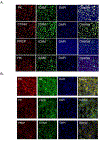
Panel A. Expression of prekallikrein, C1 inhibitor, prolylcarboxypeptidase, and high molecular weight kininogen on HMVEC. HMVEC cultured on slides were fixed with 4% paraformaldehyde and then incubated with the primary antibody (left column) and antibody to ICAM (See Supplemental Table 1). Note: in all cases, except the co-expression study with HK, the slides were treated with a goat anti-human ICAM primary antibody. For the slide with HK, the sample was treated with a mouse anti-human antibody to ICAM (Supplemental Table 1). After overnight incubation and washing, each primary antibody was treated with an appropriate secondary antibody (Supplemental Table 1). After the addition of DAPI and coverslips, the slides were photographed on a Keyence BZ-X810 All-In-One Fluorescence microscope as non-permeabilized cells at a 20X magnification. Panel B. Co-expression kallikrein/kinin system proteins on HMVEC. HMVEC cultured on slides were prepared as above and incubated with PBS-Tween containing the primary antibodies indicated on the left two panels (See Supplemental Table 1) and prepared for immunofluorescence studies as above. The slides were photographed on a Keyence BZ-X810 All-In-One Fluorescence microscope as non-permeabilized cells at a 40X magnification.
Figure 3.
Figure 3.. C1 inhibitor regulates plasma kallikrein formation on cultured HMVEC.
HMVEC were grown to 70–80% confluence in 96 well microtiter plates. After washing and blocking with 1% gelatin in HCB, the cells were incubated with 20 nM intact 120 kDa HK and zymogen PK with 10 μM Zn2+ for 1 h. After incubation, the cells were washed and 0.4 mM S2302 was added in HCB buffer and hydrolysis was measured for 1 h. Panel A. The graphs of the left are representative progress curves over 1 h of PKa formation as detected by hydrolysis of HD-Pro-Phe-Arg-pNA in the absence or presence of 0 to 620 nM C1INH. Panel B. The graph on the right are data at 60 min from the progress curve on the left. These figures are representative data from 5 similar experiments.
Figure 4.
Figure 4.. Regulation of cleavage of HK and FXII by formed PKa on HMVEC.
Panel A. HK cleavage on HMVEC by formed PKa was monitored for 60 min. Twenty nM intact HK and zymogen PK were incubated with HMVEC in HBC buffer containing gelatin and 10 μM Zn2+ in the absence (top) or presence (bottom) of 2 μM C1INH. At the indicated time (1–60 min), the adhered cells were washed and then solubilized for SDS-PAGE. HK immunoblot was performed on the solubilized samples at each time point with a polyclonal goat anti-human antibody to HK domain 6. Lane 0’ represents starting HK; Lane 1’ represents the addition of HK and PK, immediately followed by washing X3 and solubilization of the washed HMVEC with sample buffer at about 1 min. The subsequent time points were prepared similarly. The graphs to the right of the immunoblots represent the % of intact HK (green), 56 kDa light chain of HK (red) or 46 kDa light chain of HK (blue) at each indicated time. Panel B. Bradykinin formation in the supernatant over the cells was measured after 1 h of 20 nM PK activation in the presence of 20 nM HK on HMVEC in the absence or presence of 0.5 to 2.5 μM C1INH. Panel C. Proteolysis of HMVEC-bound FXII was examined. Sixty-two nM zymogen FXII was incubated with HMVEC in the absence or presence of 20 nM HK and 20 nM PK in the absence or presence of 100 μM antipain, 10 mM cysteine, or 2 μM C1INH. After 1 h incubation, the FXII bound to the cells and in their supernatant at 60 min were analyzed by immunoblot. An arrow indicates the presence of the cleaved FXII heavy chain in the supernatant immunoblot. Panel D. The cleavage time course of HMVEC-bound FXII in the presence of HK and PK is shown. Each immunoblot is a representative figure of 2 or more experiments.
Figure 5.
Figure 5.. Specificity of prekallikrein activation by prolylcarboxypeptidase on cultured HMVEC.
Panel A. The specificity for PK activation when bound to HK on cultured HMVEC was performed with inhibitors to PKa. Panel A (left): a monoclonal antibody (M202-H03 Inhibitor) and Panel A (right): a recombinant protein (EPI-KAL2 Inhibitor) were used to inhibit HMVEC-generated PKa, respectively. Panel B. Investigations determined if a commercial PRCP inhibitor [1–100 μM commercial PRCP inhibitor (PrCP Inhibitor, Calbiochem)] blocked HMVEC-generated PKa [Panel B (left)]. Panel B (right). Next, studies determined if PrCP inhibitor directly inhibited pure PKa linked to microtiter plates. Panel C. Additionally, studies determined if 0.01–3 μM chloroquine, a PRCP inhibitor, also blocked HMVEC-generated PKa [Panel C (left)]. Panel C (right). as above, studies also determined if chloroquine directly inhibited pure PKa linked to microtiter plates. Panel D. The specificity of PRCP inhibitors (PrCP Inhibitor, chloroquine) to inhibit PK autoactivation when bound to HK on plastic microtiter plate wells also was determined.
Figure 6.
Figure 6.. The influence of PRCP levels without or with C1 inhibitor on generated plasma kallikrein activity on HMVEC.
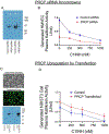
Panel A. An Immunoblot for PRCP of two experiments where HMVEC PRCP were knocked down by siRNA to PRCP. Panel B. Generated plasma kallikrein activity as measured on HMVEC on PRCP siRNA- versus Control siRNA-knocked down cells in the absence or presence of 250–1000 nM C1INH. This figure is mean±SEM of 4 independent experiments. Panel C. Top: Phase contrast photograph of the cultured NIH3T3 fibroblasts. Middle: Immunofluorescent photomicrograph of NIH3T3 cells transfected with plasmid pCMV3-PRCP-GFPSpark. Bottom: Two immunoblots of NIH3T3 fibroblasts transfected plasmid pCMV3-PRCP-GFPSpark for PRCP. Panel D. Generated plasma kallikrein activity on NIH3T3 fibroblasts that were transfected with pCMV3-PRCP-GFPSpark versus non-transfected cells in the absence or presence of 250–1000 nM C1INH. This figure is mean±SEM of 4 independent experiments.
Figure 7.
Figure 7.. Model for how PRCP and C1INH regulate BK formation by generated PKa.
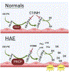
In “Normals”, there is a constitutive level of PKa formation when bound to HK on endothelium by PRCP. Formed PKa cleaves its receptor HK to form cHK and liberate BK. This pathway is regulated by ambient plasma C1INH on PKa. In “HAE” or other C1INH deficiency states and/or up-regulation of PRCP, PKa formed on the endothelium is less regulated and will cleave HK producing cHK on endothelium and in plasma and liberating BK into plasma and to bind to its receptors. When the BK degrading mechanism are exhausted, the excess BK induces angioedema.

References

    1. Schmaier AH. The contact activation and kallikrein/kinin systems: pathophysiologic and physiologic activities. J Thromb Haemost. 2016; 14:28–39. - PubMed
    1. Ivanov I, Matafonov A, Sun MF, Cheng Q, Dickeson SK, Verhamme IM et al. Proteolytic properties of single-chain factor XII: a mechanism for triggering contact activation. Blood. 2017; 129:1527–1537. - PMC - PubMed
    1. Wiggins RC. Kinin release from high molecular weight kininogen by the action of Hageman factor in the absence of kallikrein. J Biol Chem. 1983;258:8963–70. - PubMed
    1. Girey GJ, Talamo RC, Colman RW. The kinetics of the release of bradykinin by kallikrein in normal human plasma. J Lab Clin Med. 1972;80:496–505. - PubMed
    1. Joseph K, Tholanikunnel BG, Kaplan AP. Heat shock protein 90 catalyzes activation of the prekallikrein-kininogen complex in the absence of factor XII. Proc Natl Acad Sci U S A. 2002; 99:896–900. - PMC - PubMed

Publication types