

Multifunctional polydopamine - Zn2+-curcumin coated additively manufactured ceramic bone grafts with enhanced biological properties
- PMID: 37400297
- PMCID: PMC10699649
- DOI: 10.1016/j.bioadv.2023.213487
Multifunctional polydopamine - Zn2+-curcumin coated additively manufactured ceramic bone grafts with enhanced biological properties
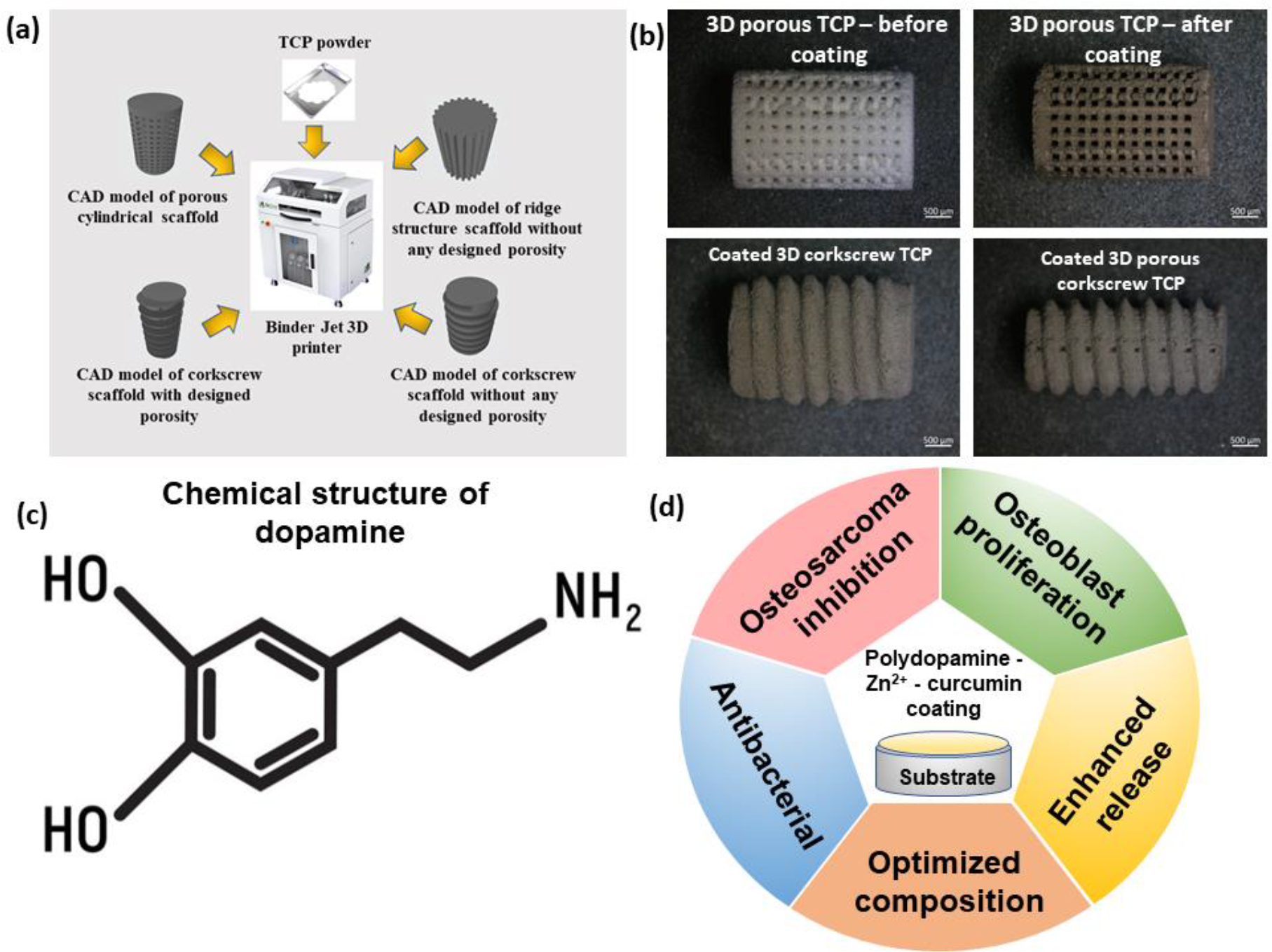
Abstract
The lack of site-specific chemotherapeutic agents after osteosarcoma surgeries often induces severe side effects. We propose the utilization of curcumin as an alternative natural chemo-preventive drug for tumor-specific delivery systems with 3D printed tricalcium phosphate (TCP) based artificial bone grafts. The poor bioavailability and hydrophobic nature of curcumin restrict its clinical use. We have used polydopamine (PDA) coating with Zn2+ functionalization to enhance the curcumin release in the biological medium. The obtained PDA-Zn2+ complex is characterized by X-ray photoelectron spectroscopy (XPS). The presence of PDA-Zn2+ coating leads to ~2 times enhancement in curcumin release. We have computationally predicted and validated the optimized surface composition by a novel multi-objective optimization method. The experimental validation of the predicted compositions indicates that the PDA-Zn2+ coated curcumin immobilized delivery system leads to a ~12 folds decrease in osteosarcoma viability on day 11 as compared to only TCP. The osteoblast viability shows ~1.4 folds enhancement. The designed surface shows the highest ~90 % antibacterial efficacy against gram-positive and gram-negative bacteria. This unique strategy of curcumin delivery with PDA-Zn2+ coating is expected to find application in low-load bearing critical-sized tumor-resection sites.
Keywords: Additive manufacturing; Antibacterial surface; Calcium phosphate; Curcumin; Drug delivery; Polydopamine coating.
Copyright © 2023 Elsevier B.V. All rights reserved.
Conflict of interest statement
Declaration of competing interest The authors do not have any possible conflict of interest. The content is solely the responsibility of the authors and does not necessarily represent the official views of the National Institute of Health.
Figures


References
-
- Rutkowski P, Antiangiogenic agents combined with systemic chemotherapy in refractory osteosarcoma, Lancet. Oncol. 22 (2021) 1206–1207. - PubMed
-
- Lahr CA, Landgraf M, Wagner F, Cipitria A, Moreno-Jiménez I, Bas O, Schmutz B, Meinert C, Cavalcanti ADS, Mashimo T, Miyasaka Y, Holzapfel BM, Shafiee A, McGovern JA, Hutmacher DW, A humanised rat model of osteosarcoma reveals ultrastructural differences between bone and mineralised tumour tissue, Bone. 158 (2022) 116018. 10.1016/j.bone.2021.116018. - DOI - PubMed
-
- Damiati LA, Tsimbouri MP, Hernandez V-L, Jayawarna V, Ginty M, Childs P, Xiao Y, Burgess K, Wells J, Sprott MR, Meek RMD, Li P, Oreffo ROC, Nobbs A, Ramage G, Su B, Salmeron-Sanchez M, Dalby MJ, Materials-driven fibronectin assembly on nanoscale topography enhances mesenchymal stem cell adhesion, protecting cells from bacterial virulence factors and preventing biofilm formation, Biomaterials. 280 (2022) 121263. 10.1016/j.biomaterials.2021.121263. - DOI - PubMed
-
- Calabrese G, De Luca G, Franco D, Morganti D, Rizzo MG, Bonavita A, Neri G, Fazio E, Neri F, Fazio B, Crea F, Leonardi AA, Lo Faro MJ, Guglielmino S, Conoci S, Structural and antibacterial studies of novel ZnO and ZnxMn(1−x)O nanostructured titanium scaffolds for biomedical applications, Biomater. Adv. 145 (2023) 213193. 10.1016/j.bioadv.2022.213193. - DOI - PubMed
MeSH terms
Substances
Grants and funding
LinkOut - more resources
Full Text Sources
