. 2023 Aug;33(8):677-691.
doi: 10.1016/j.nmd.2023.06.007. Epub 2023 Jun 22.

Canine models of Charcot-Marie-Tooth: MTMR2, MPZ, and SH3TC2 variants in golden retrievers with congenital hypomyelinating polyneuropathy

Affiliations

Canine models of Charcot-Marie-Tooth: MTMR2, MPZ, and SH3TC2 variants in golden retrievers with congenital hypomyelinating polyneuropathy

Shawna Cook et al. Neuromuscul Disord. 2023 Aug.

Abstract

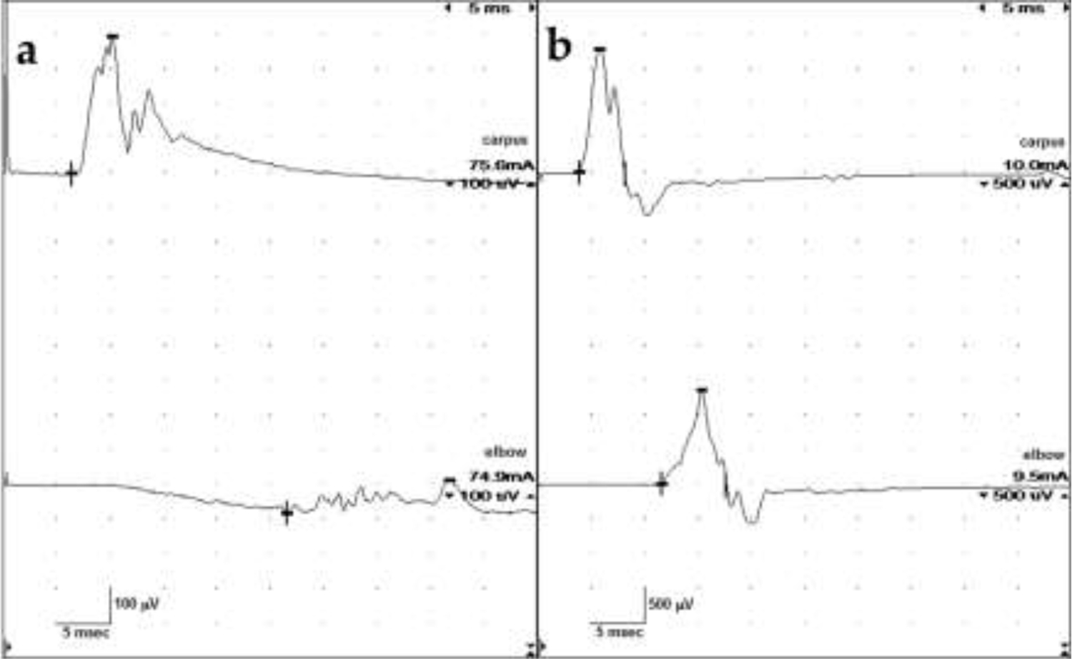
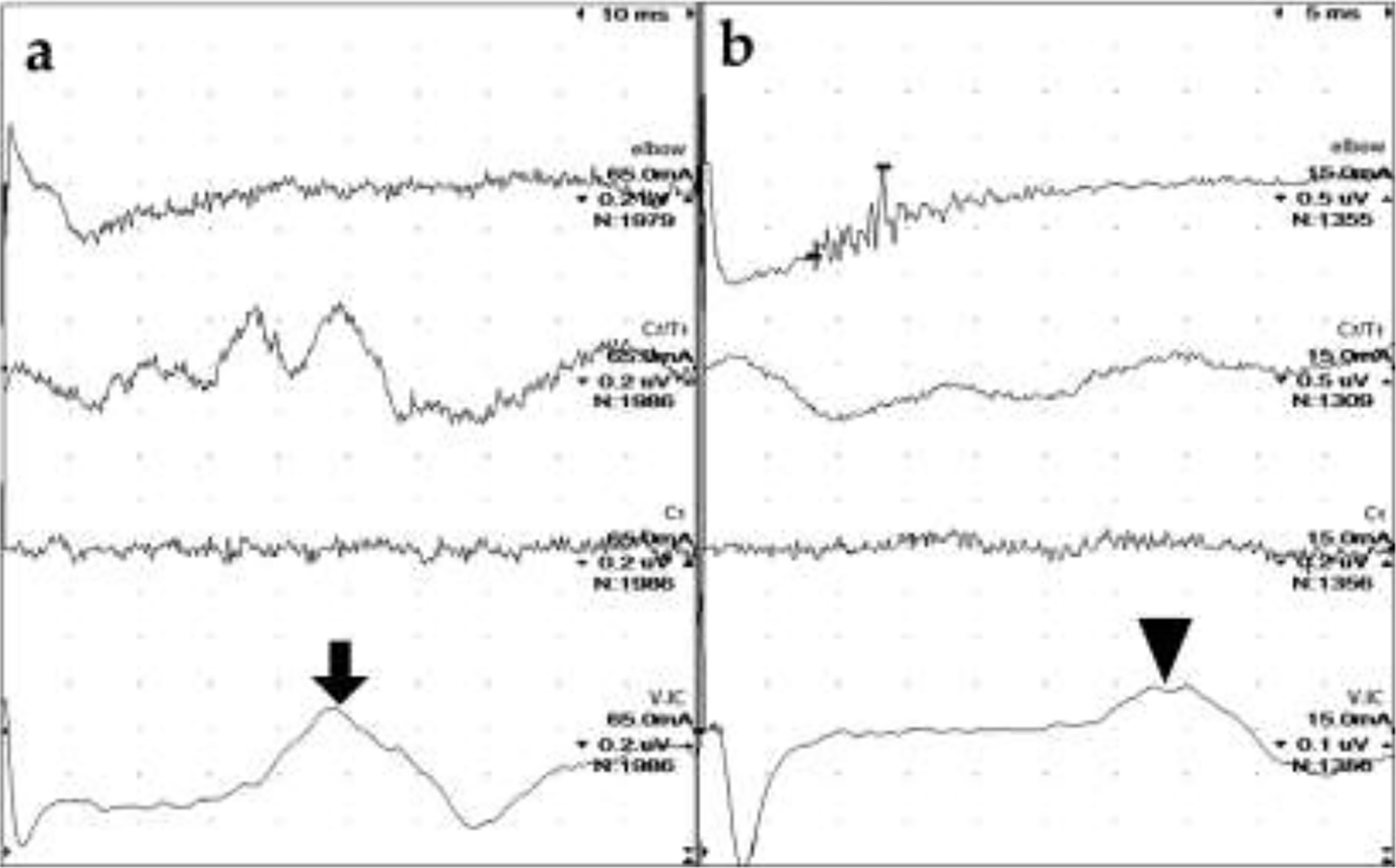
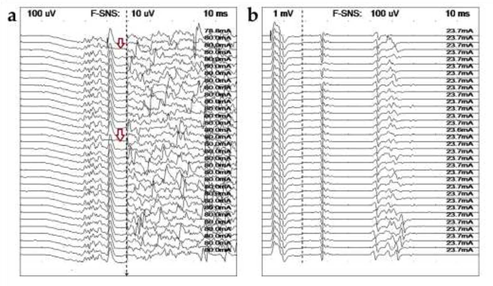
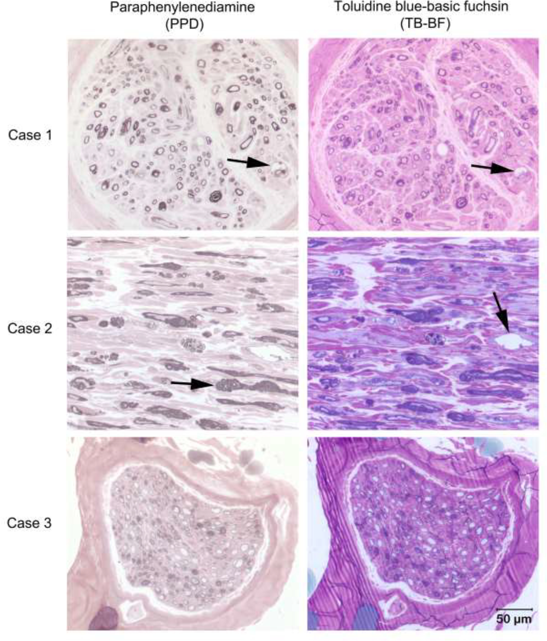
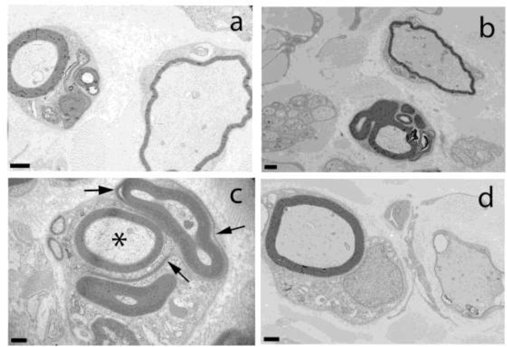
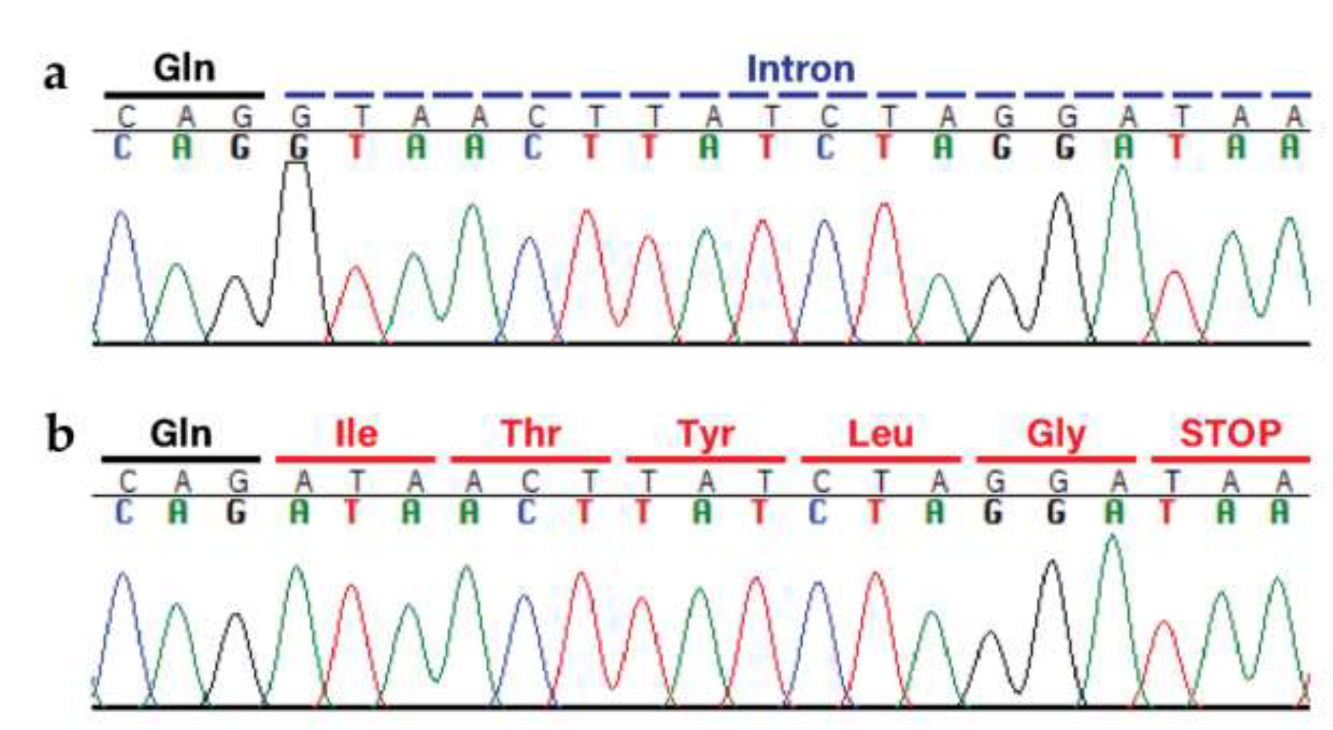
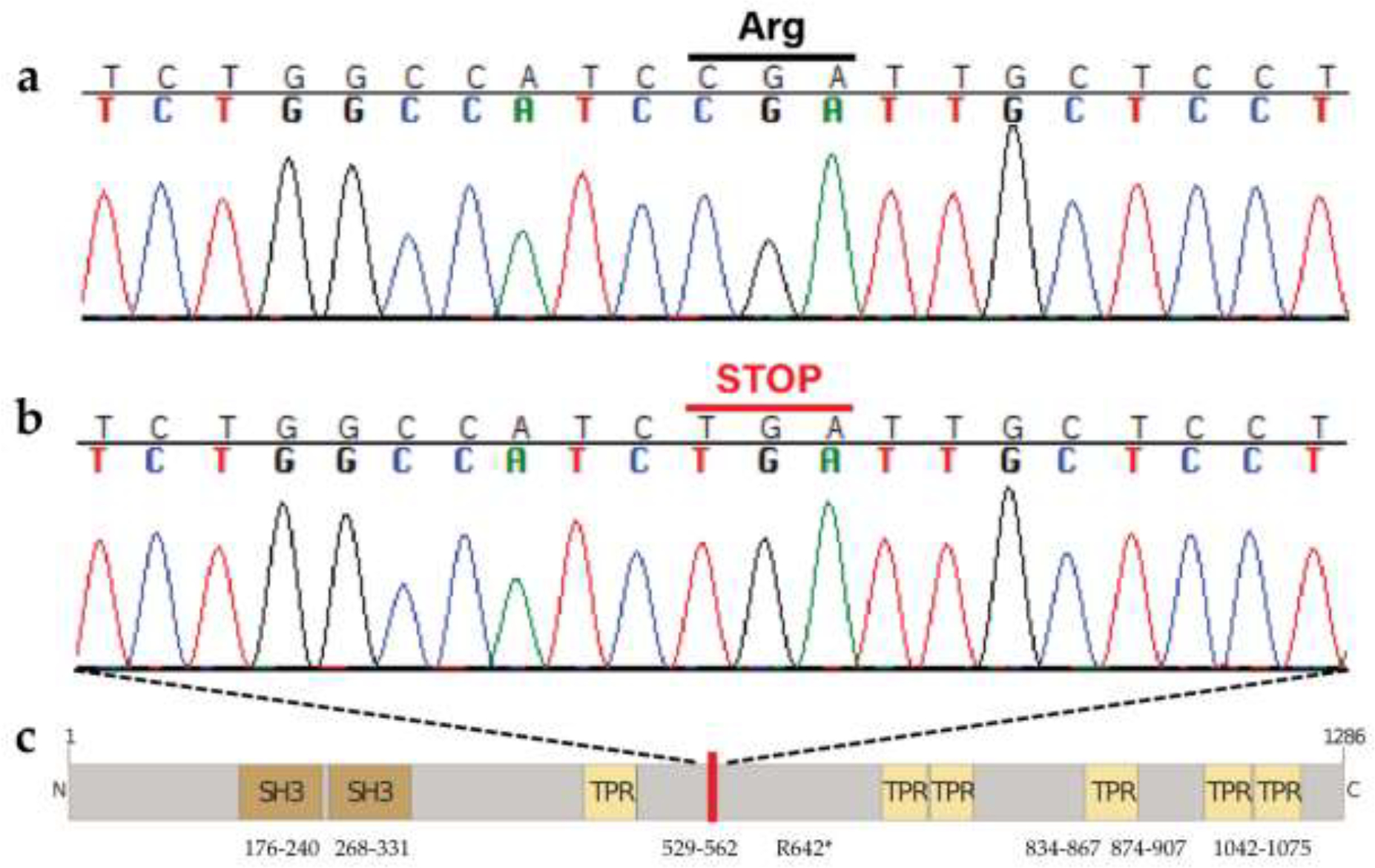
Congenital hypomyelinating polyneuropathy (HPN) restricted to the peripheral nervous system was reported in 1989 in two Golden Retriever (GR) littermates. Recently, four additional cases of congenital HPN in young, unrelated GRs were diagnosed via neurological examination, electrodiagnostic evaluation, and peripheral nerve pathology. Whole-genome sequencing was performed on all four GRs, and variants from each dog were compared to variants found across >1,000 other dogs, all presumably unaffected with HPN. Likely causative variants were identified for each HPN-affected GR. Two cases shared a homozygous splice donor site variant in MTMR2, with a stop codon introduced within six codons following the inclusion of the intron. One case had a heterozygous MPZ isoleucine to threonine substitution. The last case had a homozygous SH3TC2 nonsense variant predicted to truncate approximately one-half of the protein. Haplotype analysis using 524 GR established the novelty of the identified variants. Each variant occurs within genes that are associated with the human Charcot-Marie-Tooth (CMT) group of heterogeneous diseases, affecting the peripheral nervous system. Testing a large GR population (n = >200) did not identify any dogs with these variants. Although these variants are rare within the general GR population, breeders should be cautious to avoid propagating these alleles.

Keywords: Animal model; Dysmyelination; Electrodiagnostic testing; Genetic; Genocopies; Histopathology.


Conflict of interest statement

Declaration of Competing Interest None for any authors or co-authors on “Canine models of Charcot-Marie-Tooth: MTMR2, MPZ, and SH3TC2 variants in Golden Retrievers with congenital hypomyelinating polyneuropathy” by Cook, et al. declare any conflicts of interest.

Figures

Fig. 1
Fig. 1
Ulnar motor nerve conduction velocity studies. (a) Case 2. Amplitudes were severely reduced. The stimulus intensity required to obtain these potentials was increased (reference <5 mA). The potentials are temporally dispersed with a conduction velocity of 7 m/s. (b) Case 3. Amplitudes are also reduced but the stimulus applied was only slightly elevated. CMAP configurations were normal but somewhat jagged in appearance, with no definitive temporal dispersion noted. Nerve conduction velocities are slow at 21 m/s.
Fig. 2
Fig. 2
Radial sensory nerve conduction studies. (a) Case 2. No definitive SNAP (elbow), CD (C7/T1) or spinal evoked potential (C1) were apparent despite high sensitivity (0.2 μV/div) and a large number of signals averaged (nearly 2000). A cortical somatosensory response (arrow) was recorded by electrodes placed at the vertex (V) and intercanthus (IC). (b) Case 3. The SNAP is very temporally dispersed with a slow conduction velocity of 31 m/s. No CD or spinal evoked potentials were recorded but a cortical somatosensory response (arrowhead) is present. Note: the sweep speed in (a) is double that of (b) so the cortical SEP is later in Case 2 (nearly 50 ms) compared to Case 3 (nearly 35 ms). Sensitivities and stimulus intensities are also different. CD = cord dorsum
Fig. 3
Fig. 3
Ulnar late wave studies. (a) Case 2. Recorded from the thoracic interosseous muscle following stimulation at the elbow, no definitive late waves could be identified. Loss of the largest peak in the M-wave (CMAP) can be seen in two tracings (arrows). (b) Same study in Case 3 recorded after stimulating at the carpus, the minimum F wave latencies are prolonged, 50 ms. It is unclear whether the earlier potentials are H waves (most likely) or A waves.
Fig. 4
Fig. 4
Light Microscopic evaluation of the peroneal nerve from Cases 1–3. Transverse (Cases 1 and 3) and longitudinal (Case 2) sections from resin embedded peroneal nerve stained with paraphenylenediamine (PPD in left column) for myelin and toluidine blue-basic fuchsin (TB-BF in right column). In all three cases most myelin sheaths are inappropriately thin for the axon calibers. In Cases 1 and 2 thickened myelin sheaths (arrow in Case 2 PPD stain) and redundant myelin loops consistent with tomacula are noted along with myelin ballooning (arrows in Cases 1 PPD and TB-BF and 2 TB-BF). Bar in right lower corner = 50 μm for all images.
Fig. 5
Fig. 5
Electron Microscopic evaluation of the peroneal nerve from Case 2. Ultrastructural analysis of the peroneal nerve from Case 2 demonstrating multiple outpouchings of myelin and redundant myelin loops in large and small nerve fibers (a-c). In c, note disintegrating myelin loops (arrows) and intact axon (asterisk). Inappropriately thin myelin sheaths for the axon diameters are shown in a, b, and d. Bar in lower left corner = 1000 nm for a, 1 μm for b, 0.5 μm for c, and 1 μm for d.
Fig. 6
Fig. 6
The identified MTMR2 variant XM_038568229.1:c.1479+1G>A. a Wild-type sequence electropherogram from an unaffected dog with the original glutamine at the end of exon 12 (marked in black) and the following intron (marked in blue). b Mutant sequence electropherogram from Case 1 (Case 2 looks identical), demonstrating the single base change, loss of the splice donor site, and subsequent transcription (with translated codons marked in red).
Fig. 7
Fig. 7
The identified MPZ variant XP_038441854.1:c.Ile145Thr. a Wild-type sequence electropherogram from an unaffected dog, with the isoleucine marked in black. b Mutant sequence electropherogram from the affected GR (Case 3), with the heterozygous codon marked in red. The variant changes the isoleucine on one strand to a threonine.
Fig. 8
Fig. 8
The identified SH3TC2 variant XM_038568229.1:c.1479G>A. a Wild-type sequence electropherogram from an unaffected dog with the original arginine indicated, and b mutant sequence electropherogram from the affected GR (Case 4) with the premature stop codon, introduced halfway through the amino acid sequence, at position 642 out of 1286. c Predicted protein structure and domains, with the red bar marking the location of the stop codon. Amino acid positions are indicated.

References

    1. Bizzi A, Castelli G, Bugiani M, Barker PB, Herskovits EH, Danesi U, et al. Classification of Childhood White Matter Disorders Using Proton MR Spectroscopic Imaging. Am J Neuroradiol 2008;29:1270–5. 10.3174/ajnr.A1106. - DOI - PMC - PubMed
    1. Carroll SL. The Molecular and Morphologic Structures That Make Saltatory Conduction Possible in Peripheral Nerve. J Neuropathol Exp Neurol 2017;76:255–7. 10.1093/jnen/nlx013. - DOI - PubMed
    1. Simons M, Nave K-A. Oligodendrocytes: Myelination and Axonal Support. Cold Spring Harb Perspect Biol 2016;8:a020479. 10.1101/cshperspect.a020479. - DOI - PMC - PubMed
    1. Salzer JL. Schwann Cell Myelination. Cold Spring Harb Perspect Biol 2015;7:a020529. 10.1101/cshperspect.a020529. - DOI - PMC - PubMed
    1. Nelis E, Haites N, Van Broeckhoven C. Mutations in the peripheral myelin genes and associated genes in inherited peripheral neuropathies. Hum Mutat 1999;13:11–28. 10.1002/(SICI)1098-1004(1999)13:1&lt;11::AID-HUMU2&gt;3.0.CO;2-A. - DOI - PubMed

Publication types