

Clinical evaluation of resin infiltration treatment masking effect on hypomineralised enamel surfaces
- PMID: 37400849
- PMCID: PMC10318730
- DOI: 10.1186/s12903-023-03140-6
Clinical evaluation of resin infiltration treatment masking effect on hypomineralised enamel surfaces
Abstract
Background: Resin infiltration is a micro-invasive treatment for molar incisor hypomineralisation (MIH). In this study it was aimed to evaluate the masking effect of resin infiltration treatment (ICON) on hypomineralised enamel surface of permanent anterior teeth by using laser fluorescence, spectrophotometer, and cross-polarisation photography.
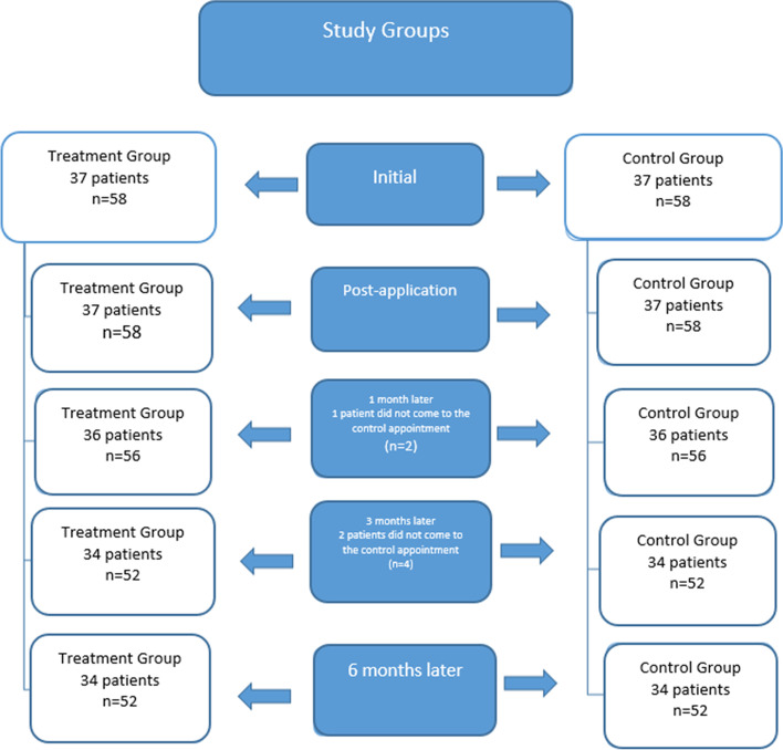
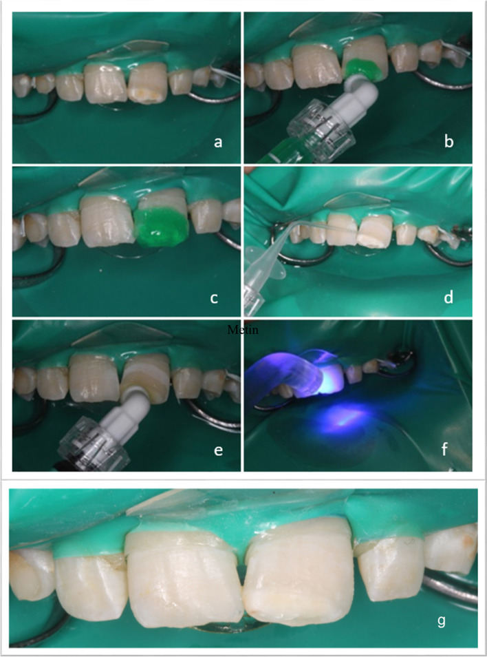
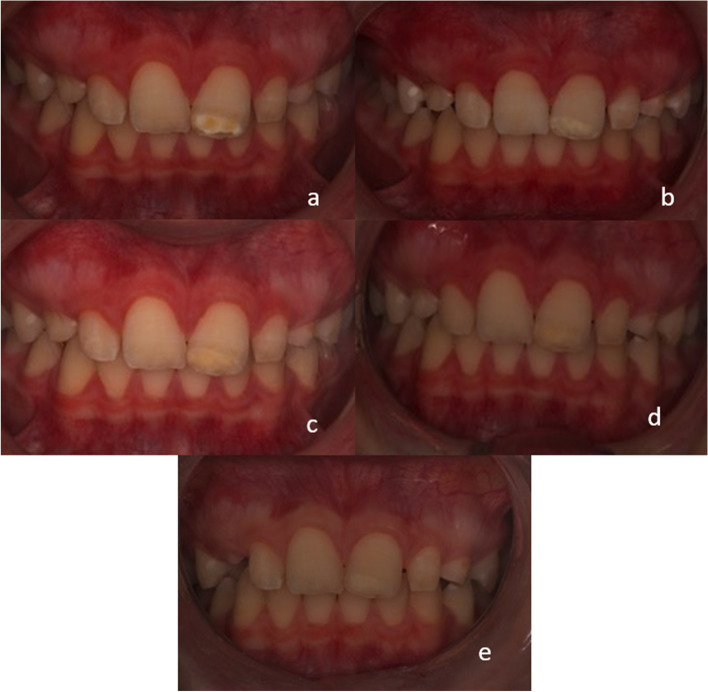
Methods: A total of 116 permanent central incisors in 37 patients were included in the study. The resin infiltration treatment (Icon®) was applied to the teeth with MIH; the healthy teeth received no treatment (control). Hypomineralised enamel lesions were evaluated by ICDAS II criteria. DIAGNOdent Pen was used to assess the lesions and healthy enamel surface quantitatively. Colour changes in enamel lesions were evaluated by using a spectrophotometer (VITA EasyShare). Each enamel lesion was imaged using a cross-polarization technique before and after treatment. All photos were assessed using Image J to evaluate the changes in lesion size. Enamel lesions were evaluated before; immediately after; 1; 3; and 6 months after treatment. Statistical significance was set as p < 0.05.
Results: After the resin infiltration, significant decreases were found in the mean DIAGNOdent values for the treatment group (p < 0.05). The colour differences before and after treatment significantly differed in all follow-ups (p < 0.05). In the treatment group, lesion areas decreased significantly after treatment (p < 0.05).
Conclusions: The resin infiltration treatment has a masking effect on MIH lesions without cavities, with stable outcomes after six months. The cross-polarization photography technique may be use to evaluate the lesion size instead of photography with flash.
Trial registration: NCT04685889 (registered 28 December 2020).
Keywords: Cross polarisation; Enamel; Laser fluorescence; Lesion; Molar incisor hypomineralization; Resin infiltration.
© 2023. The Author(s).
Conflict of interest statement
The authors declare that they have no competing interests.
Figures
References
-
- Fearne J, Anderson P, Davis GR: 3D X-ray microscopic study of the extent of variations in enamel density in first permanent molars with idiopathic enamel hypomineralisation. Br Dent J 2004, 196(10):634–638; discussion 625. - PubMed
-
- Dantas-Neta NB, Soares Figueiredo M, Lima CCB, Bendo CB. Matos de Andrade ÉM, Lima MdDM, Pordeus IA, Paiva SM: Factors associated with molar–incisor hypomineralisation in schoolchildren aged 8–10 years: a case–control study. Int J Pediatr Dent. 2018;28(6):570–577. doi: 10.1111/ipd.12412. - DOI - PubMed
Publication types
MeSH terms
Associated data
LinkOut - more resources
Full Text Sources
Medical
