Review
. 2023 Jul 3;18(1):43.
doi: 10.1186/s13024-023-00636-1.

Extrasynaptic NMDA receptors in acute and chronic excitotoxicity: implications for preventive treatments of ischemic stroke and late-onset Alzheimer's disease

Affiliations
Review

Extrasynaptic NMDA receptors in acute and chronic excitotoxicity: implications for preventive treatments of ischemic stroke and late-onset Alzheimer's disease

Shan P Yu et al. Mol Neurodegener. .

Abstract

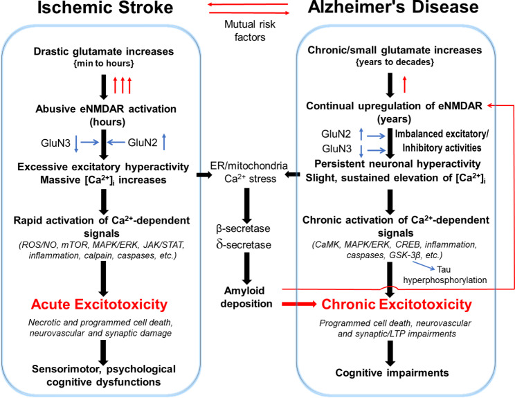
Stroke and late-onset Alzheimer's disease (AD) are risk factors for each other; the comorbidity of these brain disorders in aging individuals represents a significant challenge in basic research and clinical practice. The similarities and differences between stroke and AD in terms of pathogenesis and pathophysiology, however, have rarely been comparably reviewed. Here, we discuss the research background and recent progresses that are important and informative for the comorbidity of stroke and late-onset AD and related dementia (ADRD). Glutamatergic NMDA receptor (NMDAR) activity and NMDAR-mediated Ca2+ influx are essential for neuronal function and cell survival. An ischemic insult, however, can cause rapid increases in glutamate concentration and excessive activation of NMDARs, leading to swift Ca2+ overload in neuronal cells and acute excitotoxicity within hours and days. On the other hand, mild upregulation of NMDAR activity, commonly seen in AD animal models and patients, is not immediately cytotoxic. Sustained NMDAR hyperactivity and Ca2+ dysregulation lasting from months to years, nevertheless, can be pathogenic for slowly evolving events, i.e. degenerative excitotoxicity, in the development of AD/ADRD. Specifically, Ca2+ influx mediated by extrasynaptic NMDARs (eNMDARs) and a downstream pathway mediated by transient receptor potential cation channel subfamily M member (TRPM) are primarily responsible for excitotoxicity. On the other hand, the NMDAR subunit GluN3A plays a "gatekeeper" role in NMDAR activity and a neuroprotective role against both acute and chronic excitotoxicity. Thus, ischemic stroke and AD share an NMDAR- and Ca2+-mediated pathogenic mechanism that provides a common receptor target for preventive and possibly disease-modifying therapies. Memantine (MEM) preferentially blocks eNMDARs and was approved by the Federal Drug Administration (FDA) for symptomatic treatment of moderate-to-severe AD with variable efficacy. According to the pathogenic role of eNMDARs, it is conceivable that MEM and other eNMDAR antagonists should be administered much earlier, preferably during the presymptomatic phases of AD/ADRD. This anti-AD treatment could simultaneously serve as a preconditioning strategy against stroke that attacks ≥ 50% of AD patients. Future research on the regulation of NMDARs, enduring control of eNMDARs, Ca2+ homeostasis, and downstream events will provide a promising opportunity to understand and treat the comorbidity of AD/ADRD and stroke.

Keywords: Alzheimer’s disease; Ca2+ homeostasis; Excitotoxicity; Extrasynaptic NMDARs; GluN2B subunit; GluN3A subunit; Glutamate; Ischemic stroke; Memantine; NMDA receptors.


Conflict of interest statement

The authors declare that they have no competing interests.

Figures

Fig. 1
Fig. 1
Composition of NMDA receptors and the regulatory role of the GluN3 subunit. Functional NMDA receptors are transmembrane heterotetramers embedded in the phospholipid bilayer of glutamatergic neurons, containing two GluN1 and two GluN2 subunits. The binding of the ligand leads to the opening of the receptor cation channel in an Mg2+- and voltage-depolarization manner. The NMDAR activity and its mediated Ca2+ influx have significant impacts on synaptic transmission, neuronal plasticity, psychological/cognitive functions, and cell fates. A GluN3 (GluN3A and 3B) subunit can replace one GluN2 in the triheteromeric complex, resulting in restrained single-channel opening activities and smaller whole-cell currents compared to GluN1/GluN2 receptors. The NMDA current traces are our unpublished data, which were recorded in an Mg2+-free extracellular solution
Fig. 2
Fig. 2
Age-dependent subunit alternations of synaptic and extrasynaptic NMDA receptors and functional consequences. NMDA receptors are mainly located in the post-synaptic membrane inside and outside of the synaptic cleft. Synaptic NMDARs are directly involved in excitatory neurotransmission and synaptic plasticity, while extrasynaptic NMDARs have regulatory roles in these activities. Glutamate concentrations are markedly different between the cleft and extrasynaptic spaces. Glutamate released by astrocytes and microglia (not shown) are likely the main components of extrasynaptic glutamate, together with that spillover from the synaptic cleft. The distribution and topography of NMDARs are subjected to age-dependent alterations. In addition to a developmental switch of increased GluN2A/GluN2B ratio [340, 341], the GluN3 expression also undergoes an age-dependent change, from the initial locations of both synaptic and extrasynaptic sites to extrasynaptic/peri-synaptic site in the mature brain. This developmental change is likely associated with the functional needs of NMDAR regulation at different life stages. For example, high levels of GluN3A in immature brains is neuroprotective; while in the adult brain, the absence of GluN3 in the synaptic site allows synapse maturation and elevated plasticity. In the adult brain, GluN3 remains to exist in the extrasynaptic membrane as an endogenous neuroprotective mechanism against brain damage and neurodegeneration. In the aging/aged or degenerative brain, loss or weakening of this regulatory mechanism due to either increased GluN2B expression or deficiency of GluN3 will lead to enhanced eNMDA activity and Ca2+ dysregulation, which aggravate acute and chronic excitotoxicity associated with ischemic stroke and late-onset AD.
Fig. 3
Fig. 3
Distinctive Pro- and anti-survival mechanisms downstream from activation of synaptic and extrasynaptic NMDARs. This simplified graph illustrates a few main signaling pathways associated with the activation of synaptic and extrasynaptic NMDA receptors, respectively. Of note that although additional genes not mentioned in the text are shown in the graph, not all related signals can be included in the graph. For example, chronic stress of neuronal hyperactivity and Ca2+ elevations induce recurrent inflammation that is not shown here. In general, activation of sNMDARs leads to pro-survival effects beneficial for neuronal viability and synaptic plasticity, while activation of eNMDARs causes detrimental consequences associated with acute and chronic excitotoxicity. It is worth mentioning, however, that many signaling genes such as CaMK and MAPK kinases can play opposite actions most likely in subtype-dependent manners. In the pro-survival mechanism, the Wnt regulation of the expression of CaMKIV is an upstream protective signaling in neurodegenerative conditions [342]. Cyclic-AMP response element binding protein (CREB) plays a key function in medicating sNMDAR activation and expressions of pro-survival genes such as BDNF, MAPK, and Akt. CREB phosphorylation is mediated acutely by CaMKIV while long-term regulation may be controlled upstream by ERK1/2 [343, 344]. Activation of CREB via CaMKIV phosphorylation of CREB binding protein (CBP) requires translocation of transducer of regulated CREB activity (TORC) which is downstream of Ca2+ signaling from sNMDAR activation. Jacob and the synapto-nuclear trafficking is a relatively new mechanism linking downstream signaling of sNMDARs. Caldendrin binds to Jacob’s nuclear localization signal in a Ca2+-dependent manner [194, 195]. In contrast to these CREB-activating signals of sNMDARs, eNMDARs suppress CREB activity via the inactivation of the Ras-ERK1/2 pathway and the nuclear translocation of Jacob, which promotes CREB dephosphorylation. Calcineurin-dependent dephosphorylation of TORC and subsequent CREB activation is also downstream of sNMDAR transmission [345]. Activation of sNMDARs suppresses apoptotic cascades via suppression of the BH3-only domain gene Puma and p53, thereby limiting cytochrome c release. Downstream effectors of apoptosis including Apaf1, Caspase 3, and Caspase 9 are also suppressed [346, 347]. Contrary to these pro-survival pathways, pro-death pathways are mediated by downstream activities of eNMDARs [189, 348]. Interactions between the pro-survival and pro-death pathways may occur so that the suppression of CREB activity may result from inactivating the ERK1/2 pathway [79, 195]. Another shared pathway between synaptic and extrasynaptic NMDARs is the FOXO pathway. FOXO activity is suppressed by PI3K downstream of sNMDARs while activation of eNMDARs enhances FOXO nuclear import and the consequent transcription of FOXO3α, Bim, and Fas which lead to cell death via multiple mechanisms including excitotoxicity [205, 349]. Synaptic NMDAR activity enhances the transcription of PGC-1α, while excessive expression and activity of eNMDARs suppress CREB-dependent PGC-1α transcription [350]. In general, CaMKII is downstream of eNMDAR activity and acts as a carrier of Ca2+-regulated protease calpain to promote apoptotic cell death [351]. Moreover, Ca2+ dyshomeostasis resulting from NMDAR subunit composition such as GluN2B and GluN3A expression changes and its interaction with intracellular Ca2+ reservoirs in the ER and mitochondria play an important role in the maintenance of cellular bioenergetics, glucose metabolism, and normal mitophagy [352]. Ca2+ dyshomeostasis is thus a major trigger of the generation of ROS and increased apoptosis via the imbalance of mitochondria-initiated apoptotic genes including tBid, Bax/Bcl2, Bak/BclxL, Bad, Apaf1, cytochrome c, and caspases [353, 354]. The NMDAR-TRPM interaction is a novel cell death mechanism downstream to NMDAR and TRPM activation, which stimulates the formation of the NMDAR/TRPM complex in the extrasynaptic location. Excitotoxicity is then triggered by the complex in a “Ca2+-independent” fashion, mediated by mitochondrial dysfunction, reduced activation of ERK1/2, shut-off of the transcription factor CRAB, and cell death [229]
Fig. 4
Fig. 4
Acute and chronic excitotoxicity and shared mechanisms between ischemic stroke and sporadic AD. The sketch diagram illustrates similarities and differences between ischemic stroke and AD. Both brain disorders suffer from overactivations of eNMDARs that are subjected to regulations by glutamate concentration, expression of NMDAR subunits (e.g. GluN2 and GluN3), and other modulatory mechanisms. The vast and rapid Ca2+ influx upon cerebral ischemia and much mild but lasting Ca2+ stress in AD trigger distinctive Ca2+-dependent signaling pathways, leading to acute and chronic excitotoxicity, respectively. Depending on the severity and region of damage, ischemic stroke causes transient or permanent deficits of locomotor/sensorimotor activities and psychological/psychiatric/cognitive functions. Cerebral ischemia is also known for causing mitochondria dysfunction and ER Ca2+ stress that may be responsible for post-stroke AD-like pathology. On the other hand, chronic excitotoxicity in AD is induced by long-lasting small Ca2+ increases and deteriorating signaling pathways that lead to synaptic and neural network interruptions in specific regions critical for cognition, followed by Aβ deposition via increased activities of β- and δ-secretases [355]. This chronic excitotoxicity may cause late-onset AD in Aβ-dependent or -independent manner, which remains to be further investigated
Fig. 5
Fig. 5
NMDAR GluN3A deficiency induced sporadic AD. The graph shows age-dependent events and corresponding experimental evidence in the NMDAR GluN3A knockout mouse. A GluN3A deficiency caused by genetic mutation or functional dysfunction can result in slight but persistent neuronal hyperactivity and [Ca2+]i elevations, subsequently leading to chronic inflammation, metabolism burden, and slowly evolved degenerative excitotoxicity. The synaptic impairment and programmed neuronal cell death in the hippocampus and cortex are correlated to progressive cognitive decline. Interestingly and important to note that significant endogenous Aβ plague formation in neurons and blood vessels occurs after, but not before, cognition decline and other functional deficits [127]
Fig. 6
Fig. 6
Hypothetic timelines of late-onset AD and common comorbidity of stroke. Late-onset AD is a slow and progressive disease; its early pathophysiological cascades cultivate years to decades before clinical diagnosis and likely precede significant Aβ deposition which is a pathological event emerging in patients’ brains of around 50 years old [48]. Different from the most popular diagram showing the events after Aβ deposition [356], this graph emphasizes possible triggering mechanisms before Aβ and tau pathology. In this hypothetic model, neuronal hyperactivity and Ca2+-associated chronic excitotoxicity exist well before neuronal loss, functional deficits, increased APP processing, and Aβ/tau pathology. Meanwhile, these underlying mechanisms significantly increase the risk of stroke attacks accompanied by acute excitotoxicity. Accordingly, a preventive disease-modifying intervention such as MEM treatment is necessary in the preclinical phase, which can also serve as a preconditioning therapy against stroke that strikes more than 50% of AD patients

References

    1. Imran TF, Posner D, Honerlaw J, Vassy JL, Song RJ, Ho YL, Kittner SJ, Liao KP, Cai T, O’Donnell CJ, et al. A phenotyping algorithm to identify acute ischemic stroke accurately from a national biobank: the million veteran program. Clin Epidemiol. 2018;10:1509–21. doi: 10.2147/CLEP.S160764. - DOI - PMC - PubMed
    1. Vijayan M, Reddy PH. Stroke, vascular dementia, and Alzheimer’s Disease: Molecular Links. J Alzheimers Dis. 2016;54:427–43. doi: 10.3233/JAD-160527. - DOI - PMC - PubMed
    1. Mijajlovic MD, Pavlovic A, Brainin M, Heiss WD, Quinn TJ, Ihle-Hansen HB, Hermann DM, Assayag EB, Richard E, Thiel A, et al. Post-stroke dementia - a comprehensive review. BMC Med. 2017;15:11. doi: 10.1186/s12916-017-0779-7. - DOI - PMC - PubMed
    1. Henon H, Pasquier F, Leys D. Poststroke dementia. Cerebrovasc Dis. 2006;22:61–70. doi: 10.1159/000092923. - DOI - PubMed
    1. Association A. 2020 Alzheimer’s disease facts and figures. Alzheimers Dement 2020, PMID: 32157811. - PubMed

Publication types