. 2023 Jul 3;27(1):64.
doi: 10.1186/s40824-023-00402-w.

Non-interfacial self-assembly of synthetic protocells

Affiliations

Non-interfacial self-assembly of synthetic protocells

Xiaolin Xu et al. Biomater Res. .

Erratum in

Abstract

Background: Protocell refers to the basic unit of life and synthetic molecular assembly with cell structure and function. The protocells have great applications in the field of biomedical technology. Simulating the morphology and function of cells is the key to the preparation of protocells. However, some organic solvents used in the preparation process of protocells would damage the function of the bioactive substance. Perfluorocarbon, which has no toxic effect on bioactive substances, is an ideal solvent for protocell preparation. However, perfluorocarbon cannot be emulsified with water because of its inertia.

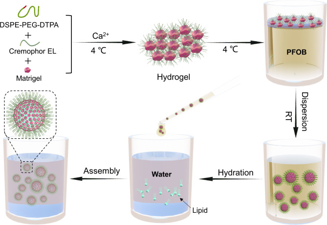
Methods: Spheroids can be formed in nature even without emulsification, since liquid can reshape the morphology of the solid phase through the scouring action, even if there is no stable interface between the two phases. Inspired by the formation of natural spheroids such as pebbles, we developed non-interfacial self-assembly (NISA) of microdroplets as a step toward synthetic protocells, in which the inert perfluorocarbon was utilized to reshape the hydrogel through the scouring action.

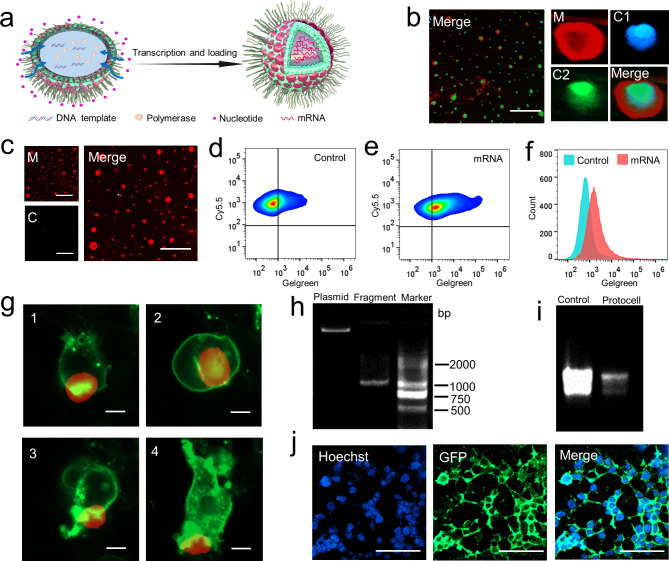
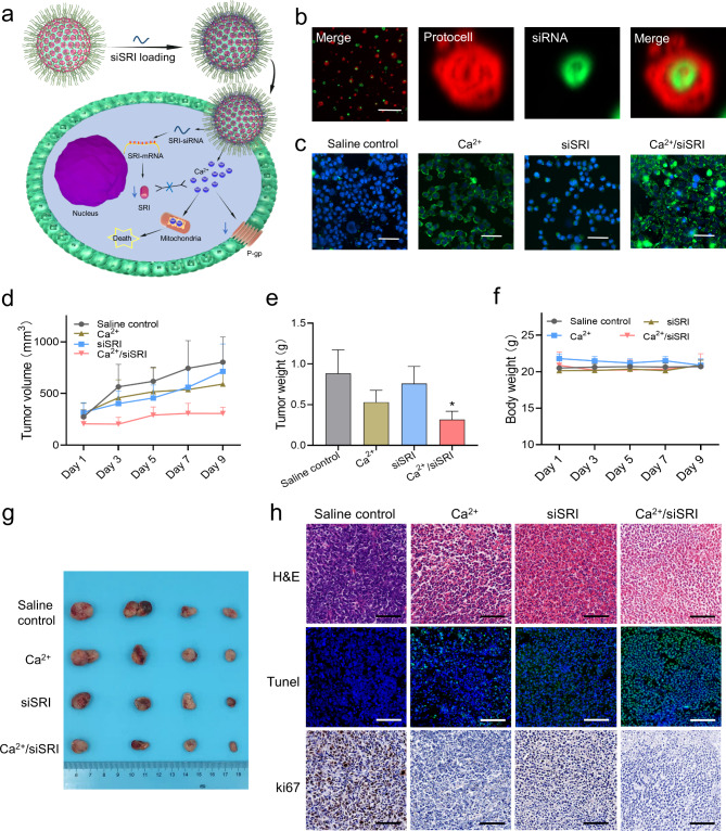
Results: The synthetic protocells were successfully obtained by using NISA-based protocell techniques, with the morphology very similar to native cells. Then we simulated the cell transcription process in the synthetic protocell and used the protocell as an mRNA carrier to transfect 293T cells. The results showed that protocells delivered mRNAs, and successfully expressed proteins in 293T cells. Further, we used the NISA method to fabricate an artificial cell by extracting and reassembling the membrane, proteins, and genomes of ovarian cancer cells. The results showed that the recombination of tumor cells was successfully achieved with similar morphology as tumor cells. In addition, the synthetic protocell prepared by the NISA method was used to reverse cancer chemoresistance by restoring cellular calcium homeostasis, which verified the application value of the synthetic protocell as a drug carrier.

Conclusion: This synthetic protocell fabricated by the NISA method simulates the occurrence and development process of primitive life, which has great potential application value in mRNA vaccine, cancer immunotherapy, and drug delivery.

Keywords: Calcium homeostasis; Cancer immunotherapy; Non-interfacial self-assembly; Protocell; mRNA vaccine.


Conflict of interest statement

The authors declare no competing financial interest.

Figures

Fig. 1
Fig. 1
Schematic illustration depicting the fabrication of NISA-based synthetic protocells
Fig. 2
Fig. 2
Characterization of synthetic protocells. (a) Microscopic photograph of hydrogel dispersing in PFOB. (b) Microscopic photograph of the synthetic protocells. (c) The particle size distribution of the synthetic protocells. (d) Atomic force microscopy image of the collapsed synthetic protocell. Scale bar, 10 μm. e-h. Flow cytometry data for synthetic protocells with no fluorescence labeling (e), Cy5.5 labeled lipids (f), FITC labeled hydrogel (g), Cy5.5 labeled lipids and FITC labeled hydrogel (h). i. Confocal images of synthetic protocells. M reflects the membrane, C reflects the core, R1 and R2 are representative synthetic protocells. Scale bar, 100 μm. j. Confocal images of a synthetic protocell. Graphs at the bottom show corresponding line profiles of fluorescence intensities across the synthetic protocell. Scale bar, 10 μm. k. 3D confocal fluorescence reconstructions of a synthetic protocell. Scale bar, 10 μm. l. 3D confocal fluorescence reconstructions of a dried synthetic protocell. Scale bar, 10 μm
Fig. 3
Fig. 3
mRNA transcription in synthetic protocells and expression in native cells. (a) Schematic illustration of mRNA transcription in synthetic protocells. (b) Confocal images of synthetic protocell post-transcription. M reflects the membrane, C1 and C2 reflect the core in the blue and green channels respectively. Scale bar, 100 μm. (c) Confocal images of synthetic protocell post-transcription, with no nucleotide addition during the transcription. Scale bar, 100 μm. d-f. Flow cytometry data for synthetic protocells post-transcription (d), no nucleotides addition group as control (e), histogram reflects the difference in mRNA transcription (f). g. Dynamic changes of 293T cells incubating with synthetic protocells in 4 h. Red reflects synthetic protocells, green reflects 293T cells, and 1, 2, 3, 4 are dynamic in order. Scale bar, 10 μm. h. Agarose gel electrophoresis of GFP fragment amplified from the plasmid. i. Agarose gel electrophoresis of GFP-mRNA transcribed in synthetic protocells, compared with GFP-mRNA transcribed by transcription kit. j. Confocal images of 293T cells incubated with GFP-mRNA loaded synthetic protocell for 24 h. Scale bar, 100 μm
Fig. 4
Fig. 4
Fabrication of artificial cancer cells via NISA method. (a) Schematic illustration of artificial cancer cell fabricated by NISA method. (b) The expression of CD44 mRNA was higher in A2780/PTX compared with its sensitive counterpart A2780 detected by qRT-PCR. (c) The expression of CD44 protein was higher in A2780/PTX compared with A2780 determined by Western blot. (d) Laser confocal micrograph of A2780 and A2780/PTX, CD44 protein was highly expressed in A2780/PTX analyzed by immunofluorescence. Scale bar, 5 μm. (e) Laser confocal micrograph of protocell A2780 and protocell A2780/PTX and the expression of CD44 protein was analyzed by immunofluorescence. Scale bar, 5 μm
Fig. 5
Fig. 5
Fabricating synthetic protocells by NISA method, and restoring calcium homeostasis in chemoresistant cancer cells. (a) Schematic illustration of synthetic protocells restoring calcium homeostasis and overcoming drug resistance. (b) Confocal images of fam-siRNA loaded synthetic protocells. Scale bar, 100 μm. (c) Confocal images reflect the calcium ion concentration in various groups. Scale bar, 100 μm. (d) Tumor volume of various groups during treatment. (e) Tumor weight from various groups after treatment. (f) Body weight from various groups during treatment. (g) Pictures of tumors harvested from the treated mice after treatment. (h) H&E, Tunnel, and Ki67 stained A549/PTX tumor slices from various groups. Scale bar, 100 μm

References

    1. Ashley CE, Carnes EC, Phillips GK, et al. The targeted delivery of multicomponent cargos to cancer cells by nanoporous particle-supported lipid bilayers. Nat Mater. 2011;10(5):389–97. - PMC - PubMed
    1. Dzieciol AJ, Mann S. Designs for life: protocell models in the laboratory. Chem Soc Rev. 2012;41(1):79–85. - PubMed
    1. Ianeselli A, Tetiker D, Stein J, et al. Non-equilibrium conditions inside rock pores drive fission, maintenance and selection of coacervate protocells. Nat Chem. 2022;14(1):32–9. - PMC - PubMed
    1. Xu C, Hu S, Chen X. Artificial cells: from basic science to applications. Mater Today (Kidlington) 2016;19(9):516–32. - PMC - PubMed
    1. Jiang W, Wu Z, Gao Z, et al. Artificial cells: past, Present and Future. ACS Nano. 2022;16(10):15705–33. - PubMed