. 2023 Aug;13(8):e3064.
doi: 10.1002/brb3.3064. Epub 2023 Jul 3.

Aged mice are less susceptible to motion sickness and show decreased efferent vestibular activity compared to young adults

Affiliations

Aged mice are less susceptible to motion sickness and show decreased efferent vestibular activity compared to young adults

David Lorincz et al. Brain Behav. 2023 Aug.

Abstract

Introduction: The efferent vestibular system (EVS) is a feedback circuit thought to modulate vestibular afferent activity by inhibiting type II hair cells and exciting calyx-bearing afferents in the peripheral vestibular organs. In a previous study, we suggested EVS activity may contribute to the effects of motion sickness. To determine an association between motion sickness and EVS activity, we examined the effects of provocative motion (PM) on c-Fos expression in brainstem efferent vestibular nucleus (EVN) neurons that are the source of efferent innervation in the peripheral vestibular organs.

Methods: c-Fos is an immediate early gene product expressed in stimulated neurons and is a well-established marker of neuronal activation. To study the effects of PM, young adult C57/BL6 wild-type (WT), aged WT, and young adult transgenic Chat-gCaMP6f mice were exposed to PM, and tail temperature (Ttail ) was monitored using infrared imaging. After PM, we used immunohistochemistry to label EVN neurons to determine any changes in c-Fos expression. All tissue was imaged using laser scanning confocal microscopy.

Results: Infrared recording of Ttail during PM indicated that young adult WT and transgenic mice displayed a typical motion sickness response (tail warming), but not in aged WT mice. Similarly, brainstem EVN neurons showed increased expression of c-Fos protein after PM in young adult WT and transgenic mice but not in aged cohorts.

Conclusion: We present evidence that motion sickness symptoms and increased activation of EVN neurons occur in young adult WT and transgenic mice in response to PM. In contrast, aged WT mice showed no signs of motion sickness and no change in c-Fos expression when exposed to the same provocative stimulus.

Keywords: aging; c-Fos; motion sickness; vestibular efferent.


Conflict of interest statement

The authors declare no conflicts of interest.

Figures
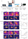
FIGURE 1
FIGURE 1
Experimental design and tail temperature (T tail) response to provocative motion (PM). (a) Experimental design and timeline of experiments; schematic was created by BioRender.com. (b–d; h–j; and n–p) Infrared images of T tail responses to PM of C57BL/6; ChAT‐gCaMP6f; and aged C57BL/6 mice, respectively. Images were taken during first 5 min of PM; between 5 and 10 min of PM (at peak tail temp), and after 15 min of PM. (e–g; k–m; and q–s) Infrared images of control responses (no PM) of C57BL/6; ChAT‐gCaMP6f; and aged C57BL/6 mice, respectively, at the same time intervals as experimental groups. Plot (i) Mean ± SEM T tail responses of C57BL/6 experimental group during PM (black trace; n = 14) and control (no PM) group (gray trace; n = 3). Vertical dashed lines for all plots indicate the start and end point of PM for experimental groups only. Plot (ii) Mean ± SEM T tail plots show response of the ChAT‐gCaMP6f experimental group during PM (black trace; n = 5) and the T tail of the control group PM (gray trace; n = 2). An outlier (red dashed line) differed from the mean response to PM (n = 1). The outlier trace suggested a summed response of a quicker onset, nonspecific, stress response added to a typical PM stress response. Plot (iii) Mean ± SEM T tail plots show response of aged C57BL/6 experimental group during PM (black trace; n = 5) and the T tail of the control group (gray trace; n = 3). An outlier (red dashed line) differed in response to mean PM (n = 1) and resembled more closely the young adult C57BL/6 experimental response in Plot i.
FIGURE 2
FIGURE 2
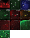
Fluorescent immunolabeling of EVN neurons and c‐Fos protein in the mouse brainstem tissue visualized by confocal microscopy. (a) Low‐magnification image of a C57BL/6 mouse brainstem slice at the level of the EVN. ChAT + Alexa594 (red) was used to label the EVN nuclei bilaterally (arrows) and other cholinergic structures. Scale bar: 1000 μm. (b) Higher power micrograph of ChAT + Alexa488 (green) labeling EVN neurons in C57BL/6 mice. (c) ChAT + Alexa405 (blue) labeling in aged C57BL/6 mice showing fluorescent EVN cells. Areas denoted by dashed lines indicate the tight clustering of EVN neurons dorsal to the genu of the facial nerve (g7n). Double labeling of genetically expressed GCaMP (green, d) and ChAT (red, e) in ChAT‐gCaMP6f transgenic EVN neurons (arrows point to extensive dendritic projections toward the MVN). (f) Merged image of GCaMP and ChAT labeling showing one to one correspondence. (g, h) c‐Fos antibody labeling in the red channel (Alexa594) was verified in the MVN of C57BL/6 and ChAT‐gCaMP6f mice. Arrowheads denote examples of the numerous c‐Fos‐labeled MVN neurons. (i, j) c‐Fos antibody labeling (Alexa488; green) was verified in the MVN of aged C57BL/6 mice. (j) Higher magnification image of aged C57BL/6 mouse showing c‐Fos within the nucleus (arrowhead) and granular appearance of lipofuscin autofluorescence in the cytoplasm (arrows). An asterisk (*) indicates nonlabeled nucleus of lipofuscin labeled cell. Scale bar: 10 μm; (b–i) 50 μm. 4V, fourth ventricle; 7n, facial motor nucleus; Abd, Abducens Nucleus; bv, blood vessel; EVN, efferent vestibular nucleus; gn7, genu of the facial nerve; MVN, medial vestibular nucleus.
FIGURE 3
FIGURE 3
c‐Fos expression in EVN neurons following provocative motion (PM). (a) EVN neurons labeled with ChAT antibody (green) and (b) c‐Fos antibody (red) labeling in the control (no PM) group of C57BL/6 mice. Arrowheads—weakly labeled c‐Fos‐positive EVN neurons. (c) Merged image of panels (a) and (b). (d) ChAT (green) and (e) c‐Fos (red) labeling in the experimental group of C57BL/6 mice after PM. Arrowheads denote c‐Fos‐labeled EVN neurons. (f) Merged image of panels (d) and (e). Graph A shows percentage of c‐Fos‐positive EVN neurons in the control (n = 3) and PM group (n = 5). **Significant difference between the control and PM group (p = .0071). (g) EVN neurons labeled with GFP antibody (green) and (h) c‐Fos (red) labeling in the control group of ChAT‐gCaMP6f mice. Arrowheads—weakly labeled c‐Fos‐positive EVN neurons. (c) Merged image of panels (g) and (h). (j) GFP (green) and (k) c‐Fos (red) labeling in the experimental group of ChAT‐gCaMP6f mice after PM. Arrowheads denote c‐Fos‐labeled EVN neurons. (l) Merged image of panels (j) and (k). Graph B shows percentage of c‐Fos‐positive EVN neurons in the control (n = 4) and PM group (n = 5). ****Significant difference between the control and PM group (p = .0001). (m) EVN neurons labeled with ChAT antibody (blue) and (n) c‐Fos (green) labeling in the control group of aged C57BL/6 mice. Arrowheads denote c‐Fos‐positive EVN neurons enlarged in the inset. Arrows denote lipofuscin autofluorescence. (o) Merged image of panels (m) and (n). (p) ChAT (blue) and (q) c‐Fos (green) labeling in the experimental group of aged C57BL/6 mice after PM. Arrow denotes lipofuscin expressing EVN cell shown in the inset. (r) Merged image of panels (p) and (q). Graph C shows the percentage of c‐Fos‐positive EVN neurons in the control (n = 3) and PM group (n = 5). Scale bar: 50μm.
FIGURE 4
FIGURE 4
Summary of tail temperature (T tail) responses to PM and their associated c‐Fos expression. (a) Summary of T tail responses to PM of all experimental groups and their controls and (b) the c‐Fos expression in all groups. c‐Fos was significantly different between the young adult C57BL/6 and aged C57BL/6 experimental groups (p < .0001) and the ChAT‐gCaMP6f and aged C57BL/6 experimental groups (p < .0001). There was no significant difference between young adult C57BL/6 and ChAT‐gCaMP6f experimental groups (p = .1730) or aged C57BL/6 control group and aged C57BL/6 experimental group.

Similar articles

References

    1. Agrawal, Y. , Merfeld, D. M. , Horak, F. B. , Redfern, M. S. , Manor, B. , Westlake, K. P. , Holstein, G. R. , Smith, P. F. , Bhatt, T. , Bohnen, N. I. , & Lipsitz, L. A. (2020). Aging, vestibular function, and balance: Proceedings of a national institute on aging/national institute on deafness and other communication disorders workshop. The Journals of Gerontology: Series A, 75(12), 2471–2480. - PMC - PubMed
    1. Benchaoui, H. , Siedek, E. , De La Puente‐Redondo, V. , Tilt, N. , Rowan, T. , & Clemence, R. (2007). Efficacy of maropitant for preventing vomiting associated with motion sickness in dogs. Veterinary Record, 161(13), 444–447. - PubMed
    1. Bergström, B. (1973a). Morphology of the vestibular nerve: II. The number of myelinated vestibular nerve fibers in man at various ages. Acta Oto‐laryngologica, 76(1–6), 173–179. - PubMed
    1. Bergström, B. (1973b). Morphology of the vestibular nerve: I. Anatomical studies of the vestibular nerve in man. Acta Oto‐Laryngologica, 76(1‐6), 162–172. - PubMed
    1. Borison, H. L. (1989). Area postrema: Chemoreceptor circumventricular organ of the medulla oblongata. Progress in Neurobiology, 32(5), 351–390. - PubMed

Publication types