. 2023 Sep 15;4(3):102402.
doi: 10.1016/j.xpro.2023.102402. Epub 2023 Jul 3.

Breast cancer PDxO cultures for drug discovery and functional precision oncology

Affiliations

Breast cancer PDxO cultures for drug discovery and functional precision oncology

Sandra D Scherer et al. STAR Protoc. .

Erratum in

Abstract

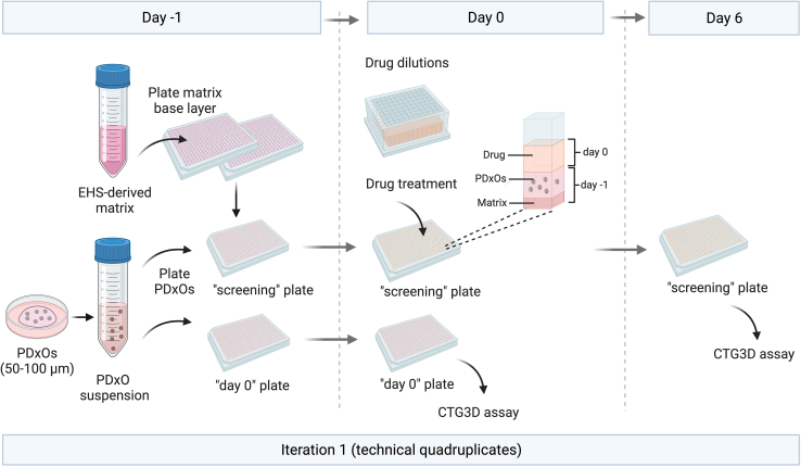
Patient-derived xenografts (PDXs) have clinical value but are time-, cost-, and labor-intensive and thus ill-suited for large-scale experiments. Here, we present a protocol to convert PDX tumors into PDxOs for long-term cultures amenable to moderate-throughput drug screens, including in-depth PDxO validation. We describe steps for PDxO preparation and mouse cell removal. We then detail PDxO validation and characterization and drug response assay. Our PDxO drug screening platform can predict therapy response in vivo and inform functional precision oncology for patients. For complete details on the use and execution of this protocol, please refer to Guillen et al.1.

Keywords: Cancer; Cell Culture; Cell-based Assays; Model Organisms; Organoids.


Conflict of interest statement

Declaration of interests The University of Utah may license the PDX and PDxO models described here to for-profit companies, which may result in tangible property royalties to members of the Welm lab who developed the models (S.D.S., A.J.B., L.Z., C.H.Y., E.C.S., K.P.G., B.E.W., and A.L.W.).

Figures

None
Graphical abstract
Figure 1
Figure 1
PDxO preparation from PDX tumor tissue (A–C) To establish PDxOs from PDX tumors, necrotic tissue areas are removed and healthy tumor tissue is cut into 3 × 3 mm fragments. Tumor fragments are then transferred into a C tube and digested in a GentleMACS Dissociator using human dissociation enzymes. The digested material is assessed under the microscope, and processed depending on the contents and structure: (A) If the digested material contains a mixture of organoids and single cells, differential centrifucation (DC) is performed to remove single cells, then the organoids are embedded into a Matrigel dome. (B) If the digested material contains only organoids, the material is embedded immediately. (C) For digestions containing only single cells, aggregation for 16 h on Ultra Low Attachment (ULA) plates is recommended prior to embedding. Scale bars represents 5 mm (left) or 500 μm (right).
Figure 2
Figure 2
Differential centrifugation of PDxO preparation that contain single cells and organoids For organoid preparations that contain organoids DC is performed to separate organoids from single cells. The organoid preparation is resuspended in 15 mL of media and a series of quick spins is performed, started with a 1 min centrifugation. The resulting supernatant is discarded, while the pellet is resuspended and spun again at s shorter time (40 s, then 30 s). After each spin, the material in the supernatant and the resuspended pellet is assessed under the microscope to determine if more DC steps are necessary. Scale bar represents 500 μm.
Figure 3
Figure 3
Embedding PDxOs in 3D Matrigel domes To embed PDxOs, a 50 μL Matrigel drop is placed into a well of a 6-well plate. Using a pipet tip the Matrigel is immediately spread out to form a cicular base layer. During 5 min of incubation of the plate at 37°C the PDxO:Matrigel mix is prepared. 200 μL of the mixture is added onto the baselayer, and incubated for 5 min at 37°C. The plate is then flipped upside down and placed back into the incubator for 10 min. After that, subtype-specific media is added to the well.
Figure 4
Figure 4
Aggregation of single cells in ULA plate To aggregate, cells are plated into a well of a 6-well ULA plate and incubated for 16 h. Images show pre (left)- and post (right) 16 h incubation. Scale bars represents 100 μm.
Figure 5
Figure 5
Passaging PDxOs after they are adapted to culture using dispase and TrypLE steps (>P2) Once mature, media is removed from PDxOs and dispase solution is added. Matrigel domes containing PDxOs are broken up manually, incubated at 37°C, then washed with media and centrifuged. The organoid pellet is resuspended in TrypLE to disassociate PDxOs into single cells. Cells are assessed under the microscope, counted and embedded in Matrigel. Scale bar represents 100 μm or 50 μm as indicated.
Figure 6
Figure 6
Diverse morphology of PDxOs with examples of successful cultures and unhealthy organoids (A–D) Successful PDxO cultures display phenotypes as shown in the panels on the left. Characteristics of unhealthy PDxO cultures are shown on the right. ∗Examples of PDxOs that are out of focus due to the 3D nature of the PDxO domes are marked with a white star (∗). Successful cultures can display as spherical, grape-like or metaplastic phenotype, and examples of these morphologies are shown in images marked with A. Additional characteristics of healthy PDxO cultures are consisteny density within the culture as shown in B, and consistent phenotypes across passages. Unhealthy cultures usually fail to grow into PDxO lines. In these cases PDxO cultures can have acellular or cytsic morphology as shown in images marked with C, or PDxOs can eject dead cells (D). Additionally, unhealthy cultures can have low viability with 20% or more dead cells during passaging (P2 or higher). Scale bar represents 50 μm.
Figure 7
Figure 7
Detection of mouse cells in PDxO cultures (A) Self eliminating mouse cells in PDxO cultures will disappear over time after several passages as shown for HCI-027 PDxOs from passage 1 (P1) to passage 9 (P9). Teal errors indicate the presumable presence of mouse cells. Right side: qRT-PCR data indicating elimination of mouse cells by passaging over time displayed as ΔCT (human GAPDH CT subtracted from mouse Gapdh CT). Dark gray indicates high mouse content, white indicates no mouse content. Scale bars represents 100 μm. (B) Images of two PDxO lines (HCI-008 and HCI-023BR) showing persistent mouse content at high passage number (P8 and P9 respectively) as shown by images (left) and qRT-PCR (right). Scale bars represents 100 μm. (C) Images of two PDxO cultures (HCI-075 and HCI-015NIR) that only contain mouse organoids. These PDxO lines failed establishment due to non detectable human content as indicated by GAPDH CT values. Scale bars represents 100 μm.
Figure 8
Figure 8
Elimination of mouse cells from PDxO cultures via FACS (A) Gating strategy for sorting human and mouse cells in HCI-048 PDxO culture. Cells were gated on SSC-A/FSC-A to exclude debris, and FSC-A/FSC-W to exclude duplets. Only cells positive for human CD298 or human EpCAM would be found in the gate positive for human cells based on the green fluorescent fluorophore that the antibodies were labeled with. Mouse cells will be labeled as such by red-fluorescence conjugated CD29/CD90.2 antobodies and are found in the APC gate, respectively. (B) Culture image show HCI-048 PDxOs pre- and post sorting. qRT-PCR confirms that mouse content was eliminated by FACS. Scale bars represents 100 μm.
Figure 9
Figure 9
Overview of PDxO drug testing On "day -1", 384-well plates are prepared by coating with EHS-deroived matrix as base layer. PDxOs suspensions are prepared, and plated onto the base layers of 384-well plates. For each drug testing assay, at least two plates are prepared, One “screening” plate, and one ”day 0″ plate. On the next day, drug dilutions are prepared and the drugs are added to the “screening” plate. On the same day, CTG3D assay is performed on the “day 0” to generate the baseline reading. At day 6, CTG3D assay is performed on the “screening” plate.

References

    1. Guillen K.P., Fujita M., Butterfield A.J., Scherer S.D., Bailey M.H., Chu Z., DeRose Y.S., Zhao L., Cortes-Sanchez E., Yang C.-H., et al. A human breast cancer-derived xenograft and organoid platform for drug discovery and presicion oncology. Nat. Cancer. 2022;3:232–250. doi: 10.1038/s43018-022-00337-6. - DOI - PMC - PubMed
    1. DeRose Y.S., Gligorich K.M., Wang G., Georgelas A., Bowman P., Courdy S.J., Welm A.L., Welm B.E. Patient-derived models of human breast cancer: protocols for in vitro and in vivo applications in tumor biology and translational medicine. Curr. Protoc. Pharmacol. 2013;Chapter 14:Unit14.23. doi: 10.1002/0471141755.ph1423s60. - DOI - PMC - PubMed
    1. Carlson Scholz J.A., Garg R., Compton S.R., Allore H.G., Zeiss C.J., Uchio E.M. Poliomyelitis in MuLV-infected ICR-SCID mice after injection of basement membrane matrix contaminated with lactate dehydrogenase-elevating virus. Comp. Med. 2011;61:404–411. - PMC - PubMed
    1. Clark N.A., Hafner M., Kouril M., Williams E.H., Muhlich J.L., Pilarczyk M., Niepel M., Sorger P.K., Medvedovic M. GRcalculator: an online tool for calculating and mining drug response data. BMC Cancer. 2017;17:698. doi: 10.1186/s12885-017-3689-3. - DOI - PMC - PubMed
    1. Pinto M.P., Jacobsen B.M., Horwitz K.B. An immunohistochemical method to study breast cancer cell subpopulations and their growth regulation by hormones in three-dimensional cultures. Front. Endocrinol. 2011;2:15. doi: 10.3389/fendo.2011.00015. - DOI - PMC - PubMed

Publication types