NRF1 knockdown alleviates lipopolysaccharide-induced pulmonary inflammatory injury by upregulating DKK3 and inhibiting the GSK-3β/β-catenin pathway
- PMID: 37402316
- PMCID: PMC10711350
- DOI: 10.1093/cei/uxad071
NRF1 knockdown alleviates lipopolysaccharide-induced pulmonary inflammatory injury by upregulating DKK3 and inhibiting the GSK-3β/β-catenin pathway
Abstract
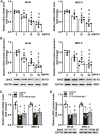
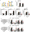
Excessive inflammatory injury is the main cause of the incidence of severe neonatal pneumonia (NP) and associated deaths. Although dickkopf-3 (DKK3) exhibits anti-inflammatory activity in numerous pathological processes, its role in NP is still unknown. In this study, human embryonic lung WI-38 and MRC-5 cells were treated with lipopolysaccharide (LPS) to induce inflammatory injury of NP in vitro. The expression of DKK3 was downregulated in LPS-stimulated WI-38 and MRC-5 cells. DKK3 overexpression decreased LPS-induced inhibition of cell viability, and reduced LPS-induced apoptosis of WI-38 and MRC-5 cells. DKK3 overexpression also reduced LPS-induced production of pro-inflammatory factors such as ROS, IL-6, MCP-1, and TNF-α. Nuclear respiratory factors 1 (NRF1) knockdown was found to upregulate DKK3 and inactivate the GSK-3β/β-catenin pathway in LPS-injured WI-38 and MRC-5 cells. NRF1 knockdown also suppressed LPS-induced inhibition on cell viability, repressed LPS-induced apoptosis, and inhibited the accumulation of ROS, IL-6, MCP-1, and TNF-α in LPS-injured WI-38 and MRC-5 cells. DKK3 knockdown or re-activation of the GSK-3β/β-catenin pathway reversed the inhibitory effects of NRF1 knockdown on LPS-induced inflammatory injury. In conclusion, NRF1 knockdown can alleviate LPS-triggered inflammatory injury by regulating DKK3 and the GSK-3β/β-catenin pathway.
Keywords: DKK3; GSK-3β/β-catenin; NRF1; inflammatory injury; neonatal pneumonia.
© The Author(s) 2023. Published by Oxford University Press on behalf of the British Society for Immunology. All rights reserved. For permissions, please e-mail: journals.permissions@oup.com.
Conflict of interest statement
The authors declare no conflicts of interest.
Figures


Similar articles
-
DKK3 overexpression attenuates cardiac hypertrophy and fibrosis in an angiotensin-perfused animal model by regulating the ADAM17/ACE2 and GSK-3β/β-catenin pathways.J Mol Cell Cardiol. 2018 Jan;114:243-252. doi: 10.1016/j.yjmcc.2017.11.018. Epub 2017 Dec 5. J Mol Cell Cardiol. 2018. PMID: 29196099
-
GSK-3Beta-Dependent Activation of GEF-H1/ROCK Signaling Promotes LPS-Induced Lung Vascular Endothelial Barrier Dysfunction and Acute Lung Injury.Front Cell Infect Microbiol. 2017 Aug 4;7:357. doi: 10.3389/fcimb.2017.00357. eCollection 2017. Front Cell Infect Microbiol. 2017. PMID: 28824887 Free PMC article.
-
miR-709 modulates LPS-induced inflammatory response through targeting GSK-3β.Int Immunopharmacol. 2016 Jul;36:333-338. doi: 10.1016/j.intimp.2016.04.005. Epub 2016 May 25. Int Immunopharmacol. 2016. PMID: 27232654
-
Novel anti-inflammatory role for glycogen synthase kinase-3beta in the inhibition of tumor necrosis factor-alpha- and interleukin-1beta-induced inflammatory gene expression.J Biol Chem. 2006 Jun 23;281(25):16985-16990. doi: 10.1074/jbc.M602446200. Epub 2006 Apr 6. J Biol Chem. 2006. PMID: 16601113
-
The Wnt/β-catenin pathway regulates inflammation and apoptosis in ventilator-induced lung injury.Biosci Rep. 2023 Mar 31;43(3):BSR20222429. doi: 10.1042/BSR20222429. Biosci Rep. 2023. PMID: 36825682 Free PMC article.
Cited by
-
Proteomic Risk Score of Increased Respiratory Susceptibility: A Multi-Cohort Study.Am J Respir Crit Care Med. 2024 Sep 10;211(1):64-74. doi: 10.1164/rccm.202403-0613OC. Online ahead of print. Am J Respir Crit Care Med. 2024. PMID: 39254293
-
Advances and insights for DKK3 in non-cancerous diseases: a systematic review.PeerJ. 2025 Feb 13;13:e18935. doi: 10.7717/peerj.18935. eCollection 2025. PeerJ. 2025. PMID: 39959827 Free PMC article.
-
Elamipretide(SS-31) Attenuates Idiopathic Pulmonary Fibrosis by Inhibiting the Nrf2-Dependent NLRP3 Inflammasome in Macrophages.Antioxidants (Basel). 2023 Nov 21;12(12):2022. doi: 10.3390/antiox12122022. Antioxidants (Basel). 2023. PMID: 38136142 Free PMC article.
References
-
- McAllister DA, Liu L, Shi T, Chu Y, Reed C, Burrows J, et al. . Global, regional, and national estimates of pneumonia morbidity and mortality in children younger than 5 years between 2000 and 2015: a systematic analysis. Lancet Glob Health 2019, 7, e47–57. doi:10.1016/S2214-109X(18)30408-X. - DOI - PMC - PubMed
MeSH terms
Substances
LinkOut - more resources
Full Text Sources
Medical
Miscellaneous

