. 2023 Jul 4;20(1):53.
doi: 10.1186/s12987-023-00448-x.

Spontaneously hypertensive rats can become hydrocephalic despite undisturbed secretion and drainage of cerebrospinal fluid

Affiliations

Spontaneously hypertensive rats can become hydrocephalic despite undisturbed secretion and drainage of cerebrospinal fluid

Sara Diana Lolansen et al. Fluids Barriers CNS. .

Abstract

Background: Hydrocephalus constitutes a complex neurological condition of heterogeneous origin characterized by excessive cerebrospinal fluid (CSF) accumulation within the brain ventricles. The condition may dangerously elevate the intracranial pressure (ICP) and cause severe neurological impairments. Pharmacotherapies are currently unavailable and treatment options remain limited to surgical CSF diversion, which follows from our incomplete understanding of the hydrocephalus pathogenesis. Here, we aimed to elucidate the molecular mechanisms underlying development of hydrocephalus in spontaneously hypertensive rats (SHRs), which develop non-obstructive hydrocephalus without the need for surgical induction.

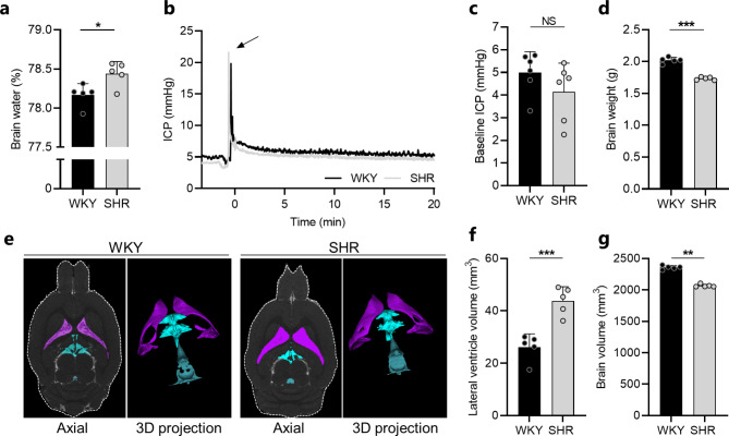
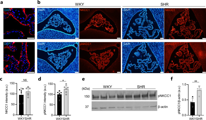
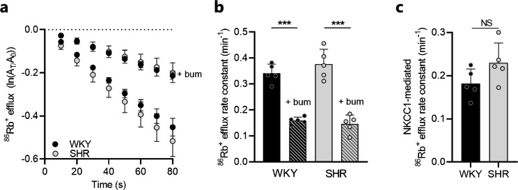
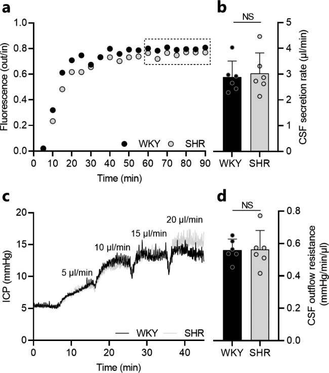
Methods: Magnetic resonance imaging was employed to delineate brain and CSF volumes in SHRs and control Wistar-Kyoto (WKY) rats. Brain water content was determined from wet and dry brain weights. CSF dynamics related to hydrocephalus formation in SHRs were explored in vivo by quantifying CSF production rates, ICP, and CSF outflow resistance. Associated choroid plexus alterations were elucidated with immunofluorescence, western blotting, and through use of an ex vivo radio-isotope flux assay.

Results: SHRs displayed brain water accumulation and enlarged lateral ventricles, in part compensated for by a smaller brain volume. The SHR choroid plexus demonstrated increased phosphorylation of the Na+/K+/2Cl- cotransporter NKCC1, a key contributor to choroid plexus CSF secretion. However, neither CSF production rate, ICP, nor CSF outflow resistance appeared elevated in SHRs when compared to WKY rats.

Conclusion: Hydrocephalus development in SHRs does not associate with elevated ICP and does not require increased CSF secretion or inefficient CSF drainage. SHR hydrocephalus thus represents a type of hydrocephalus that is not life threatening and that occurs by unknown disturbances to the CSF dynamics.

Keywords: Cerebrospinal fluid; Choroid plexus; Hydrocephalus; Intracranial pressure; Outflow resistance; Spontaneously hypertensive rat; Ventriculomegaly.


Conflict of interest statement

The authors declare no competing interests.

Figures

Fig. 1
Fig. 1
SHRs display ventricular enlargement and excessive brain water accumulation. a Brain water content quantified from WKY rats (n = 5) and SHRs (n = 5) using the wet and dry brain weight. b Representative ICP traces for one WKY rat and one SHR. Arrow indicates injection of 5 µl aCSF to assure proper ICP reading. c Baseline ICP in WKY rats (n = 6) and SHRs (n = 6) quantified from a stable 20 min time period. d Brain weight of WKY rats (n = 5) and SHRs (n = 5) assessed post mortem. e Representative MRI brain sections (2D axial plane and 3D projections) of one WKY rat and one SHR demonstrating the lateral ventricle volume (purple), third ventricle volume (turquoise), and fourth ventricle volume (blue). f Lateral ventricle volumes of WKY rats (n = 5) and SHRs (n = 5) quantified from MRI. g Brain volumes of WKY rats (n = 5) and SHRs (n = 5) quantified by MRI. Error bars represent standard deviation and statistical significance was tested with an unpaired two-tailed t-test or a Mann-Whitney test depending on normality. *P < 0.05, **P < 0.01, *** P < 0.001, NS = not significant
Fig. 2
Fig. 2
The SHR choroid plexus demonstrates upregulation of pNKCC1. a Confocal microscopy images demonstrating apical localization of NKCC1 (red, top panel) and pNKCC1 (red, lower panel) in SHR choroid plexus. Cell nuclei are stained with DAPI (blue). Scale bar: 50 μm. b Representative immunofluorescence images of the choroid plexus from WKY rats (left panels) and SHRs (right panels) stained for NKCC1 (red, top panels), pNKCC1 (red, lower panels), and cell nuclei (DAPI, blue). Scale bar: 100 μm. c-d Quantification of NKCC1 (c) or pNKCC1 (d) fluorescence intensity in choroid plexus from WKY rats and SHRs, n = 6 in each group. e Western blot of pNKCC1 in choroid plexus from WKY rats and SHRs. β-actin served as loading control. f quantification of pNKCC1 abundance in choroid plexus from WKY rats (n = 4) and SHRs (n = 4) normalized to β-actin. Error bars represent standard deviation and statistical significance was tested with an unpaired two-tailed t-test or a Mann-Whitney test. * P < 0.05, **P < 0.01, NS = not significant
Fig. 3
Fig. 3
Choroid plexus NKCC1 transport activity is unaltered in SHRs. a Loss of 86Rb+ from the choroid plexus as a function of time in WKY rats (n = 5) and SHRs (n = 5) in presence or absence of 20 µM bumetanide (bum). The y-axis is the natural logarithm of the choroid plexus 86Rb+ amount left at time T (AT) divided by the initial amount at time 0 (A0). b Efflux rate constant for 86Rb+ in WKY rats (n = 5) and SHRs (n = 5) in presence or absence of 20 µM bumetanide (bum). c NKCC1-mediated 86Rb+ efflux rate constant (bumetanide-sensitive fractions) in WKY rats (n = 5) and SHRs (n = 5). Error bars represent standard deviation and statistical significance was tested with one-way ANOVA followed by Sidak’s multiple comparisons test or an unpaired-two tailed t-test. ***P < 0.001, NS = not significant
Fig. 4
Fig. 4
The CSF secretion rate and outflow resistance is not elevated in SHRs. a Representative time course traces of the fluorescence ratio of dextran (outflow/inflow) during ventriculo-cisternal perfusion of one WKY rat and one SHR. Squared insert indicates the region used for quantification of CSF secretion rates. b CSF secretion rate in WKY rats (n = 6) and SHRs (n = 6) quantified from the fluorescence ratio of dextran. c Representative ICP traces of one WKY rat and one SHR undergoing measurements of CSF outflow resistance by infusion of aCSF at rates indicated in the figure. e CSF outflow resistance in WKY rats (n = 6) and SHRs (n = 6). Error bars represent standard deviation and statistical significance was tested with an unpaired two-tailed t-test. NS = not significant

Similar articles

Cited by

References

    1. Hochstetler A, Raskin J, Blazer-Yost BL. Hydrocephalus: historical analysis and considerations for treatment. Eur J Med Res. 2022;27:168. - PMC - PubMed
    1. Singh R, Prasad RS, Singh RC, Trivedi A, Bhaikhel KS, Sahu A. Evaluation of Pediatric Hydrocephalus: Clinical, Surgical, and Outcome Perspective in a Tertiary Center. Asian J Neurosurg. 2021;16:706–13. - PMC - PubMed
    1. Williams MA, Nagel SJ, Luciano MG, Relkin N, Zwimpfer TJ, Katzen H, et al. The clinical spectrum of hydrocephalus in adults: report of the first 517 patients of the adult Hydrocephalus Clinical Research Network registry. J Neurosurg. 2019;132:1773–84. - PubMed
    1. Laurence KM, Coates S. The natural history of Hydrocephalus: detailed analysis of 182 unoperated cases. Arch Dis Child. 1962;37:345–62. - PMC - PubMed
    1. di Curzio DL. Animal models of Hydrocephalus. Open J Mod Neurosurg. 2018;08:57–71.