. 2023 Oct;15(10):866-880.
doi: 10.1111/1753-0407.13439. Epub 2023 Jul 4.

Protective effects of nattokinase against microvasculopathy and neuroinflammation in diabetic retinopathy

Affiliations

Protective effects of nattokinase against microvasculopathy and neuroinflammation in diabetic retinopathy

Zijing Huang et al. J Diabetes. 2023 Oct.

Abstract

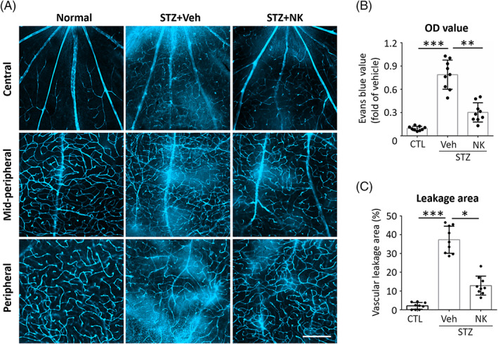
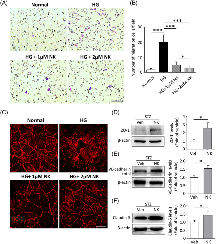
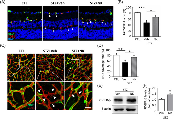
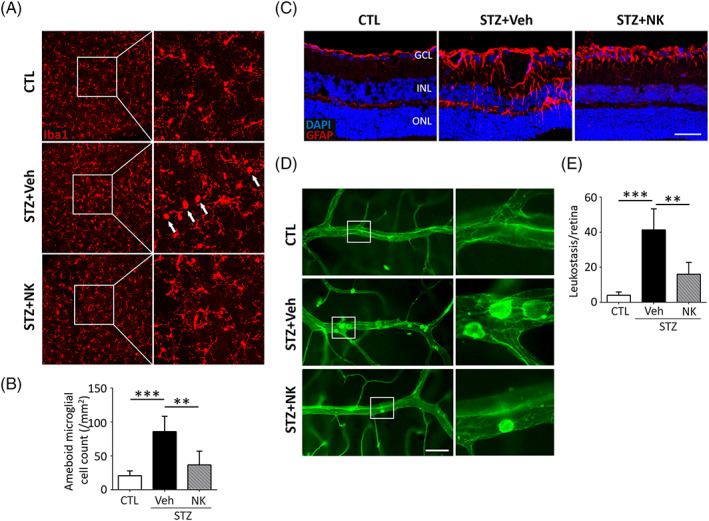
Aims: Diabetic retinopathy (DR) is a significant global public health concern. Alternative, safe, and cost-effective pharmacologic approaches are warranted. We aimed to investigate the therapeutic potential of nattokinase (NK) for early DR and the underlying molecular mechanism.

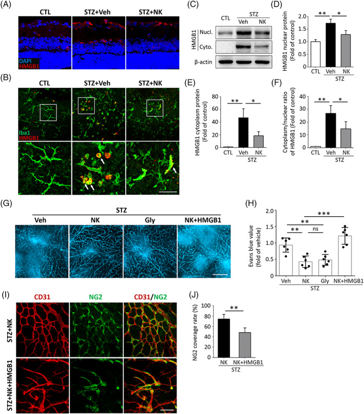
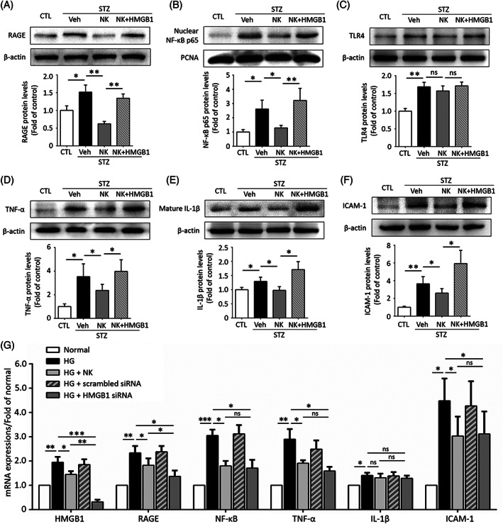
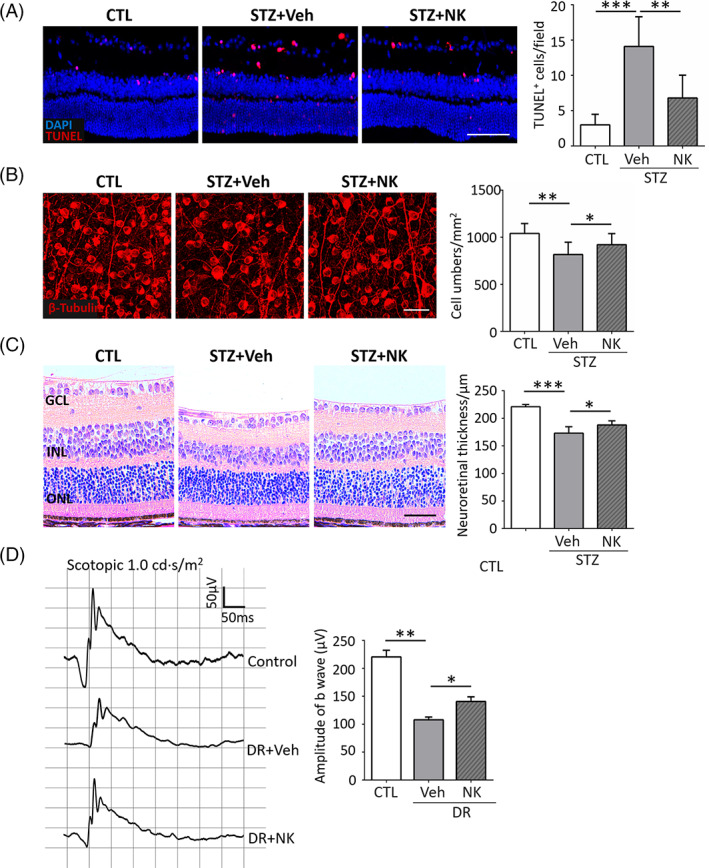
Methods: A mouse model of diabetes induced by streptozotocin was utilized and NK was administered via intravitreal injection. Microvascular abnormities were evaluated by examining the leakage from blood-retinal barrier dysfunction and loss of pericytes. Retinal neuroinflammation was examined through the assessment of glial activation and leukostasis. The level of high mobility group box 1 (HMGB1) and its downstream signaling molecules was evaluated following NK treatment.

Results: NK administration significantly improved the blood-retinal barrier function and rescued pericyte loss in the diabetic retinas. Additionally, NK treatment inhibited diabetes-induced gliosis and inflammatory response and protected retinal neurons from diabetes-induced injury. NK also improved high glucose-induced dysfunction in cultured human retinal micrangium endothelial cells. Mechanistically, NK regulated diabetes-induced inflammation partially by modulating HMGB1 signaling in the activated microglia.

Conclusions: This study demonstrated the protective effects of NK against microvascular damages and neuroinflammation in the streptozotocin-induced DR model, suggesting that NK could be a potential pharmaceutical agent for the treatment of DR.

目的: 糖尿病视网膜病变(DR)是一个重要的全球公共卫生问题。需要寻求安全、有效且经济的替代性药物治疗方案。本研究的目的是探讨纳豆激酶(NK)对早期DR的治疗潜力及其潜在的分子机制。 方法: 使用链脲佐啶诱导的糖尿病小鼠模型,通过玻璃体腔内注射给予NK。通过检查血液视网膜屏障功能障碍导致的渗漏和毛细血管周细胞丢失来评估微血管异常。通过评估胶质细胞活化和白细胞粘附来评估视网膜神经炎症。在NK处理后,评估高迁移率族蛋白B1(HMGB1)及其下游信号分子的水平。 结果: NK处理显著改善了糖尿病大鼠血-视网膜屏障功能并逆转了糖尿病视网膜中毛细血管周细胞的丢失。此外,NK处理抑制了糖尿病诱导的胶质细胞活化和炎症反应,并保护视网膜神经元免受糖尿病损伤。NK还改善了高糖诱导的人视网膜微血管内皮细胞功能障碍。在机制上,NK通过调节活化的小胶质细胞中的HMGB1信号,部分调节糖尿病诱导的炎症。 结论: 本研究表明,NK对链脲佐啶诱导的DR模型中的微血管损伤和神经炎症具有保护作用,这表明NK可能是治疗DR的潜在药物。.

Keywords: diabetic retinopathy; high mobility group box 1; microvasculopathy; nattokinase; neuroinflammation; 微血管病变; 神经炎症; 糖尿病视网膜病变; 纳豆激酶; 高迁移率族蛋白B1.


Conflict of interest statement

The authors declare that they have no competing interests.

Figures

FIGURE 1
FIGURE 1
NK attenuated microvascular leakage in mouse models of diabetic retinopathy and ischemic retinopathy. (A–C) Representative images and statistical analysis of Evans blue assays of the STZ‐induced diabetic retinopathy model. In the diabetic retina, the breakdown of blood retinal barrier caused leakage of the dye into the retinal parenchyma, as compared to the presence of sharply outlined vessels in the normal retina. Treatment of NK (STZ + NK) markedly reduced vascular leakage as compared to the control treatment (STZ + Veh). n = 9 retinas in each group. Scale bar: 100 μm. Data are shown as mean ± SD. **p < .01. ***p < .001. CTL, control; NK, nattokinase; OD, optical density; STZ, streptozotocin; Veh, vehicle.
FIGURE 2
FIGURE 2
NK improved endothelial dysfunction via modulation of tight junctions. (A, B) Representative images and statistical analysis of transwell migration assays in cultured human retinal micrangium endothelial cells (HRMECs). High glucose (HG, 50 mM) increased HRMEC migration as compared to the normal glucose conditions, which was largely inhibited by pretreatment of NK (HG + NK) in a dose‐dependent manner. Purple, migrated cells; White, pores of the membrane. Scale bar: 100 μm. N = 6 wells and 3 fields per well were randomly chosen for analysis. (C) Representative images of the ZO‐1 immunostaining on HRMECs. NK improved the integrity of tight junction protein ZO‐1 under HG condition in a dose‐dependent manner as compared to the control treatment (Veh). Scale bar: 25 μm. (D–F) Western blotting results showed upregulated levels of endothelial junctional proteins, including ZO‐1, VE‐cadherin, and claudin‐5, in the STZ‐induced diabetic retina with NK treatment. β‐actin was used as the loading control. Data are shown as mean ± SD. *p < .05. ***p < .001. NK, nattokinase; STZ, streptozotocin; VE, vascular endothelial; Veh, vehicle.
FIGURE 3
FIGURE 3
NK prevented pericyte loss in the STZ‐induced diabetic retina. (A, B) Representative images and statistical analysis of coimmunostaining of CD31 and NG2 on retinal cryosections. In normal retina, the vessel integrity was maintained as most of CD31+ endothelial cells were covered with NG2+ pericytes, whereas STZ‐induced diabetes resulted in an increase in the number and proportion of CD31+NG2 cells (arrows), indicating the loss of pericyte coverage of vessels. NK treatment significantly increased the number and percentage of CD31+NG2+ cells (arrowheads) as compared to the control treatment (Veh). n = 6 retinas from 6 mice for analysis. Scale bar: 100 μm. (C, D) Representative images and statistical analysis of co‐immunostaining of CD31 and NG2 on retinal flat‐mounts. Arrowheads indicated the microvessels with pericyte loss, which was partly rescued by treatment of NK as compared to the control treatment (Veh). Scale bar: 50 μm. (E, F) Western blotting analysis showed upregulation of PDGFR‐β, a pericyte marker, in the NK‐treated diabetic retina. β‐Actin was used as the loading control. Data are shown as mean ± SD. *p < .05. **p < .01. ***p < .001. CTL, control; NG2, nerve/glial antigen 2; NK, nattokinase; PDGFR‐β, platelet derived growth factor receptor beta; STZ, streptozotocin; Veh, vehicle.
FIGURE 4
FIGURE 4
NK orchestrated glial activation and alleviated leukostasis in the STZ‐induced diabetic retina. (A, B) Representative images and statistical analysis of immunostaining of the Iba1+ microglia on retinal whole mounts. Microglia presented an activated “ameboid” profile, featured with enlarged somas and short lamellipodia (arrows) in the diabetic retinas as compared to a ramified microglia phenotype that presented small cell bodies and long processes in the normal retinas (CTL). NK treatment reduced the number of ameboid microglia in the diabetic retinas as compared to the control treatment (Veh). n = 6 retinas from 6 mice for analysis. Scale bar: 50 μm. (C) Immunostaining of GFAP on retinal astrocytes and Müller cells. In the normal retina, GFAP was mainly localized to astrocytes in the superficial layer. In the diabetic retina, intensive GFAP labeling in astrocytes and Müller cells in the inner layer was detected, whereas NK significantly reduced GFAP immunoreactivity as compared to the control treatment (Veh). Scale bar: 100 μm. (D, E) Representative images and statistical analysis of leukostasis assay showing increased number of adhered leukocytes in the STZ‐induced diabetic retina, which was abolished by NK treatment, as compared to the control treatment (Veh). n = 6 retinas from 6 mice for analysis. Scale bar: 50 μm. Data are shown as mean ± SD. *p < .05, **p < .01. ***p < .001. CTL, control; GFAP, glial fibrillary acidic protein; Iba1, ionized calcium‐binding adapter molecule 1; NK, nattokinase; STZ, streptozotocin; Veh, vehicle.
FIGURE 5
FIGURE 5
HMGB1 was involved in the NK‐mediated protective effect against experimental diabetic retinopathy. (A) Representative images and statistical analysis of immunostaining assay showed an increased fluorescence intensity of HMGB1 in the diabetic retina, which was markedly reversed by NK administration, as compared to the control treatment (Veh). n = 6 retinas from 6 mice for analysis. Scale bar: 50 μm. (B) Double immunostaining assay showed that HMGB1 was mainly colocalized with the ameboid microglia. (C–F) Western blotting analysis using the NE‐PERTM Nuclear and Cytoplasmic Extraction Reagents showed that NK inhibited the cytoplasmic protein levels and nuclear‐cytoplasmic translocation of HMGB1. (G, H) Evans blue assay in retinal whole mounts showed comparable vasoprotective effect of NK and glycyrrhizin (Gly), a specific HMGB1 inhibitor, in the diabetic retina. Coadministration of recombinant HMGB1 abolished the effect of NK against vascular leakage. n = 6 retinas in each group. Scale bar: 100 μm. (I‐J) The prosurvival effect of NK on NG2+ pericytes was reduced by cotreatment of recombinant HMGB1. Scale bar: 100 μm. Data are presented as mean ± SD. *p < .05, **p < .01, ***p < .001. CTL, control; Cyto, cytoplasmic; HMGB1, high mobility group box 1; NG2, nerve/glial antigen 2; NK, nattokinase; Ns, no significance; Nucl, nuclear; STZ, streptozotocin; Veh, vehicle.
FIGURE 6
FIGURE 6
NK modulated HMGB1/RAGE/NF‐κB signaling and its downstream inflammatory cytokines in the diabetic retina and cultured HRMECs. (A–F) Representative images of western blotting assays and statistical analysis. NK treatment down‐regulated the levels of RAGE (A) and NF‐κB (B) downstream of HMGB1, but not toll‐like receptor 4 (TLR4) (C), in the diabetic retina, as compared to the control treatment (Veh). NK treatment significantly reduced the expression of several pro‐inflammatory cytokines, including TNF‐α (D), IL‐1β (E), and ICAM‐1 (F), as compared to the control treatment (Veh). Proliferating cell nuclear antigen (PCNA) was used as the loading control for nuclear NF‐κB p65 expression. (G) qPCR analysis for the HMGB1 signaling and its downstream molecules in cultured HRMECs. Both NK and HMGB1 siRNA treatment could downregulated the levels RAGE/NF‐κB and pro‐inflammatory cytokines, except for IL‐1β. The western blotting and qPCR assays were independently repeated for three times in the analysis. Data are shown as mean ± SD. *p < .05, **p < .01, ***p < .001. CTL, control; HG, high glucose; HMGB1, high mobility group box 1; HRMEC, human retinal micrangium endothelial cell; ICAM‐1, intercellular adhesion molecule 1; IL‐1β, interleukin‐1β; NF‐κB, nuclear factor kappa B; NK, nattokinase; Ns, no significance; qPCR, quantitative polymerase chain reaction; RAGE, receptor for advanced glycation end‐products; STZ, streptozotocin; TNF‐α, tumor necrosis factor‐alpha; Veh, vehicle.
FIGURE 7
FIGURE 7
NK rescued retinal cells from apoptosis and improved neural functions in the diabetic retina. (A) TUNEL assay on retinal cryosections showed that NK significantly prevented retinal neurons from apoptosis as compared to the control treatment (Veh). Scale bar: 100 μm. n = 6 retinas from 6 mice for analysis. (B) Immunostaining of β‐tubulin on retinal whole mounts showed that NK increased the number of retinal ganglion cells under diabetic conditions. Scale bar: 50 μm. (C) Representative and statistical analysis of H&E staining. The decrease in neuroretinal thickness in DR mice was partially rescued by NK treatment. Scale bar: 100 μm. (D) Representative and statistical analysis of ERG in normal and DR mice. The amplitudes of b wave in the NK‐treated mice significantly increased as compared to that of the vehicle‐treated mice (Veh). n = 9 retinas from 9 mice in each group. Data are shown as mean ± SD. *p < .05, **p < .01, ***p < .001. CTL, control; DR, diabetic retinopathy; ERG, electroretinogram; H&E, hematoxylin and eosin; NK, nattokinase; STZ, streptozotocin; TUNEL, terminal‐deoxynucleoitidyl transferase mediated nick end‐labeling; Veh, vehicle.

Similar articles

Cited by

References

    1. Cheung N, Mitchell P, Wong TY. Diabetic retinopathy. Lancet. 2010;376(9735):124‐136. doi: 10.1016/S0140-6736(09)62124-3 - DOI - PubMed
    1. Yau JW, Rogers SL, Kawasaki R, et al. Global prevalence and major risk factors of diabetic retinopathy. Diabetes Care. 2012;35(3):556‐564. doi: 10.2337/dc11-1909 - DOI - PMC - PubMed
    1. Bergers G, Hanahan D. Modes of resistance to anti‐angiogenic therapy. Nat Rev Cancer. 2008;8(8):592‐603. doi: 10.1038/nrc2442 - DOI - PMC - PubMed
    1. Chung EJ, Roh MI, Kwon OW, Koh HJ. Effects of macular ischemia on the outcome of intravitreal bevacizumab therapy for diabetic macular edema. Retina. 2008;28(7):957‐963. doi: 10.1097/IAE.0b013e3181754209 - DOI - PubMed
    1. Hammes HP, Lin J, Renner O, et al. Pericytes and the pathogenesis of diabetic retinopathy. Diabetes. 2002;51(10):3107‐3112. doi: 10.2337/diabetes.51.10.3107 - DOI - PubMed