. 2023 Aug 31;14(4):e0078923.
doi: 10.1128/mbio.00789-23. Epub 2023 Jul 5.

Impact of HIV-1 Vpu-mediated downregulation of CD48 on NK-cell-mediated antibody-dependent cellular cytotoxicity

Affiliations

Impact of HIV-1 Vpu-mediated downregulation of CD48 on NK-cell-mediated antibody-dependent cellular cytotoxicity

Lorie Marchitto et al. mBio. .

Abstract

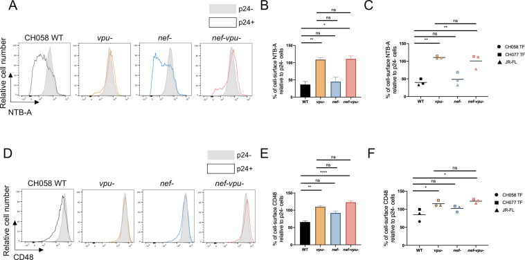
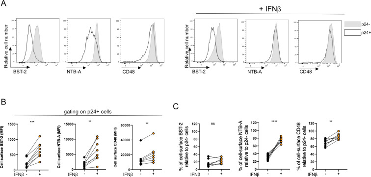
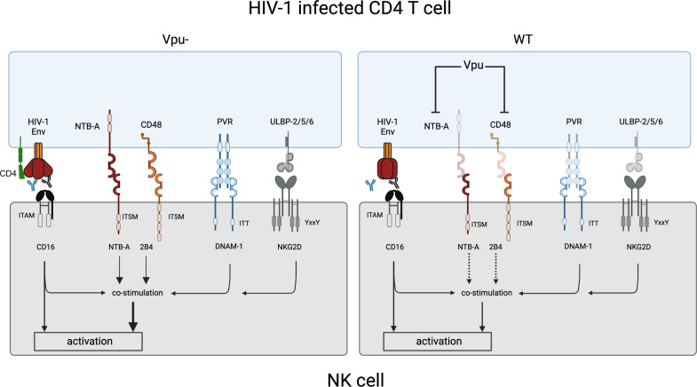
HIV-1 evades antibody-dependent cellular cytotoxicity (ADCC) responses not only by controlling Env conformation and quantity at the cell surface but also by altering NK cell activation via the downmodulation of several ligands of activating and co-activating NK cell receptors. The signaling lymphocyte activation molecule (SLAM) family of receptors, which includes NTB-A and 2B4, act as co-activating receptors to sustain NK cell activation and cytotoxic responses. These receptors cooperate with CD16 (FcγRIII) and other activating receptors to trigger NK cell effector functions. In that context, Vpu-mediated downregulation of NTB-A on HIV-1-infected CD4 T cells was shown to prevent NK cell degranulation via an homophilic interaction, thus contributing to ADCC evasion. However, less is known on the capacity of HIV-1 to evade 2B4-mediated NK cell activation and ADCC. Here, we show that HIV-1 downregulates the ligand of 2B4, CD48, from the surface of infected cells in a Vpu-dependent manner. This activity is conserved among Vpu proteins from the HIV-1/SIVcpz lineage and depends on conserved residues located in its transmembrane domain and dual phosphoserine motif. We show that NTB-A and 2B4 stimulate CD16-mediated NK cell degranulation and contribute to ADCC responses directed to HIV-1-infected cells to the same extent. Our results suggest that HIV-1 has evolved to downmodulate the ligands of both SLAM receptors to evade ADCC. IMPORTANCE Antibody-dependent cellular cytotoxicity (ADCC) can contribute to the elimination of HIV-1-infected cells and HIV-1 reservoirs. An in-depth understanding of the mechanisms used by HIV-1 to evade ADCC might help develop novel approaches to reduce the viral reservoirs. Members of the signaling lymphocyte activation molecule (SLAM) family of receptors, such as NTB-A and 2B4, play a key role in stimulating NK cell effector functions, including ADCC. Here, we show that Vpu downmodulates CD48, the ligand of 2B4, and this contributes to protect HIV-1-infected cells from ADCC. Our results highlight the importance of the virus to prevent the triggering of the SLAM receptors to evade ADCC.

Keywords: ADCC; BST-2; CD4; CD48; HIV-1; NK cell; NTB-A; Nef; SLAM; Vpu.


Conflict of interest statement

The authors declare no conflict of interest.

Figures

Fig 1
Fig 1
HIV-1 downregulation of NTB-A and CD48. Primary CD4+ T cells were infected with indicated IMCs. At 48 h post infection, cells were stained with anti-NTB-A and anti-CD48 Abs, followed with the appropriate secondary Abs. (A and C) The graphs shown represent the percentage of median fluorescence intensities (MFI) of (A) NTB-A or (C) CD48 detected on the surface of p24+ cells (black bars) relative to the bystander p24 cells (gray bars) for at least four independent experiments. (B and D) Graph representing the mean percentage of surface expression for (B) NTB-A and (D) CD48 for each tested virus. Error bars indicate means ± standard errors of the means (SEM). Statistical significance was tested using unpaired t-tests or Mann-Whitney U tests based on statistical normality (*P < 0.05; **P < 0.01; ***P < 0.001; ****P < 0.0001; ns, non-significant).
Fig 2
Fig 2
HIV-1 Vpu downregulates CD48. Primary CD4+ T cells infected with CH058 TF either WT or defective for Nef, and/or Vpu expression were stained for NTB-A and CD48 cell surface expression 48 h post infection. (A and D) Histograms representing cell surface (A) NTB-A and (D) CD48. (A and D) Uninfected bystander p24 cells are shown in gray, while infected p24+ cells are shown in black (WT), orange (vpu−), blue (nef−), or red (nef-vpu−). (B and E) Bar graphs representing cell surface (B) NTB-A or (E) CD48 for at least six independent experiments. (C and F) Cells were also infected with CH058 TF, CH077 TF, and JR-FL IMC either WT or defective for Nef and/or Vpu expression. Dot plots indicate the mean cell-surface percentage for (C) NTB-A and (F) CD48 for each virus. Error bars indicate means ± standard errors of the means (SEM). Statistical significance was tested using a (A–F) Ordinary one-way ANOVA or Kruskal-Wallis tests based on statistical normality (*P < 0.05; **P < 0.01; ****P < 0.0001; ns, non-significant).
Fig 3
Fig 3
Vpu-mediated downregulation of CD48 is conserved among different primate lentivirus lineages. (A) HEK293T cells were co-transfected with a plasmid expressing the hCD48 (0.6 µg) and pCGCG vector expressing eGFP alone or together with CH058 TF vpu or nef (0, 0.15, 0.3, or 0.6 µg). Cell-surface CD48 was assessed 48 h post transfection and used to calculate the percentage of receptor downregulation. The data represent three independent experiments. (B–F) HEK293T cells were co-transfected with plasmids expressing (B and C) hCD48 (0.6 µg), (D) hNTB-A (1 µg), (E) hBST-2 (1 µg), or (F) hCD4 (1 µg) and pCGCG vector expressing eGFP alone or together with indicated Vpu (0.6 µg). Cell surface levels of NTBA, CD48, BST-2, and CD4 were assessed 48 h post transfection. (B) Histograms representing CD48 surface levels on HEK293T cells co-transfected with hCD48 and pCGCG vector expressing eGFP alone or together with indicated vpu (C–F) Bar graphs indicate the percentage of cell-surface levels of each surface protein detected on cells transfected with Vpu vectors relative to cells transfected with the control vector. Error bars indicate means ± standard errors of the means (SEM). (G) Correlation between the percentage of cell-surface CD48 and NTB-A, BST-2, or CD4. Statistical significance was tested using (A) one-way ANOVA test and (G) Spearman and Pearson rank correlation test (*P < 0.05; **P < 0.01; ns, non-significant).
Fig 4
Fig 4
Vpu transmembrane domain and phosphoserine motif are important for CD48 downregulation. Primary CD4+ T cells infected with CH058 TF viruses expressing wild-type vpu (WT), vpu S52A/S56A, vpu A14L/A18L, or defective for vpu expression (vpu−) were stained for NTB-A and CD48 cell-surface expression 48 h post infection. (A and C) Histograms representing cell surface (A) NTB-A and (C) CD48. (A and C) Uninfected bystander p24 cells are shown in gray, while infected p24+ cells are shown in black (WT), orange (vpu−), blue (vpu S52A/S56A), or red (vpu A14L/A18L). (B and D) Bar graph representing the percentage of cell-surface (B) NTB-A or (D) CD48 for at least five independent experiments. Error bars indicate means ± standard errors of the means (SEM). Statistical significance was tested using (A–D) Statistical significance was tested using an ordinary one-way ANOVA (*P < 0.05; **P < 0.01; ***P < 0.001; ****P < 0.0001; ns, non-significant).
Fig 5
Fig 5
Upregulation of BST-2 by IFN-β treatment affects CD48 downregulation. Primary CD4+ T cells infected with CH058 TF WT virus were treated or not with IFN-β 24 h post infection, and cell-surface BST-2, NTB-A, and CD48 levels were assessed 48 h post infection. (A) Histograms representing cell surface BST-2, NTB-A, and CD48 levels when (left) untreated or (right) treated with IFN-β. Uninfected bystander p24 cells are shown in gray, while infected p24+ cells are depicted in black dot line. (B and C) Dots representing (B) median fluorescence intensity (MFI) detected on p24+ infected cells or (C) the percentage of cell-surface BST-2, NTB-A, or CD48 in p24+ cells relative to p24 cells in eight independent experiments. Error bars indicate means ± standard errors of the means (SEM). (B and C) Statistical significance was tested using paired t-tests or Wilcoxon tests based on statistical normality (**P < 0.01; ***P < 0.001; ****P < 0.0001; ns, non-significant).
Fig 6
Fig 6
NTB-A and 2B4 trigger NK cell degranulation to a similar extent. P815 cells were incubated with indicated mAbs or a matched IgG isotype. P815 cells were then mixed with purified NK cells and incubated for 4 h. CD3CD56+ cells were evaluated for percentage of cell-surface CD107a. Contour plots depict NK cells stimulation. Bar graphs represent the percentage of CD107a expression among CD3CD56+ cells. (A) Stimulation with P815 cells coated with isotype control Abs or anti-CD16 Abs, +/−anti-NTB-A Abs, and/or anti-2B4 Abs. (B) Stimulation with P815 cells coated with isotype control Abs or anti-CD16 Abs, +/−anti-NKG2D, +/−anti-NTB-A Abs, and/or anti-2B4 Abs. (C) Stimulation with P815 cells coated with isotype control Abs or anti-CD16 Abs, +/−anti-DNAM-1, +/−anti-NTB-A Abs, and/or anti-2B4 Abs. Statistical differences relative to stimulation with anti-CD16 Abs alone are represented above each antibody tested. Statistical significance was tested using paired t-tests or Wilcoxon tests based on statistical normality (*P < 0.05; **P < 0.01; ns, non-significant).
Fig 7
Fig 7
NTB-A and 2B4 engagement enhances ADCC against HIV-1-infected cells. Primary CD4+ T cells infected with CH058 TF WT or vpu− viruses were used as target cells, while autologous PBMCs were used as effector cells to perform ADCC killing assay. PBMCs were pre-incubated or not with anti-NTB-A and/or anti-2B4 Abs or their matched IgG isotype controls prior to incubation with target cells. Bar graph represents the percentage of ADCC obtained with the bNAb 3BNC117 in seven independent experiments. Statistical significance was tested using one-way ANOVA test (*P < 0.05; **P < 0.01; ns, non-significant).
Fig 8
Fig 8
Vpu-mediated downmodulation of NTB-A and CD48 contribute to evade ADCC. CD4 downmodulation by Vpu contributes to evade ADCC mediated by non-neutralizing Abs (depicted in blue) by preventing Env-CD4 interaction. The binding of CD16 to non-neutralizing or broadly neutralizing antibodies (depicted in black) bound to HIV-1 Env can directly activate NK cell effector functions but also in cooperation with others activating and coactivating receptors such as NKG2D, DNAM-1, 2B4, and NTB-A (left panel). The downmodulation of NTB-A and CD48 by Vpu on HIV-1-infected cells contributes to reduce the cooperativity between the SLAMs (NTB-A and 2B4), the activating receptors (DNAM-1 and NKG2D) and CD16 to stimulate NK cell effector functions, thus contributing to evade ADCC (right panel).

Similar articles

Cited by

References

    1. Bonsignori M, Pollara J, Moody MA, Alpert MD, Chen X, Hwang KK, Gilbert PB, Huang Y, Gurley TC, Kozink DM, Marshall DJ, Whitesides JF, Tsao CY, Kaewkungwal J, Nitayaphan S, Pitisuttithum P, Rerks-Ngarm S, Kim JH, Michael NL, Tomaras GD, Montefiori DC, Lewis GK, DeVico A, Evans DT, Ferrari G, Liao HX, Haynes BF. 2012. Antibody-dependent cellular cytotoxicity-mediating antibodies from an HIV-1 vaccine efficacy trial target multiple epitopes and preferentially use the Vh1 Gene family. J Virol 86:11521–11532. doi: 10.1128/JVI.01023-12 - DOI - PMC - PubMed
    1. Jegaskanda S, Weinfurter JT, Friedrich TC, Kent SJ. 2013. Antibody-dependent cellular cytotoxicity is associated with control of pandemic H1N1 influenza virus infection of macaques. J Virol 87:5512–5522. doi: 10.1128/JVI.03030-12 - DOI - PMC - PubMed
    1. Beltrán D, López-Vergès S. 2014. NK cells during Dengue disease and their recognition of dengue virus-infected cells. Front Immunol 5:192. doi: 10.3389/fimmu.2014.00192 - DOI - PMC - PubMed
    1. Banks ND, Kinsey N, Clements J, Hildreth JEK. 2002. Sustained antibody-dependent cell-mediated cytotoxicity (ADCC) in SIV-infected macaques correlates with delayed progression to AIDS. AIDS Res Hum Retroviruses 18:1197–1205. doi: 10.1089/08892220260387940 - DOI - PubMed
    1. Alpert MD, Harvey JD, Lauer WA, Reeves RK, Piatak M, Carville A, Mansfield KG, Lifson JD, Li W, Desrosiers RC, Johnson RP, Evans DT. 2012. ADCC develops over time during persistent infection with live-attenuated SIV and is associated with complete protection against SIV(Mac)251 challenge. PLoS Pathog 8:e1002890. doi: 10.1371/journal.ppat.1002890 - DOI - PMC - PubMed

Publication types

Substances