. 2023 Jul 19;25(28):18698-18710.
doi: 10.1039/d3cp01321a.

Living on the edge: light-harvesting efficiency and photoprotection in the core of green sulfur bacteria

Affiliations

Living on the edge: light-harvesting efficiency and photoprotection in the core of green sulfur bacteria

Alexander Klinger et al. Phys Chem Chem Phys. .

Abstract

Photosynthetic green sulfur bacteria are able to survive under extreme low light conditions. Nevertheless, the light-harvesting efficiencies reported so far, in particular for Fenna-Matthews-Olson (FMO) protein-reaction center complex (RCC) supercomplexes, are much lower than for photosystems of other species. Here, we approach this problem with a structure-based theory. Compelling evidence for a light-harvesting efficiency around 95% is presented for native (anaerobic) conditions that can drop down to 47% when the FMO protein is switched into a photoprotective mode in the presence of molecular oxygen. Light-harvesting bottlenecks are found between the FMO protein and the RCC, and the antenna of the RCC and its reaction center (RC) with forward energy transfer time constants of 39 ps and 23 ps, respectively. The latter time constant removes an ambiguity in the interpretation of time-resolved spectra of RCC probing primary charge transfer and provides strong evidence for a transfer-to-the trap limited kinetics of excited states. Different factors influencing the light-harvesting efficiency are investigated. A fast primary electron transfer in the RC is found to be more important for a high efficiency than the site energy funnel in the FMO protein, quantum effects of nuclear motion, or variations in the mutual orientation between the FMO protein and the RCC.


Conflict of interest statement

There are no conflicts to declare.

Figures

Fig. 1
Fig. 1. Structure of the FMO-RCC supercomplex and light harvesting. (A) Arrangement of BChl a (all but 802 and 803) and Chl a (802 and 803) pigments in the cryo-EM structural model of the FMO-RCC supercomplex containing the trimeric FMO protein and the dimeric RCC. The numbering of the pigments in the RCC is according to the protein data bank file 6M32 and that in the FMO protein follows the usual convention in the literature. Pigments that are colored equally are assigned to the same exciton domain in the calculation of optical spectra and energy transfer as explained in the main text. Numbers on the arrows are energy transfer time constants obtained in the 5-compartment model. The colored dashed lines depict the 4 excited states compartments denoted as FMO (black), RCC-Ant1 (purple), RCC-Ant2 (brown) and RCC-RC (blue). The 5th compartment contains the first charge-separated state formed by electron transfer from the special pair, which is encircled by a red-dashed line. Graphics of the molecules were made using VMD. (B) Sum of exciton state populations (black lines) and population of the charge separated state (red lines) obtained in different models as a function of time, assuming initial population of the FMO protein by transfer from the external baseplate, as described in the text.
Fig. 2
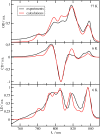
Fig. 2. Optical spectra of the FMO protein. Comparison of experimental (black lines) linear absorption (upper panel), circular dichroism (middle panel) and linear dichroism (lower panel) spectra at T = 6 K with calculations (red lines), using site energies obtained from a genetic algorithm and excitonic couplings calculated with a reaction field factor fRF = 1.15. The numerical values of the site energies and the excitonic couplings are given in the ESI, Table S1 and Section S1.3.1, respectively.
Fig. 3
Fig. 3. Optical spectra of the RCC. Comparison of experimental (black lines) linear absorption spectra (upper panel) at T = 77 K, circular dichroism (middle panel) and linear dichroism (lower panel) spectra at T = 6 K with calculations (red lines), using site energies obtained from a genetic algorithm and excitonic couplings calculated with a reaction field factor fRF = 1.15. The numerical values of the site energies and the excitonic couplings are given in the ESI, Table S2 and Section S1.3.1, respectively.
Fig. 4
Fig. 4. Light harvesting under anaerobic and aerobic conditions. Lightharvesting and trapping by primary charge transfer calculated in the 5-compartment model. The solid lines show the populations of the compartments under anaerobic (no quenching) conditions and the dashed lines are obtained assuming oxidative stress, where quenching of excitation energy occurs in the FMO protein, as described in the text.
Fig. 5
Fig. 5. Different factors affecting the light-harvesting efficiency. Population of excited states (black lines) and charge separated state P+ obtained in the 5-compartment model with the original parameters are compared to those obtained by inverting the site energy funnel in the FMO protein, a classical description of nuclear motion, and assuming a slow electron transfer as explained in the text.
Fig. 6
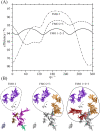
Fig. 6. Robustness of light harvesting. (A) Energy transfer efficiency calculated for different fictitious situations, in which the FMO protein is monomeric (FMO 2), dimeric (FMO 2 + 3) or trimeric (FMO 1 + 2 + 3) and it is rotated around the symmetry axis of the trimer by an angle φ relative to the RCC. (B) Illustration of the configurations considered in (A). Only the RCC-Ant1 subunit of the RCC is shown and for FMO 2 + 3 and FMO 1 + 2 + 3 the RCC-Ant1 is shown in grey for better visibility.

Similar articles

Cited by

References

    1. Blankenship R. E., Molecular Mechanisms of Photosynthesis, Wiley, 2021
    1. Croce R. van Amerongen H. Science. 2020;369:eaay2058. doi: 10.1126/science.aay2058. - DOI - PubMed
    1. Scholes G. D. Fleming G. R. Olaya-Castro A. van Grondelle R. Nat. Chem. 2011;3:763–773. doi: 10.1038/nchem.1145. - DOI - PubMed
    1. Overmann J. Cypionka H. Pfennig N. Limnol. Oceanogr. 1992;37:150–155. doi: 10.4319/lo.1992.37.1.0150. - DOI
    1. Marschall E. Jogler M. Henßge U. Overmann J. Environ. Microbiol. 2010;12:1348–1362. doi: 10.1111/j.1462-2920.2010.02178.x. - DOI - PubMed

Substances