Case Reports
. 2023 Jul;24(7):647-659.
doi: 10.3348/kjr.2023.0173.

Correct Closure of the Left Atrial Appendage Reduces Stagnant Blood Flow and the Risk of Thrombus Formation: A Proof-of-Concept Experimental Study Using 4D Flow Magnetic Resonance Imaging

Affiliations
Case Reports

Correct Closure of the Left Atrial Appendage Reduces Stagnant Blood Flow and the Risk of Thrombus Formation: A Proof-of-Concept Experimental Study Using 4D Flow Magnetic Resonance Imaging

Min Jae Cha et al. Korean J Radiol. 2023 Jul.

Abstract

Objective: The study was conducted to investigate the effect of correct occlusion of the left atrial appendage (LAA) on intracardiac blood flow and thrombus formation in patients with atrial fibrillation (AF) using four-dimensional (4D) flow magnetic resonance imaging (MRI) and three-dimensional (3D)-printed phantoms.

Materials and methods: Three life-sized 3D-printed left atrium (LA) phantoms, including a pre-occlusion (i.e., before the occlusion procedure) model and correctly and incorrectly occluded post-procedural models, were constructed based on cardiac computed tomography images from an 86-year-old male with long-standing persistent AF. A custom-made closed-loop flow circuit was set up, and pulsatile simulated pulmonary venous flow was delivered by a pump. 4D flow MRI was performed using a 3T scanner, and the images were analyzed using MATLAB-based software (R2020b; Mathworks). Flow metrics associated with blood stasis and thrombogenicity, such as the volume of stasis defined by the velocity threshold (|V̅| < 3 cm/s), surface-and-time-averaged wall shear stress (WSS), and endothelial cell activation potential (ECAP), were analyzed and compared among the three LA phantom models.

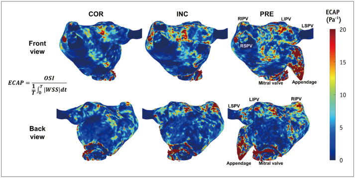
Results: Different spatial distributions, orientations, and magnitudes of LA flow were directly visualized within the three LA phantoms using 4D flow MRI. The time-averaged volume and its ratio to the corresponding entire volume of LA flow stasis were consistently reduced in the correctly occluded model (70.82 mL and 39.0%, respectively), followed by the incorrectly occluded (73.17 mL and 39.0%, respectively) and pre-occlusion (79.11 mL and 39.7%, respectively) models. The surface-and-time-averaged WSS and ECAP were also lowest in the correctly occluded model (0.048 Pa and 4.004 Pa-1 , respectively), followed by the incorrectly occluded (0.059 Pa and 4.792 Pa-1 , respectively) and pre-occlusion (0.072 Pa and 5.861 Pa-1 , respectively) models.

Conclusion: These findings suggest that a correctly occluded LAA leads to the greatest reduction in LA flow stasis and thrombogenicity, presenting a tentative procedural goal to maximize clinical benefits in patients with AF.

Keywords: 4D flow MRI; Atrial fibrillation; Hemodynamics; Left atrial appendage occlusion; Stasis; Thrombogenicity.


Conflict of interest statement

The authors have no potential conflicts of interest to disclose.

Figures

Fig. 1
Fig. 1. Graphical illustration of pre-occlusion (PRE), incorrectly occluded (INC), and correctly occluded (COR) left atrium (LA) phantoms and summarized results of the hemodynamic analysis using 4D flow magnetic resonance imaging (MRI). The potential source of thrombosis is reduced by left atrial appendage occlusion (LAAO), and more in COR state than in INC state.
Fig. 2
Fig. 2. Process of producing three life-sized 3D-printed phantoms of the left atrium (LA). A: Computed tomography (CT) image of the LA of the target patient. B: 3D image of the LA was reconstructed based on preprocedural cardiac CT images. C: 3D LA phantom was printed based on the image (pre-occlusion [PRE]). D: Ex vivo left atrial appendage occlusion (LAAO) was performed accurately and intentionally imprecisely in a PRE 3D LA phantom, arrow shows blockage of the appendage with the occluder. E: Internal volume comparison based on LAAO conditions, arrows show the internal volume changes caused by the occluder. F: 3D models of the correctly- and incorrectly occluded LA phantoms were printed based on the images, yellow arrowheads and white arrows indicate geometric differences near the appendage due to the occluder. RSPV = right superior pulmonary vein, RIPV = right inferior pulmonary vein, LIPV = left inferior pulmonary vein, LSPV = left superior pulmonary vein, INC = incorrectly occluded, COR = correctly occluded
Fig. 3
Fig. 3. Schematic of the closed-loop flow circuit. A custom closed-loop flow circuit was developed to simulate and collect the data of patient-specific blood flow using specialized equipment and magnetic resonance imaging (MRI). The red and blue arrows at the bottom center of the figure indicate the direction of flow exiting and entering the phantom, respectively. DAQ = data acquisition, LA = left atrium, RSPV = right superior pulmonary vein, RIPV = right inferior pulmonary vein, LIPV = left inferior pulmonary vein, LSPV = left superior pulmonary vein, MV = mitral valve
Fig. 4
Fig. 4. Path line analysis of 4D flow magnetic resonance imaging among correctly occluded (COR), incorrectly occluded (INC), and pre-occlusion (PRE) left atrium (LA) phantoms. A-C: Color-coded path lines for the entire LA. D-F: Color-coded path lines for the region near the appendage. G-I: Path lines originating from the right superior pulmonary vein (RSPV) (red), right inferior pulmonary vein (RIPV) (green), left superior pulmonary vein (LSPV) (yellow), and left inferior pulmonary vein (LIPV) (blue). Each LA phantom showed a different flow pattern in terms of distribution, orientation, and magnitude.
Fig. 5
Fig. 5. Comparison of left atrium (LA) flow stasis among correctly occluded (COR), incorrectly occluded (INC), and pre-occlusion (PRE) LA phantoms. A: Volume of LA stasis (|V⃗|) during a cardiac cycle. B: Average volume of stasis with different velocity thresholds ranging from 3–5 cm/s. C: The total flow rate variation during the cardiac cycle. LPM = liter per minute
Fig. 6
Fig. 6. Surfaces of the initial positions of fluid particles at a particle residence time (PRT) of 5 cardiac cycles (4 seconds) in correctly occluded (COR), incorrectly occluded (INC), and pre-occlusion (PRE) left atrium phantoms. The internal volume of the iso-PRT surface is the lowest in the COR model, followed by the INC and PRE models. The red circles indicate appendages and neighboring regions. RSPV = right superior pulmonary vein, RIPV = right inferior pulmonary vein, LIPV = left inferior pulmonary vein, LSPV = left superior pulmonary vein
Fig. 7
Fig. 7. Endothelial cell activation potential contours in the front and back projections of the 3D structures of correctly occluded (COR), incorrectly occluded (INC), and pre-occlusion (PRE) left atrium phantoms. Surface-averaged endothelial cell activation potential (ECAP) is lowest for COR and highest for PRE. OSI = oscillatory shear index, WSS = wall shear stress, RSPV = right superior pulmonary vein, RIPV = right inferior pulmonary vein, LIPV = left inferior pulmonary vein, LSPV = left superior pulmonary vein

References

    1. Pellman J, Sheikh F. Atrial fibrillation: mechanisms, therapeutics, and future directions. Compr Physiol. 2015;5:649–665. - PMC - PubMed
    1. Go AS, Hylek EM, Phillips KA, Chang Y, Henault LE, Selby JV, et al. Prevalence of diagnosed atrial fibrillation in adults: national implications for rhythm management and stroke prevention: the AnTicoagulation and Risk Factors in Atrial Fibrillation (ATRIA) Study. JAMA. 2001;285:2370–2375. - PubMed
    1. Holmes DR, Jr, Lakkireddy DR, Whitlock RP, Waksman R, Mack MJ. Left atrial appendage occlusion: opportunities and challenges. J Am Coll Cardiol. 2014;63:291–298. - PubMed
    1. Kirchhof P, Benussi S, Kotecha D, Ahlsson A, Atar D, Casadei B, et al. 2016 ESC Guidelines for the management of atrial fibrillation developed in collaboration with EACTS. Eur Heart J. 2016;37:2893–2962. - PubMed
    1. Blackshear JL, Odell JA. Appendage obliteration to reduce stroke in cardiac surgical patients with atrial fibrillation. Ann Thorac Surg. 1996;61:755–759. - PubMed

Publication types