

Artery of Percheron infarction presented with isolated downgaze paralysis: A case report
- PMID: 37404218
- PMCID: PMC10315925
- DOI: 10.1016/j.radcr.2023.06.015
Artery of Percheron infarction presented with isolated downgaze paralysis: A case report
Abstract
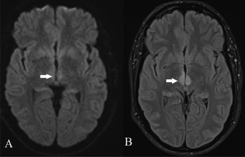
Isolated downgaze paralysis is the most infrequent expression of vertical gaze abnormalities. Vertical eye movements are controlled by nuclei and circuits located in the thalamic-mesencephalon region, and more particularly the rostral interstitial nucleus of the medial longitudinal fasciculus (riMLF). The Artery of Percheron (AP) is a rare vascular anatomic variation that supplies the paramedian region of the thalami and the rostral portion of the mesencephalon. We present a unique case of isolated downgaze paralysis caused by AP ischemia.
Keywords: Artery of Percheron; Downgaze paralysis; Midbrain; Stroke; Thalamus.
© 2023 The Authors. Published by Elsevier Inc. on behalf of University of Washington.
Figures


Similar articles
-
Pure downgaze palsy from isolated infarction of the left paramedian thalamic-mesencephalon junction.Neurol Sci. 2024 Sep;45(9):4633-4634. doi: 10.1007/s10072-024-07612-7. Epub 2024 May 26. Neurol Sci. 2024. PMID: 38796823
-
Parinaud's syndrome: electro-oculographic and anatomical analyses of six vascular cases with deductions about vertical gaze organization in the premotor structures.Brain. 1982 Dec;105 (Pt 4):667-96. doi: 10.1093/brain/105.4.667. Brain. 1982. PMID: 7139250
-
Vertical gaze palsy and selective unilateral infarction of the rostral interstitial nucleus of the medial longitudinal fasciculus (riMLF).J Neurol Neurosurg Psychiatry. 1990 Jan;53(1):67-71. doi: 10.1136/jnnp.53.1.67. J Neurol Neurosurg Psychiatry. 1990. PMID: 2303833 Free PMC article.
-
Vertical gaze paralysis and intermittent unresponsiveness in a patient with a thalamomesencephalic stroke.J Neuroophthalmol. 1995 Dec;15(4):230-5. J Neuroophthalmol. 1995. PMID: 8748560 Review.
-
Nuclear, internuclear, and supranuclear ocular motor disorders.Handb Clin Neurol. 2011;102:319-31. doi: 10.1016/B978-0-444-52903-9.00018-2. Handb Clin Neurol. 2011. PMID: 21601072 Review.
Cited by
-
The clinical significance of artery of Percheron in cerebral ischemia: a summary overview.Ann Med Surg (Lond). 2025 May 30;87(7):4310-4315. doi: 10.1097/MS9.0000000000003439. eCollection 2025 Jul. Ann Med Surg (Lond). 2025. PMID: 40851980 Free PMC article. Review.
-
Navigating the clinical landscape of artery of Percheron infarction: A systematic review.eNeurologicalSci. 2024 Aug 21;37:100521. doi: 10.1016/j.ensci.2024.100521. eCollection 2024 Dec. eNeurologicalSci. 2024. PMID: 39257866 Free PMC article. Review.
References
Publication types
LinkOut - more resources
Full Text Sources
