

Intraoperative use of ultra-low-field, portable magnetic resonance imaging - first report
- PMID: 37404510
- PMCID: PMC10316138
- DOI: 10.25259/SNI_124_2023
Intraoperative use of ultra-low-field, portable magnetic resonance imaging - first report
Abstract
Background: Intraoperative use of portable magnetic resonance imaging (pMRI) has become a valuable tool in a surgeon's arsenal since its inception. It allows intraoperative localization of tumor extent and identification of residual disease, hence maximizing tumor resection. Its utility has been widespread in high-income countries for the past 20 years, but in lower-middle-income countries (LMIC), it is still not widely available due to several reasons, including cost constraints. The use of intraoperative pMRI may be a cost-effective and efficient substitute for conventional MRI machines. The authors present a case where a pMRI device was used intraoperatively in an LMIC setting.
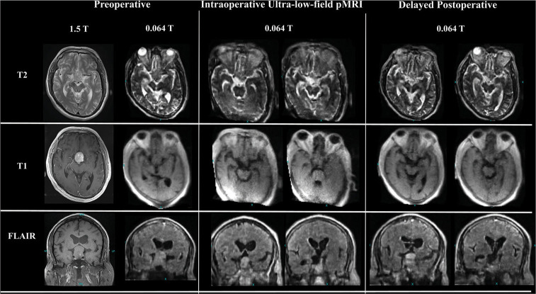
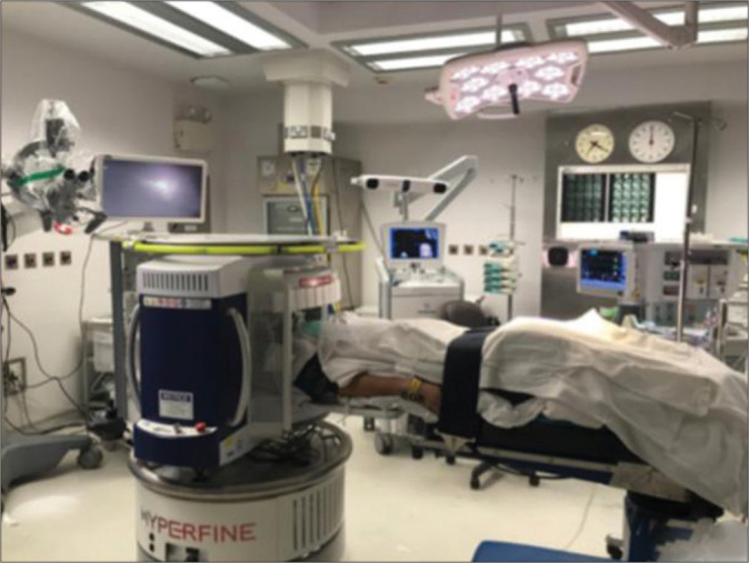
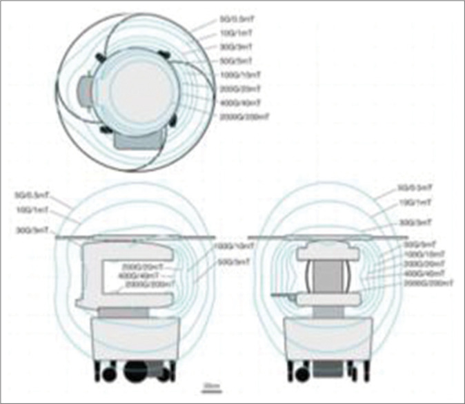
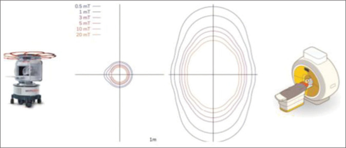
Case description: The authors performed a microscopic transsphenoidal resection of a sellar lesion with intraoperative imaging using the pMRI system on a 45-year-old man with a nonfunctioning pituitary macroadenoma. Without the need for an MRI suite or other MRI-compatible equipment, the scan was conducted within the confinements of a standard operating room. Low-field MRI showed some residual disease and postsurgical changes, comparable to postoperative high-field MRI.
Conclusion: To the best of our knowledge, our report provides the first documented successful intraoperative transsphenoidal resection of a pituitary adenoma using an ultra-low-field pMRI device. The device can potentially enhance neurosurgical capacity in resource-constrained settings and improve patient outcomes in developing country.
Keywords: Brain tumors; Intraoperative magnetic resonance imaging (Intraoperative MRI); Portable magnetic resonance imaging (Portable MRI); Ultra-low field magnetic resonance imaging (Ultra-low-field MRI).
Copyright: © 2023 Surgical Neurology International.
Conflict of interest statement
There are no conflicts of interest.
Figures
References
-
- Berkmann S, Schlaffer S, Nimsky C, Fahlbusch R, Buchfelder M. Follow-up and long-term outcome of nonfunctioning pituitary adenoma operated by transsphenoidal surgery with intraoperative high-field magnetic resonance imaging. Acta Neurochir (Wien) 2014;156:2233–43. - PubMed
-
- Brochier S, Galland F, Kujas M, Parker F, Gaillard S, Raftopoulos C, et al. Factors predicting relapse of nonfunctioning pituitary macro adenomas after neurosurgery: A study of 142 patients. Eur J Endocrinol. 2010;163:193–200. - PubMed
-
- Hlavica M, Bellut D, Lemm D, Schmid C, Bernays RL. Impact of ultra-low-field intraoperative magnetic resonance imaging on extent of resection and frequency of tumor recurrence in 104 surgically treated nonfunctioning pituitary adenomas. World Neurosurg. 2013;79:99–109. - PubMed
-
- Malkin RA. Barriers for medical devices for the developing world. Expert Rev Med Devices. 2007;4:759–63. - PubMed
Publication types
LinkOut - more resources
Full Text Sources
Miscellaneous