. 2023 Jul;18(3):1280-1287.
doi: 10.1016/j.jds.2023.02.013. Epub 2023 Mar 6.

Action of econazole on Ca2+ levels and cytotoxicity in OC2 human oral cancer cells

Affiliations

Action of econazole on Ca2+ levels and cytotoxicity in OC2 human oral cancer cells

Jue-Long Wang et al. J Dent Sci. 2023 Jul.

Abstract

Background/purpose: Econazole is an antifungal drug. Antifungal activity of econazole against non-dermatophyte molds was reported. Econazole inhibited Ca2+ channels and stimulated cytotoxicity in lymphoma and leukemia cells. Ca2+ cations are crucial second envoy that triggers various processes. This research was aimed to investigate action of econazole on Ca2+ levels and cytotoxicity in OC2 human oral cancer cells.

Materials and methods: Cytosolic Ca2+ levels ([Ca2+]i) were detected employing fura-2 as a probe in a RF-5301PC spectrofluorophotometer (Shimadzu). Cytotoxicity was determined using 4-[3-[4-lodophenyl]-2-4(4-nitrophenyl)-2H-5-tetrazolio-1,3-benzene disulfonate] (WST-1) to detect fluorescence changes.

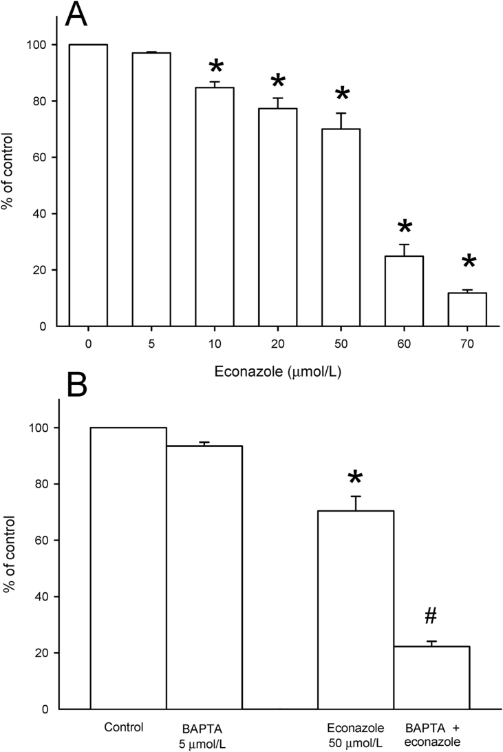
Results: Econazole at 10-50 μmol/L provoked [Ca2+]i raises. Forty % of 50 μml//L econazole-induced signal was diminished when external Ca2+ was eliminated. The Ca2+ influx provoked by econazole was suppressed by different degrees by store-induced Ca2+ influx suppressors SKF96365 and nifedipine; GF109203X (a protein C [PKC] inhibitor); an extracellular signaling pathway (ERK) 1/2 blocker PD98059, and phospholipase A2 suppressor aristolochic acid, but was enhanced by phorbol 12-myristate 13 acetate (PMA; a PKC activator) by 18%. Without external Ca2+, econazole-caused [Ca2+]i raises were abolished by thapsigargin. In contrast, econazole partially suppressed the [Ca2+]i raises caused by thapsigargin. U73122 fell short to change econazole-caused [Ca2+]i responses. Econazole (10-70 μmol/L) elicited cytotoxicity in a dose-dependent fashion. Blockade of 50 μmol/L econazole-induced [Ca2+] rises with BAPTA/AM enhanced econazole-induced cytotoxicity by 72%.

Conclusion: Econazole evoked [Ca2+]i raises and provoked cytotoxicity in a concentration-dependent manner in OC2 human oral cancer cells. In Ca2+-containing solution, BAPTA/AM enhanced 50 μmol/L econozole-induced cytotoxicity.

Keywords: Cellular calcium; Cytotoxicity; Econazole; Fura-2; OC2 human oral cancer cells.


Conflict of interest statement

The authors have no conflicts of interest relevant to this article.

Figures

Fig. 1
Figure 1
Effect of econazole on [Ca2+]i (A) The solution contained Ca2+. (B) Assays were performed in Ca2+-deprived solution. (C) Response-concentration association of econazole-provoked [Ca2+]i elevations. Y axis (control) represented percentage of the accumulated part beneath responses (25–250 s) of [Ca2+]i elevations evoked by 50 μmol/L econazole in Ca2+-containing solution. Findings were reported as the mean ± standard error of three separate experiments. ∗P value < 0.05: significant differences in contrast to open loops. Ca2+: calcium ions; [Ca2+]i: cytosolic free Ca2+ level; sec: second.
Fig. 2
Figure 2
Effect of Mn2+ on econazole-induced Ca2+ incursion. One min prior to assays, MnCl2 (50 μmol/L) was administered to a medium containing Ca2+. The y axis represents arbitrary fluorescence collected at excitation wavelength of 360 nm. Trace a: no econazole present. Trace b: in the presence of econazole (20 μmol/L). Data were shown as the mean ± standard error of three independent assays. Sec: second; Mn2+: manganese; MnCl2: manganese (II) chloride.
Fig. 3
Figure 3
In chemical-pretreated sets, chemicals were added 60 s before addition of 20 μmol/L econazole. Results were presented as percentage of first column which was integrated district beneath the responses (25–200 s) of econazole-evoked [Ca2+]i rises (control). Data were reported as the mean of standard error of three separate assays. ∗P value < 0.05 in contrast to first column. [Ca2+]i: cytosolic free Ca2+ level; sec: second.
Fig. 4
Figure 4
(A) (B) Thapsigargin (1 μmol/L) and econazole (20 μmol/L) were administered to cells as illustrated. These analyses were conducted in Ca2+-deprived solution. Findings were reported as the mean ± standard error of three independent assays. Ca2+: calcium ions.
Fig. 5
Figure 5
Action of U73122 on econazole-caused Ca2+ discharge. The following assays were conducted in Ca2+-deprived solution. (A) ATP (10 μmol/L) was administrated to cells as shown. (B) U73122 (2 μmol/L) and ATP (10 μmol/L) fell to change resting [Ca2+]i and econazole (20 μmol/L)-provoked [Ca2+]i raises. (C) In the existence of 2 μmol/L U73343 (incubated for 1 min), 10 μmol/L ATP provoked a considerable Ca2+ response. Findings were indicated as the mean ± standard error of three separate examinations. ATP: adenosine triphosphate; Ca2+: calcium ions; [Ca2+]i: cytosolic free Ca2+ level; U73122: 1-(6-((3-methoxyestra-1,3,5(10)-trien-17-yl)amino)hexyl)-1H-pyrrole-2,5-dione; U73343: 1-[6-[[(17b)-3-methoxyestra-1,3,5(10)-trien-17-yl]amino]hexyl]-2,5-pyrrolidinedione. Min: minute.
Fig. 6
Figure 6
(A) After overnight incubation of cells with econazol (0–70 μmol/L), the viability was investigated. Results were represented as percentage of control which meant increases in cell count in the presence of econazole. Cell counts of control had 99,222 ± 180 cells/well prior to assays and 14,888 ± 145 cells/well after overnight incubation. (B) To investigate whether [Ca2+]i raises altered cytotoxicity, 5 μmol/L BAPTA/AM was administered to cells 1 h prior to econazole (50 μmol/L) treatment overnight in the presence of Ca2+. Results were represented as percentage of control which meant increases in cell count after overnight incubation without econazole or BAPTA/AM. Cytotoxicity assay was performed. Data were shown as the mean ± standard error of three independent experiments. ∗P value < 0.05 in contrast to first bar. #P value < 0.01 in contrast to third bar. BAPTA/AM: 1,2-bis(2-aminophenoxy)ethane-N,N,N',N'-tetraacetic acid tetrakis (acetoxymethyl ester); Ca2+: calcium ions; [Ca2+]i: cytosolic free Ca2+ level; H: hour.

References

    1. James Q., Rosso D., Kircik L.H. Optimizing topical antifungal therapy for superficial cutaneous fungal infections: focus on topical naftifine for cutaneous dermatophytosis. J Drugs Dermatol JDD. 2013;12:s165–s171. - PubMed
    1. Hu Z., Zhang J., Cheng X. Antifungal efficiency of miconazole and econazole and the interaction with transport protein: a comparative study. Pharm Biol. 2015;53:251–261. - PubMed
    1. Mangas-Sánchez J., Busto E., Gotor-Fernández V., Malpartida F., Gotor V.J. Asymmetric chemoenzymatic synthesis of miconazole and econazole enantiomers. The importance of chirality in their biological evaluation. Org Chem. 2011;76:2115–2122. - PubMed
    1. Wilm K., Stahl A.J. Effects of econazole nitrate on yeast cells and mitochondria. Biochem Pharmacol. 1983;32:1825–1830. - PubMed
    1. Bastide M., Jouvert S., Bastide J.M. A comparison of the effects of several antifungal imidazole derivatives and polyenes on Candida albicans: an ultrastructural study by scanning electron microscopy. Can J Microbiol. 1982;28:1119–1126. - PubMed

LinkOut - more resources