. 2023 Jun 19:13:1195063.
doi: 10.3389/fcimb.2023.1195063. eCollection 2023.

Peptidyl-prolyl cis/trans isomerase Pin1 interacts with hepatitis B virus core particle, but not with HBc protein, to promote HBV replication

Affiliations

Peptidyl-prolyl cis/trans isomerase Pin1 interacts with hepatitis B virus core particle, but not with HBc protein, to promote HBV replication

Hyeonjoong Kwon et al. Front Cell Infect Microbiol. .

Abstract

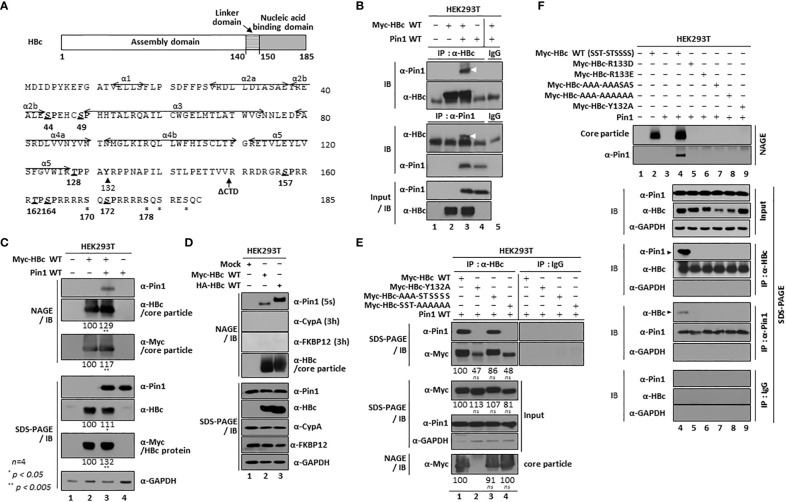
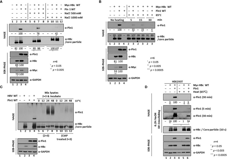
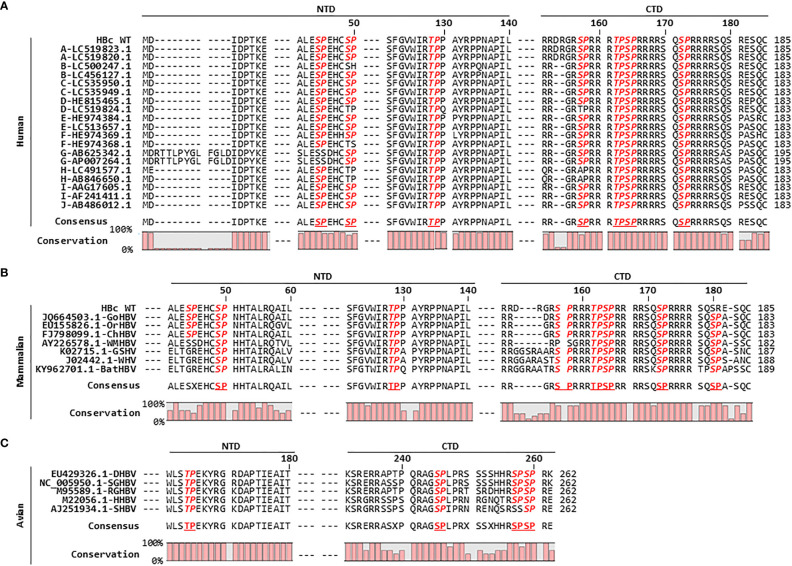
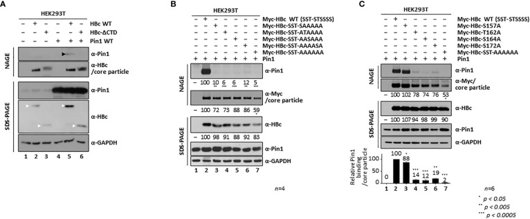
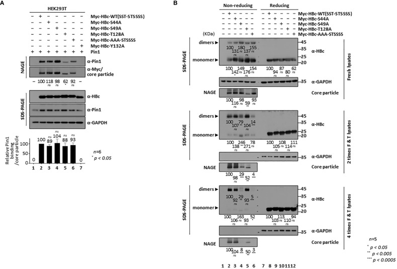
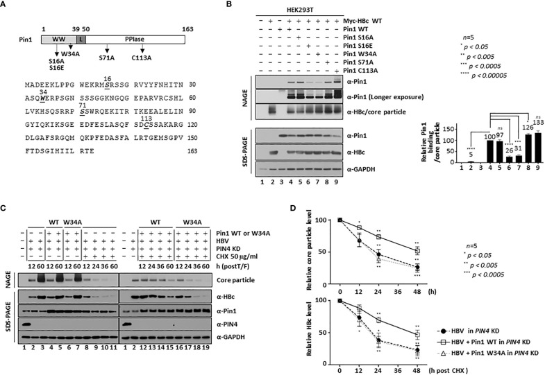
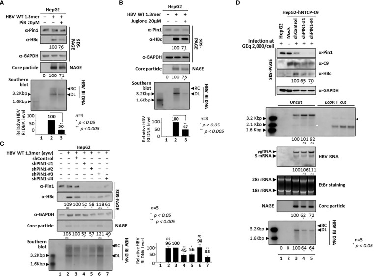
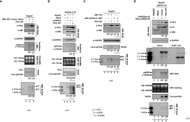
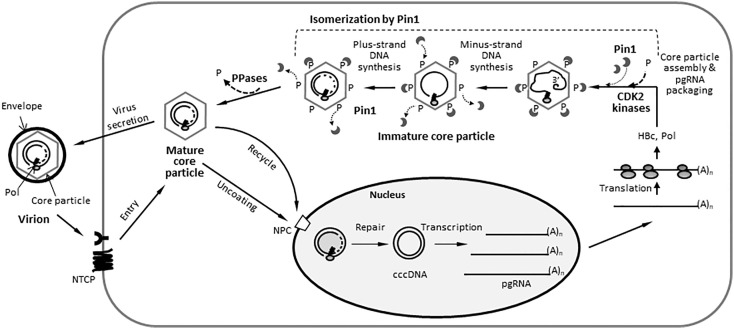
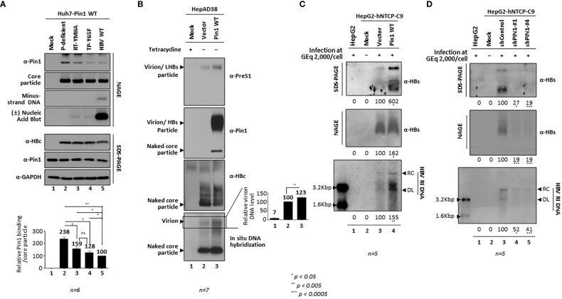
Here, we demonstrate that the peptidyl-prolyl cis/trans isomerase Pin1 interacts noncovalently with the hepatitis B virus (HBV) core particle through phosphorylated serine/threonine-proline (pS/TP) motifs in the carboxyl-terminal domain (CTD) but not with particle-defective, dimer-positive mutants of HBc. This suggests that neither dimers nor monomers of HBc are Pin1-binding partners. The 162TP, 164SP, and 172SP motifs within the HBc CTD are important for the Pin1/core particle interaction. Although Pin1 dissociated from core particle upon heat treatment, it was detected as an opened-up core particle, demonstrating that Pin1 binds both to the outside and the inside of the core particle. Although the amino-terminal domain S/TP motifs of HBc are not involved in the interaction, 49SP contributes to core particle stability, and 128TP might be involved in core particle assembly, as shown by the decreased core particle level of S49A mutant through repeated freeze and thaw and low-level assembly of the T128A mutant, respectively. Overexpression of Pin1 increased core particle stability through their interactions, HBV DNA synthesis, and virion secretion without concomitant increases in HBV RNA levels, indicating that Pin1 may be involved in core particle assembly and maturation, thereby promoting the later stages of the HBV life cycle. By contrast, parvulin inhibitors and PIN1 knockdown reduced HBV replication. Since more Pin1 proteins bound to immature core particles than to mature core particles, the interaction appears to depend on the stage of virus replication. Taken together, the data suggest that physical association between Pin1 and phosphorylated core particles may induce structural alterations through isomerization by Pin1, induce dephosphorylation by unidentified host phosphatases, and promote completion of virus life cycle.

Keywords: HBV replication; PPIase Pin1; Pin1-core particle interaction; core particle; hepatitis B virus.


Conflict of interest statement

The authors declare that the research was conducted in the absence of any commercial or financial relationships that could be construed as a potential conflict of interest.

Figures

Figure 1
Figure 1
HBV Core Particles, But Not HBc Dimers or Monomers, Interact with Pin1. (A) Schematic diagram and amino acid sequence of the HBV HBc (adw). The positions of the helices (Wynne et al., 1999) are shown by arrows. The CTD-truncated (at amino acid 149) HBc used for this study is denoted as ΔCTD. The S/TP motifs are in bold, italicized, and underlined. Y132 and other phosphoacceptor sites are marked by arrowheads and asterisks, respectively. (B) Myc-HBc WT and Pin1 WT can be coimmunoprecipitated. HEK293T cells were (co)transfected with mock (lane 1), Myc-HBc WT (lane 2), Myc-HBc WT plus Pin1 WT (lane 3), or Pin1 WT (lane 4). Two days after transfection, cell lysates were immunoprecipitated with an in-house rabbit polyclonal anti-HBc antibody (Jung et al., 2012) and then immunoblotted with a mouse monoclonal anti-Pin1 antibody, or vice versa. As a negative control, normal rabbit IgG or normal mouse IgG was used to examine lysates from Myc-HBc WT plus Pin1 WT cotransfected cells (lane 5). Immunoprecipitated proteins were subjected to SDS-PAGE, followed by immunoblotting with anti-HBc and anti-Pin1 antibodies. Lysates were also subjected to SDS-PAGE, followed by immunoblotting with anti-HBc and anti-Pin1 antibodies. Coimmunoprecipitated proteins are marked with a white arrowhead. (C) Pin1 interacts with the core particle. HEK293T cells were (co)transfected as above. Two days after transfection, cell lysates were prepared and subjected to NAGE; transferred to PVDF membranes; and immunoblotted with anti-Pin1, anti-HBc, or mouse monoclonal anti-Myc antibodies. Lysates were also subjected to SDS-PAGE, followed by immunoblotting with anti-HBc, anti-Myc, anti-Pin1, or mouse monoclonal anti-GAPDH antibodies, as described in B. GAPDH was used as a loading control. (D) The interaction between Pin1 and the core particle is more specific than that between the core particle and other PPIases. HEK293T cells were transfected with mock (lane 1), Myc-HBc WT (lane 2), or HA-HBc WT (lane 3), and cell lysates were subjected to NAGE plus immunoblotting and SDS-PAGE plus immunoblotting, as described in (B, C). Additionally, blots were probed with rabbit monoclonal anti-CypA and mouse monoclonal anti-FKBP12 antibodies. (E) The core particle, not a dimer or monomer of HBc, binds to Pin1 through the CTD phosphoacceptor S/TP motifs of HBc. HEK293T cells were cotransfected with Pin1 WT plus Myc-HBc WT (SST-STSSSS) (lane 1), or with the Myc-HBc-Y132A (lane 2), Myc-HBc-AAA-STSSSS (lane 3), or Myc-HBc-SST-AAAAAA (lane 4) mutants. Lysates were immunoprecipitated and then subjected to SDS-PAGE and immunoblotting as described in B. Core particle immunoblotting after NAGE was also performed as described in (C). (F) Pin1 does not interact with core particle-defective, dimer-positive HBc mutants. HEK293T cells were (co)transfected with mock (lane 1), Myc-HBc WT (lane 2), Pin1 WT (lane 3), Pin1 WT plus Myc-HBc WT (lane 4), Myc-HBc-R133D (lane 5), Myc-HBc-R133E (lane 6), Myc-HBc-AAA-AAASAS (lane 7), Myc-HBc-AAA-AAAAAA (lane 8), or Myc-HBc-Y132A (lane 9). Lysates were immunoprecipitated and then subjected to SDS-PAGE and immunoblotting as described in (B). Representative data are shown. Relative levels of core particles and HBc proteins were calculated using ImageJ 1.50b software. Statistical significance was evaluated using Student’s t test. ns, not significant; * P < 0.05; ** P < 0.005, relative to the corresponding control.
Figure 2
Figure 2
Dynamic On-off Interaction Between Pin1 and the Core Particle. (A, B) High salt (A) and high temperature (B) conditions weaken the Pin1/core particle interaction. HEK293T cells were (co)transfected with mock (lane 1), Myc-HBc WT (lane 2), Myc-HBc WT plus Pin1 WT (lane 3), or Pin1 WT (lane 4). Cell lysates prepared at 2 days post-transfection were treated with 500 mM or 1,000 mM NaCl (A), or heated at 65°C for 5, 15, or 30 min (B). (C) The in vitro interaction between Pin1 and core particle requires phosphorylation of the core particle. HEK293T cells were (co)transfected with mock (lane 1), HBV WT (lane 2), Pin1 WT (lane 3), or HBV WT plus Pin1 WT (lane 4). Pin1 WT-transfected cell lysates and HBV WT-transfected cell lysates were mixed and incubated for the indicated times (C, lanes 5−8), or HBV WT-transfected lysates were treated with CIAP and then mixed with Pin1 WT-transfected lysates for the indicated times (C, lanes 9−12). (D) Pin1 binds both outside and inside HBV core particles. HEK293T cells were (co)transfected with mock (lanes 1 and 4), Myc-HBc WT plus 3×FLAG (lanes 2 and 5), or Myc-HBc WT plus Pin1 WT (lanes 3 and 6). At 48 h post-transfection, lysates were either left unheated (lanes 1−3) or heated at 65°C for 2 h (lanes 4−6), followed by NAGE plus immunoblotting and SDS-PAGE plus immunoblotting as described in Figure 1 . Core particles bound to the PVDF membrane were treated with 0.2N NaOH for 1 min to open them up, crosslinked with UV, and immunoblotted with anti-HBc or anti-Pin1 antibodies. The indicated lysates were subjected to NAGE plus immunoblotting, and to SDS-PAGE plus immunoblotting, as described in Figure 1 . Representative data are shown. Relative levels of Pin1-bound core particles, core particles, and HBc proteins were calculated using ImageJ 1.50b software. Statistical significance was evaluated using Student’s t test. Ns, not significant; * P < 0.05; ** P < 0.005; *** P < 0.0005, relative to the corresponding control.
Figure 3
Figure 3
HBc Amino Acid Sequence Alignment Reveals that the HBc of Human, Mammalian, and Avian Hepadnaviruses Harbors Conserved S/TP Motifs. (A) The HBc S/TP motifs are highly conserved among ten genotypes of human HBV in the National Center for Biotechnology Information (NCBI), including human HBV [adw subtype (HBc WT)]. The partial HBc NTD and CTD amino acid sequences were aligned using CLC Main Workbench 8 software. The HBV genotype of each isolate, followed by the NCBI accession number, is indicated in the left column. (B) Mammalian hepadnavirus HBc S/TP motifs are highly conserved. Alignment of HBc amino acid sequences from gorilla HBV (GoHBV), orangutan HBV (OrHBV), chimpanzee HBV (ChHBV), woolly monkey HBV (WMHBV), ground squirrel hepatitis virus (GSHV), woodchuck hepatitis virus (WHV), and bat HBV (batHBV) is shown. (C) Avian hepadnavirus HBc S/TP motifs are highly conserved. Alignment of the HBc amino acid sequences from duck HBV (DHBV), snow goose HBV (SGHBV), ross goose HBV (RGHBV), heron HBV (HHBV), and stork HBV (SHBV) is shown. The accession numbers for the mammalian and avian hepadnaviruses are presented. Conserved S/TP motifs are bold and italicized. The consensus sequences and percentage conservation are shown at the bottom.
Figure 4
Figure 4
The HBc 162TP, 164SP, and 172SP motifs within the CTD are important for the Pin1/core particle interaction. (A) The HBc CTD is important for the Pin1/core particle interaction. HEK293T cells were (co)transfected with mock (lane 1), HBc WT (lane 2), HBc-ΔCTD (lane 3), Pin1 WT (lane 4), HBc WT plus Pin1 WT (lane 5), or HBc-ΔCTD plus Pin1 WT (lane 6). (B) A single S/TP phosphoacceptor site within the CTD of HBc is not sufficient for the Pin1/core particle interaction. HEK293T cells were cotransfected with Pin1 WT plus mock (lane 1), Myc-HBc WT (Myc-HBc-SST-STSSSS) (lane 2), Myc-HBc-SST-SAAAAA (lane 3), Myc-HBc-SST-ATAAAA (lane 4), Myc-HBc-SST-AASAAA (lane 5), Myc-HBc-SST-AAAASA (lane 6), or Myc-HBc-SST-AAAAAA (lane 7). (C) The HBc 162TP, 164SP, and 172SP motifs within the CTD are important for the Pin1/core particle interaction. HEK293T cells were cotransfected with Pin1 WT plus mock (lane 1), Myc-HBc WT (lane 2), Myc-HBc-S157A (lane 3), Myc-HBc- T162A (lane 4), Myc-HBc-S164A (lane 5), Myc-HBc-S172A (lane 6), or Myc-HBc-SST-AAAAAA (lane 7). The data in the graph represent the mean ± SD of six independent experiments. Two days after transfection, lysates were subjected to NAGE plus immunoblotting, and SDS-PAGE plus immunoblotting (A-C), as described in Figure 1 . GAPDH was used as a loading control. Representative data are shown. Relative levels of Pin1-bound core particles, core particles, and HBc proteins were calculated using ImageJ 1.50b software. Statistical significance was evaluated using Student’s t test. * P < 0.05; ** P < 0.005; *** P < 0.0005, relative to the corresponding control.
Figure 5
Figure 5
HBc NTD S/TP motifs Contribute to HBV Core Particle Stability and Assembly. (A) HBc NTD S/TP motifs are not important for the Pin1/core particle interaction. HEK293T cells were (co)transfected with Pin1 WT plus mock (lane 1), Myc-HBc WT (lane 2), Myc-HBc-S44A (lane 3), Myc-HBc-S49A (lane 4), Myc-HBc-T128A (lane 5), Myc-HBc-AAA-STSSSS (lane 6), or Myc-HBc-Y132A (lane 7). (B) HBc NTD S/TP motifs contribute to the structural stability of the HBV core particle. HEK293T cells were transfected with mock (lane 1), Myc-HBc WT (SST-STSSSS) (lane 2), Myc-HBc-S44A (lane 3), Myc-HBc-S49A (lane 4), Myc-HBc-T128A (lane 5), or Myc-HBc-AAA-STSSSS (lane 6). Two days after transfection, lysates were put through repeated freezing and thawing cycles and then subjected to nonreducing PAGE or reducing SDS-PAGE (B). Lysates were subjected to NAGE plus immunoblotting, and SDS-PAGE plus immunoblotting (A, B), as described in Figure 1 . GAPDH was used as a loading control. Representative data are shown. Relative levels of Pin1-bound core particles, core particles, HBc proteins, and dimeric and monomeric HBc proteins, were calculated using ImageJ 1.50b software. Statistical significance was evaluated using Student’s t test. ns, not significant; * P < 0.05; ** P < 0.005; *** P < 0.0005, relative to the corresponding control.
Figure 6
Figure 6
The 16Ser and 34Trp Substrate Binding Residues of Pin1 Promote Core Particle Stability. (A) Schematic diagram and amino acid sequences of Pin1. The Pin1 WW, linker, and PPIase domains are indicated. Important amino acid residues are shown in bold and italics and are underlined. The substrate binding-positive dephosphorylated mimetic S16A, the substrate binding-negative phosphorylated mimetic S16E, the substrate binding-negative W34A, and the PPIase-active S71A and PPIase-inactive C113A mutants are indicated on the diagram. (B) The substrate binding 16Ser and 34Trp residues of Pin1 are important for the Pin1/core particle interaction. HEK293T cells were transfected with mock (lane 1), Myc-HBc WT (lane 2), or Pin1 WT (lane 3) or cotransfected with HBc WT plus Pin1 WT (lane 4), Pin1 S16A (lane 5), Pin1 S16E (lane 6), Pin1 W34A (lane 7), Pin1 S71A (lane 8), or Pin1 C113A (lane 9). (C, D) Pin1 WT promotes core particle stability. HEK293T-shPIN4 KD cells were (co)transfected with mock (lane 1), HBV WT (lanes 2, 3, 8-11), Pin1 WT plus HBV WT (lanes 4, 5, 12-15), or Pin1 W34A mutant plus HBV WT (lanes 6, 7, 16-19). At 12 h post-transfection, HEK293T cells were treated with 50 μg/ml cycloheximide for 0, 12, 24, or 48 h (lanes 8-19), and lysates were prepared at the indicated times. Lysates were subjected to NAGE plus immunoblotting, and to SDS-PAGE plus immunoblotting (B, C), as described in Figure 1 . A rabbit monoclonal anti-PIN4 antibody was used to detect Par14/Par17 proteins. GAPDH was used as a loading control. Representative data are shown. Relative levels of Pin1-bound core particles, core particles, and HBc proteins were calculated using ImageJ 1.50b software. Statistical significance was evaluated using Student’s t test. ns, not significant; * P < 0.05; *** P < 0.0005; **** P < 0.00005, relative to the corresponding control.
Figure 7
Figure 7
Pin1 Inhibition or PIN1 KD Downregulates HBV Replication. (A, B) Parvulin inhibitors PiB and Juglone downregulate HBV replication. HepG2 cells were transfected with mock (lane 1) or 1.3mer HBV WT (adw) (lanes 2 and 3). At 48 h post-transfection, HepG2 cells were treated with DMSO (lane 2), 20 μM PiB (lane 3) (A), ethanol (lane 2), or 20 μM Juglone (lane 3) (B) for 24 h. (C) HBV replication is downregulated in PIN1 KD cells. HepG2 cells were transduced with lentivirus-like particles containing control shRNA (shControl) (lane 3) or PIN1-targeting shRNAs (shPIN1-#1, shPIN1-#2, shPIN1-#3, and shPIN1-#4) (lanes 4-7). Nontransduced (lane 2) and transduced (lane 3-7) HepG2 cells were transiently transfected with 1.3mer HBV WT (ayw). Lane 1 shows a negative control without transduction and transfection. At 3 days post-transfection, cell lysates were prepared, and core particle immunoblotting after NAGE and SDS-PAGE plus immunoblotting were performed as described in Figure 1 . Southern blotting was performed to detect HBV DNA synthesis. In brief, HBV DNA was extracted from isolated core particles, separated, transferred to a nylon membrane, hybridized with a random-primed 32P-labeled full-length HBV specific probe, and subjected to autoradiography. HBV replicative intermediate, partially double-stranded relaxed circular, and double-stranded linear DNAs are marked as HBV RI DNA, RC, and DL, respectively (A-C). (D) PIN1 KD decreases HBV replication in infected cells. HepG2 (lane 1), HepG2-hNTCP-C9-shControl (lane 3), HepG2-hNTCP-C9-shPIN1-#1 (lane 4), and HepG2-hNTCP-C9-shPIN1-#4 (lane 5) cells were grown in collagen-coated 6-well plates, infected with 2×10³ GEq of HBV per cell, and lysed at 9 days postinfection. Lane 2 represents a mock-infected HepG2-hNTCP-C9 cell. Lysates were subjected to NAGE plus immunoblotting and SDS-PAGE plus immunoblotting, as described in Figure 1 . A mouse monoclonal anti-C9 antibody was used to detect hNTCP-C9. GAPDH was used as a loading control. Southern blotting was performed to detect HBV DNA synthesis, as described above. For northern blotting, total RNAs were prepared at 5 days postinfection. In brief, 20 μg of total RNA was separated by 1% formaldehyde agarose gel electrophoresis, transferred to nylon membranes, hybridized, and subjected to autoradiography as described above for southern blotting. Next, cccDNA was extracted and subjected to southern blotting without linearization (Uncut) or following linearization with EcoR I (EcoR I cut). The white and black arrowheads indicate cccDNA and linearized cccDNA to 3.2 kb genome-length, respectively. The 2.1 kb cccDNA, 3.5 kb pgRNA, 2.4 and 2.1 kb S mRNAs, and 28S and 18S rRNAs are indicated. Representative data are shown. Relative levels of RI DNAs, cccDNA, HBV RNAs, core particles, and HBc proteins were calculated using ImageJ 1.50b software. Statistical significance was evaluated using Student’s t test. ns, not significant; * P < 0.05; ** P < 0.005, relative to the corresponding control.
Figure 8
Figure 8
Overexpression of Pin1 Upregulates HBV Replication. (A) HBV replication in HepG2 cells is upregulated by overexpression of Pin1. HepG2 cells were transiently (co)transfected with mock (lane 1), 1.3mer HBV WT (adw) (lane 2), or 1.3mer HBV WT (adw) plus Pin1 WT (lane 3). (B) HBV replication in HepG2.2.15 cells increases upon overexpression of Pin1. HepG2.2.15 cells were transiently transfected with mock (lane 2), pcDNA3 (lane 3), or Pin1 WT (lane 4). Mock transfected HepG2 cells were used as a negative control (lane 1). (C) Upregulated replication of HBV upon overexpression of Pin1 is independent of HBx. HepG2 cells were transiently (co)transfected with mock (lane 1), HBV WT (adw) (lane 2), HBV WT (adw) plus Pin1 WT (lane 3), HBx-deficient HBV mutant (lane 4), or HBx-deficient HBV mutant plus Pin1 WT (lane 5). Transcription of pgRNA from HBV WT and an HBx-deficient HBV mutant was controlled by the CMV IE promoter. (D) Increased HBV replication in infected cells overexpressing Pin1. HepG2 cells (lane 1) or HepG2-hNTCP-C9 cells transduced with empty vector (lane 3) or Pin1 WT (lane 4) were grown in collagen-coated 6-well plates and infected with 2×10³ GEq of HBV per cell (lanes 1, 3, and 4), as described in Figure 7D . Lane 2 shows a mock-infected control. The white and black arrowheads indicate cccDNA and linearized cccDNA to 3.2 kb genome-length, respectively. Cells were lysed at 5 (for total RNA) or 9 days postinfection. (A-D) Lysates were subjected to NAGE plus immunoblotting and to SDS-PAGE plus immunoblotting, as described in Figure 1 . GAPDH was used as a loading control. To detect HBV DNA and HBV RNA, southern blotting and northern blotting were performed, respectively, as described in Figure 7 . cccDNA was subjected to southern blotting, as described in Figure 7 . The 2.1 kb cccDNA, 3.5 kb pgRNA, 2.4 and 2.1 kb S mRNAs, and 28S and 18S rRNAs are indicated. Representative data are shown. Relative levels of RI DNAs, cccDNA, HBV RNAs, core particles, and HBc proteins were calculated using ImageJ 1.50b software. Statistical significance was evaluated using Student’s t test. ns, not significant; * P < 0.05; ** P < 0.005; ***P < 0.0005, relative to the corresponding control.
Figure 9
Figure 9
Proposed Model for the Actions of Pin1 During the HBV Life Cycle. After binding to the NTCP receptor on hepatocytes, the HBV virion is internalized and uncoated, and its RC DNA is delivered to the nucleus. The RC DNA genome is repaired to form cccDNA, which serves as the template for transcription. Viral RNAs, including pgRNA, are exported to the cytoplasm, where viral proteins such as HBc and Pol are translated. The pgRNA is copackaged with Pol into immature core particles composed of HBc proteins. At this stage, host CDK2 is supposed to phosphorylate the phosphoacceptor S/TP motifs on HBc, thereby forming Pin1-binding sites on the core particle. The pgRNA is reverse transcribed to minus-strand DNA and then plus-strand DNA, thus forming RC DNA-containing mature core particles. The mature core particles are dephosphorylated via bound Pin1 at S/TP sites on core particle by unidentified host PPases, such as PDP2, followed by envelopment by viral HBs envelope proteins. They are then secreted extracellularly as virions or recycled back to the nucleus to amplify the cccDNA pool. RC DNA, relaxed circular DNA; cccDNA, covalently closed circular DNA; pgRNA, pregenomic RNA; Pol, HBV DNA polymerase; CDK2, cyclin-dependent protein kinase 2; P, phosphates; PPase, host protein phosphatase; PDP2, pyruvate dehydrogenase phosphatase 2.
Figure 10
Figure 10
Binding of Pin1 to Intracellular Core Particles Differs According to Replication Stage and Increases Secretion of Virions. (A) Fewer Pin1 proteins bind to the mature core particle than to the immature core particle. Stable Huh7-Pin1-expressing cells were transiently transfected with mock (lane 1), the P-deficient mutant (lane 2), the RT-YMHA mutant (lane 3), the TP-Y65F mutant (lane 4), or HBV WT (lane 5). As described in Figure 1 , lysates were subjected to NAGE plus immunoblotting and to SDS-PAGE plus immunoblotting. In situ DNA hybridization was performed to detect minus-strand DNA in the core particle, and in situ nucleic acid hybridization was performed to detect plus- and minus-stranded nucleic acids in the core particle. In brief, isolated core particles on a PVDF membrane were treated with 0.2 N NaOH, hybridized to either a DIG-labeled full-length plus-strand HBV RNA probe or a random-primed 32P-labeled full-length HBV specific probe, and subjected to autoradiography. (B) Pin1 overexpression increases virion secretion in stable HepAD38 cells. HepAD38 cells were transduced with empty vector (lane 2) or with Pin1 (lane 3) transcript-containing pseudoviral particles and then selected with puromycin. After removal of tetracycline to induce HBV transcription, the HepAD38-vector control and HepAD38-Pin1 stable cells were incubated for 3 days, and culture supernatants were harvested. HepAD38-Pin1 stable cells cultured with tetracycline (lane 1) were used as a negative control. Cleared culture supernatants were precipitated by ultracentrifugation on a 20% (w/w) sucrose cushion (26,000 rpm for 3 h at 4°C). Pellets containing HBV virions, subviral particles, and naked core particles were resuspended in TNE buffer and subjected to NAGE plus immunoblotting or in situ nucleic acid blotting to detect virions and/or LHBs particles, Pin1 on virions and/or on naked core particles, naked core particles, or virion DNA. (C) Pin1 overexpression increases virion secretion by HBV-infected cells. HepG2 (lane 1), vector-transduced HepG2-hNTCP-C9 (lane 3), and Pin1-transduced HepG2-hNTCP-C9 (lane 4) cells were infected with HBV as described in Figure 7D . Lane 2 shows mock-infected HepG2-hNTCP-C9 cells. (D) Pin1 KD downregulates secretion of HBV virions from HBV-infected cells. HepG2 (lane 1), shControl-transduced (lane 3), shPIN1-#1-transduced (lane 4), and shPIN1-#4-transduced (lane 5) HepG2-hNTCP-C9 cells were infected with HBV as described. Lane 2 shows mock-infected HepG2-hNTCP-C9 cells. Pellets obtained from culture supernatants were subjected to NAGE plus immunoblotting, SDS-PAGE plus immunoblotting, and southern blotting (C, D), as described in Figures 1 , 7 . The data in the graphs represent the mean ± SD from six (A) or seven (B) independent experiments. Representative data are shown. Relative levels of Pin1-bound core particles, virion DNAs, virion HBs proteins, and virions and/or subviral particles were calculated using ImageJ 1.50b software. Statistical significance was evaluated using Student’s t test. ns, not significant; * P < 0.05; ** P < 0.005; *** P < 0.0005, relative to the corresponding control.

Similar articles

Cited by

References

    1. Basagoudanavar S. H., Perlman D. H., Hu J. (2007). Regulation of hepadnavirus reverse transcription by dynamic nucleocapsid phosphorylation. J. Virol. 81, 1641–1649. doi: 10.1128/JVI.01671-06 - DOI - PMC - PubMed
    1. Bourne C. R., Katen S. P., Fulz M. R., Packianathan C., Zlotnick A. (2009). A mutant hepatitis b virus core protein mimics inhibitors of icosahedral capsid self-assembly. Biochemistry 48, 1736–1742. doi: 10.1021/bi801814y - DOI - PMC - PubMed
    1. Cai D., Nie H., Yan R., Guo J. T., Block T. M., Guo H. (2013). A southern blot assay for detection of hepatitis b virus covalently closed circular DNA from cell cultures. Methods Mol. Biol. 1030, 151–161. doi: 10.1007/978-1-62703-484-5_13 - DOI - PMC - PubMed
    1. Chen C., Wang J. C., Zlotnick A. (2011). A kinase chaperones hepatitis b virus capsid assembly and captures capsid dynamics in vitro . PloS Pathog. 7, e1002388. doi: 10.1371/journal.ppat.1002388 - DOI - PMC - PubMed
    1. Chen C. J., Yang H. I., Su J., Jen C. L., You S. L., Lu S. N., et al. . (2006). Risk of hepatocellular carcinoma across a biological gradient of serum hepatitis b virus DNA level. Jama 295, 65–73. doi: 10.1001/jama.295.1.65 - DOI - PubMed

Publication types

MeSH terms

Substances