Review
. 2023 Jun 19:10:1178163.
doi: 10.3389/fcvm.2023.1178163. eCollection 2023.

The pivotal role of ECG in cardiomyopathies

Affiliations
Review

The pivotal role of ECG in cardiomyopathies

Elisa Silvetti et al. Front Cardiovasc Med. .

Abstract

Cardiomyopathies are a heterogeneous group of pathologies characterized by structural and functional alterations of the heart. Recent technological advances in cardiovascular imaging offer an opportunity for deep phenotypic and etiological definition. Electrocardiogram (ECG) is the first-line diagnostic tool in the evaluation of both asymptomatic and symptomatic individuals. Some electrocardiographic signs are pathognomonic or fall within validated diagnostic criteria of individual cardiomyopathy such as the inverted T waves in right precordial leads (V1-V3) or beyond in individuals with complete pubertal development in the absence of complete right bundle branch block for the diagnosis of arrhythmogenic cardiomyopathy of the right ventricle (ARVC) or the presence of low voltages typically seen in more than 60% of patients with amyloidosis. Most other electrocardiographic findings such as the presence of depolarization changes including QRS fragmentation, the presence of epsilon wave, the presence of reduced or increased voltages as well as alterations in the repolarization phase including the negative T waves in the lateral leads, or the profound inversion of the T waves or downsloping of the ST tract are more non-specific signs which can however raise the clinical suspicion of cardiomyopathy in order to initiate a diagnostic procedure especially using imaging techniques for diagnostic confirmation. Such electrocardiographic alterations not only have a counterpart in imaging investigations such as evidence of late gadolinium enhancement on magnetic resonance imaging, but may also have an important prognostic value once a definite diagnosis has been made. In addition, the presence of electrical stimulus conduction disturbances or advanced atrioventricular blocks that can be seen especially in conditions such as cardiac amyloidosis or sarcoidosis, or the presence of left bundle branch block or posterior fascicular block in dilated or arrhythmogenic left ventricular cardiomyopathies are recognized as a possible expression of advanced pathology. Similarly, the presence of ventricular arrhythmias with typical patterns such as non-sustained or sustained ventricular tachycardia of LBBB morphology in ARVC or non-sustained or sustained ventricular tachycardia with an RBBB morphology (excluding the "fascicular pattern") in arrhythmogenic left ventricle cardiomyopathy could have a significant impact on the course of each disease. It is therefore clear that a learned and careful interpretation of ECG features can raise suspicion of the presence of a cardiomyopathy, identify diagnostic "red flags" useful for orienting the diagnosis toward specific forms, and provide useful tools for risk stratification. The purpose of this review is to emphasize the important role of the ECG in the diagnostic workup, describing the main ECG findings of different cardiomyopathies.

Keywords: arrhythmogenic cardiomyopathy; diagnosis; dilated cardiomyopathy; electrocardiogram (ECG); hypertrophic cardiomyopathy; risk stratification.


Conflict of interest statement

The authors declare that the research was conducted in the absence of any commercial or financial relationships that could be construed as a potential conflict of interest. The reviewer GS declared a past co-authorship with the author LC to the handling editor.

Figures

Figure 1
Figure 1
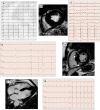
Conduction disorders in patients with DCM. ECG performed in a 60-year-old man with DCM shows LBBB and normal QRS axis (A). Basal ECG of a 78-year-old patient fulfills criteria for LAFB (B). ECG and CMR findings of a 20-year-old male with history of ventricular arrhythmias and mild LV dysfunction. Basal ECG (C) showing low QRS voltages (<0.5 mV) in limb leads and LPFB (AQRS ≈ + 110°). Post-contrast image showing subepicardial circumferential LGE pattern (D). Four-chamber CMR image of a 42-year-old man presented with pulmonary edema showing severe LV dilation (E). ECG at presentation displaying normal QRS axis, intraventricular conduction delay, signs of LVH (Sokolow–Lyon criteria) with secondary repolarization abnormalities (F). All the ECGs presented in the figure were performed at 25 mm/s with 1 mm/mV. ECG, electrocardiogram; CMR, cardiac magnetic resonance; DCM, dilated cardiomyopathy; LAFB, left anterior fascicular block; LBBB, left bundle branch block; LGE, late gadolinium enhancement; LPFB, left posterior fascicular block; LVH, left ventricular hypertrophy.
Figure 2
Figure 2
Ventricular depolarization and repolarization abnormalities in patients with DCM. A 30-year-old female with a likely pathogenic variant in FLNC gene. Twelve-lead ECG shows low QRS voltages both in limb and precordial leads (A). Basal ECG of a 46-year-old patient with DCM showing pathological infero-lateral Q waves (blue boxes, B); a premature ventricular beat with RBBB morphology and superior axis is also present. Post-contrast CMR images of patient in panel B showing subepicardial LGE involving the left ventricular lateral wall (blue arrows, C). A 38-year-old male with a pathogenic variant in LMNA gene and familiar history of DCM and sudden cardiac death. Basal ECG displaying low QRS voltages in precordial leads and specific ECG signs of “septal remodeling” (pathological Q waves, QRS fragmentation, poor R-wave progression in V1–V3 leads; red box); a premature ventricular beat with RBBB morphology and right axis deviation is also present (D). Basal ECG of a 39-year-old DSP mutation carrier shows low QRS voltages in limb leads, negative T waves in inferior leads (asterisks, E). All the ECGs presented in the figure were performed at 25 mm/s with 1 mm/mV. ECG, electrocardiogram; CMR, cardiac magnetic resonance; DCM, dilated cardiomyopathy; LGE, late gadolinium enhancement; RBBB, right bundle branch block.
Figure 3
Figure 3
ECG abnormalities in patients with myotonic dystrophy type I. (A) Twelve-lead ECG in a 51-year-old patient shows prolongation of the PR interval (400 ms). (B) First-degree atrioventricular block, right bundle branch block, and pathological lateral Q waves in a 56-year-old patient. (C) Left bundle branch morphology regular wide tachycardia in a 52-year-old patient with history of arrhythmic storm. All the ECGs presented in the figure were performed at 25 mm/s with 1 mm/mV. ECG, electrocardiogram.
Figure 4
Figure 4
ECG characteristics in patients with arrhythmogenic cardiomyopathy. A 65-year-old PKP2 mutation carrier with arrhythmogenic right ventricular cardiomyopathy. ECG shows RBBB with T-wave inversion extending to the left precordial leads and inferior leads (A). ECG and CMR findings in a case of biventricular arrhythmogenic cardiomyopathy variant in a 34-year-old female. ECG showing LPFB, diffuse negative T waves, and monomorphic premature ventricular beats with LBBB/inferior axis morphology (B). Post-contrast CMR images in long-axis four-chamber view showing right ventricular dilation and subepicardial LGE involving the apical LV wall and the apical layer of the LV lateral wall (red arrow, C). ECG and CMR findings in a case of left dominant arrhythmogenic cardiomyopathy variant in a 35-year-old male with underlying PKP2 pathogenic variant. Basal ECG showing low QRS voltages in limb leads and T-wave inversion in both inferior and precordial leads (D). Post-contrast CMR images in long-axis three-chamber view showing subepicardial LGE involving the basal and mid-apical layers of the LV infero-lateral wall (red arrows, E). All the ECGs presented in the figure were performed at 25 mm/s with 1 mm/mV. ECG, electrocardiogram; CMR, cardiac magnetic resonance; LBBB, left bundle branch block; LGE, late gadolinium enhancement; LPFB, left posterior fascicular block; LV, left ventricular; RBBB, right bundle branch block.
Figure 5
Figure 5
ECG abnormalities in patients with hypertrophic cardiomyopathy. ECG and CMR findings in a 78-year-old patient with obstructive HCM. Twelve-lead ECG showing ST-segment alterations in infero-lateral leads (A). Post-contrast CMR images in short axis showing hypertrophic septal asymmetric cardiomyopathy (B). Pathological Q waves in lateral leads and signs of LVH (Sokolow–Lyon criteria) in a 55-year-old male with non-obstructive HCM (C). ECG and CMR findings in a 56-year-old male with non-obstructive HCM. Note the low QRS voltages in limb leads at 12-lead ECG (D). LGE is found at the insertion points of the interventricular septum with hazy mid-wall enhancement in areas of hypertrophy (white arrows, E). CMR and ECG findings in a case of apical HCM in a 56-year-old female. CMR reveals obliteration of the cavity at the apex and the apical displacement of papillary muscles (F). ECG shows signs of LVH and deeply inverted T waves in precordial leads and lateral leads (G). All the ECGs presented in the figure were performed at 25 mm/s with 1 mm/mV. ECG, electrocardiogram; CMR, cardiac magnetic resonance; HCM, hypertrophic Cardiomyopathy; LGE, late gadolinium enhancement; LVH, left ventricular hypertrophy.
Figure 6
Figure 6
ECG characteristics in HCM phenocopies and sarcoidosis. ECG and CMR findings in a 45-year-old patient with Anderson–Fabry disease. Twelve-lead ECG displaying signs of LVH and deep T-wave inversion in D1-aVL and left precordial leads (A). Post-contrast CMR images in short axis showing LVH and LGE within the basal postero-lateral LV wall (white arrows, B). CMR and ECG features of a patient affected by cardiac ATTR amyloidosis. End-diastolic frame of cine CMR sequence in long-axis four-chamber view showing moderate LVH (C). Basal ECG showing low QRS voltages both in limb and precordial leads (D). ECG of a 30-year-old female with cardiac sarcoidosis. Note the atrioventricular dissociation with wide QRS escape rhythm (RBBB and LAFB morphology, E). CMR of the patient in panel E reveals transmural LGE in the basal septum (red arrow, F). All the ECGs presented in the figure were performed at 25 mm/s with 1 mm/mV. ECG, electrocardiogram; CMR, cardiac magnetic resonance; HCM, hypertrophic cardiomyopathy; LAFB, left anterior fascicular block; LGE, late gadolinium enhancement; LVH, left ventricular hypertrophy; RBBB, right bundle branch block.

References

    1. Elliott P, Andersson B, Arbustini E, Bilinska Z, Cecchi F, Charron P, et al. Classification of the cardiomyopathies: a position statement from the European Society of Cardiology Working Group on Myocardial and Pericardial Diseases. Eur Heart J. (2008) 29:270–6. 10.1093/eurheartj/ehm342 - DOI - PubMed
    1. Thiene G. Sudden cardiac death and cardiovascular pathology: from anatomic theater to double helix. Am J Cardiol. (2014) 114:1930–6. 10.1016/j.amjcard.2014.09.037 - DOI - PubMed
    1. Rapezzi C, Arbustini E, Caforio ALP, Charron P, Gimeno-Blanes J, Heliö T, et al. Diagnostic work-up in cardiomyopathies: bridging the gap between clinical phenotypes and final diagnosis. A position statement from the ESC working group on myocardial and pericardial diseases. Eur Heart J. (2013) 34:1448–58. 10.1093/eurheartj/ehs397 - DOI - PubMed
    1. Calò L, Lanza O, Crescenzi C, Parisi C, Panattoni G, Martino A, et al. The value of the 12-lead electrocardiogram in the prediction of sudden cardiac death. Eur Heart J. (in press 2023):218–26. 10.1093/eurheartjsupp/suad023 - DOI - PMC - PubMed
    1. Pinto YM, Elliott PM, Arbustini E, Adler Y, Anastasakis A, Böhm M, et al. Proposal for a revised definition of dilated cardiomyopathy, hypokinetic non-dilated cardiomyopathy, and its implications for clinical practice: a position statement of the ESC working group on myocardial and pericardial diseases. Eur Heart J. (2016) 37:1850–8. 10.1093/eurheartj/ehv727 - DOI - PubMed

LinkOut - more resources