

Segmental Lung Torsion
- PMID: 37404793
- PMCID: PMC10316302
- DOI: 10.1148/ryct.220258
Segmental Lung Torsion
Abstract
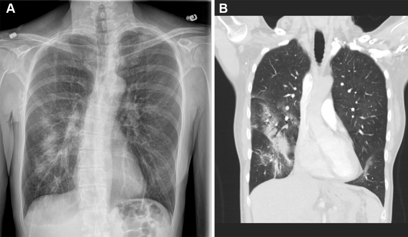
The authors report an unusual case of segmental lung torsion detected at CT pulmonary angiography in a patient with dyspnea. This case highlights the importance for clinicians and radiologists to consider and be familiar with the diagnosis of lung torsion, a rare and potentially life-threatening pathologic condition that can be successfully treated with emergent surgery if detected early. Keywords: CT, CT Angiography, Pulmonary, Thorax, Lung, Emergency Radiology Supplemental material is available for this article. © RSNA, 2023.
Keywords: CT; CT Angiography; Emergency Radiology; Lung; Pulmonary; Thorax.
© 2023 by the Radiological Society of North America, Inc.
Conflict of interest statement
Disclosures of conflicts of interest: R.E.K. No relevant relationships. Y.H. No relevant relationships. J.H. No relevant relationships. M.B. No relevant relationships.
Figures

References
-
- Felson B . Lung torsion: radiographic findings in nine cases . Radiology 1987. ; 162 ( 3 ): 631 – 638 . - PubMed
-
- Dai J , Xie D , Wang H , et al. . Predictors of survival in lung torsion: A systematic review and pooled analysis . J Thorac Cardiovasc Surg 2016. ; 152 ( 3 ): 737 – 745.e3 . - PubMed
-
- Hammer MM , Madan R . Clinical and imaging features in lung torsion and description of a novel imaging sign . Emerg Radiol 2018. ; 25 ( 2 ): 121 – 127 . - PubMed
-
- Jhala K , Madan R , Hammer M . A pictorial review of lung torsion using 3D CT cinematic rendering . Emerg Radiol 2021. ; 28 ( 1 ): 171 – 176 . - PubMed
-
- Hennink S , Wouters MW , Klomp HM , Baas P . Necrotizing pneumonitis caused by postoperative pulmonary torsion . Interact Cardiovasc Thorac Surg 2008. ; 7 ( 1 ): 144 – 145 . - PubMed
Publication types
LinkOut - more resources
Full Text Sources
