. 2023 Jun 19:14:1160065.
doi: 10.3389/fimmu.2023.1160065. eCollection 2023.

Immunogenicity and efficacy of a novel multi-patch SARS-CoV-2/COVID-19 vaccine candidate

Affiliations

Immunogenicity and efficacy of a novel multi-patch SARS-CoV-2/COVID-19 vaccine candidate

Beatriz Perdiguero et al. Front Immunol. .

Abstract

Introduction: While there has been considerable progress in the development of vaccines against SARS-CoV-2, largely based on the S (spike) protein of the virus, less progress has been made with vaccines delivering different viral antigens with cross-reactive potential.

Methods: In an effort to develop an immunogen with the capacity to induce broad antigen presentation, we have designed a multi-patch synthetic candidate containing dominant and persistent B cell epitopes from conserved regions of SARS-CoV-2 structural proteins associated with long-term immunity, termed CoV2-BMEP. Here we describe the characterization, immunogenicity and efficacy of CoV2-BMEP using two delivery platforms: nucleic acid DNA and attenuated modified vaccinia virus Ankara (MVA).

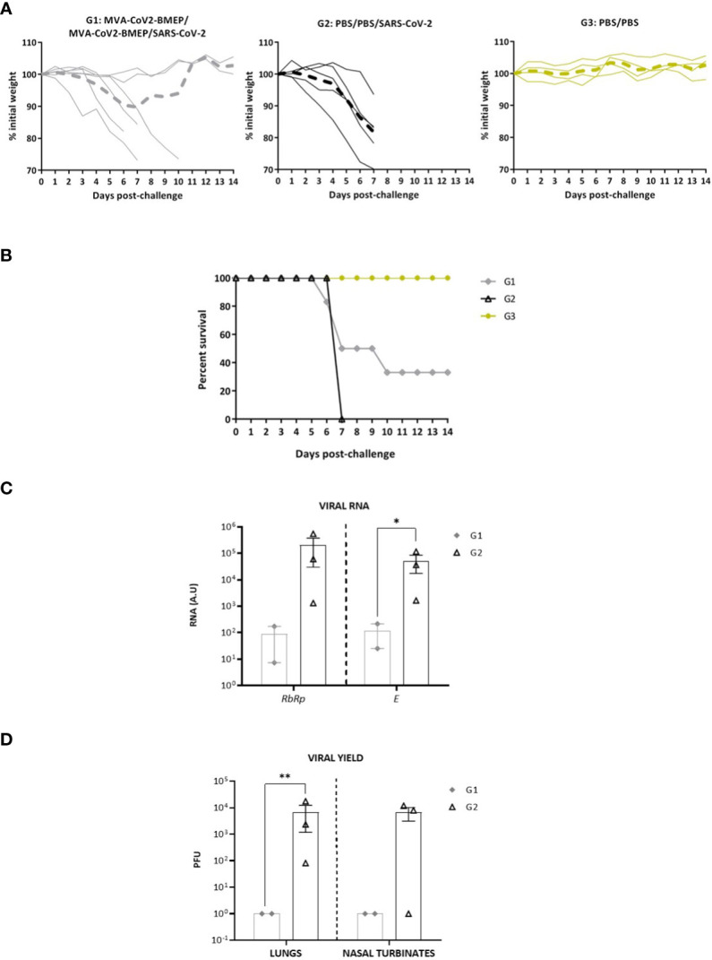
Results: In cultured cells, both vectors produced a main protein of about 37 kDa as well as heterogeneous proteins with size ranging between 25-37 kDa. In C57BL/6 mice, both homologous and heterologous prime/boost combination of vectors induced the activation of SARS-CoV-2-specific CD4 and CD8 T cell responses, with a more balanced CD8+ T cell response detected in lungs. The homologous MVA/MVA immunization regimen elicited the highest specific CD8+ T cell responses in spleen and detectable binding antibodies (bAbs) to S and N antigens of SARS-CoV-2. In SARS-CoV-2 susceptible k18-hACE2 Tg mice, two doses of MVA-CoV2-BMEP elicited S- and N-specific bAbs as well as cross-neutralizing antibodies against different variants of concern (VoC). After SARS-CoV-2 challenge, all animals in the control unvaccinated group succumbed to the infection while vaccinated animals with high titers of neutralizing antibodies were fully protected against mortality, correlating with a reduction of virus infection in the lungs and inhibition of the cytokine storm.

Discussion: These findings revealed a novel immunogen with the capacity to control SARS-CoV-2 infection, using a broader antigen presentation mechanism than the approved vaccines based solely on the S antigen.

Keywords: B and T cell immune responses; SARS-CoV-2; binding and neutralizing antibodies; efficacy; mice studies; multi-patch vaccine; poxvirus MVA.


Conflict of interest statement

The authors declare that the research was conducted in the absence of any commercial or financial relationships that could be construed as a potential conflict of interest.

Figures

Figure 1
Figure 1
In vitro characterization of the DNA vector expressing the polyvalent multi-patch CoV2-BMEP protein. (A) Schematic representation of the chimeric polyvalent multi-patch vaccine CoV2-BMEP targeting B cells. (B) 3D structure prediction of the CoV2-BMEP protein. (C, D) Time-course expression of CoV2-BMEP protein after DNA transfection by western-blotting and confocal microscopy analyses. (C) Transfected HEK-293T cells were harvested at 24, 48 and 72 hours post-transfection and pellets and supernatants (SN) were processed as described in Materials and Methods, fractionated by 12% SDS-PAGE and analyzed by western-blotting using the rabbit polyclonal anti-HA antibody. (D) Subcellular localization of CoV2-BMEP protein by confocal microscopy. Transfected HeLa cells were fixed at 24, 48 and 72 hours post-transfection and labeled with a rabbit polyclonal anti-HA antibody. After incubation with the corresponding secondary antibody conjugated with Alexa Fluor 488 (green staining), cells were stained with DAPI to detect cell nuclei and visualized by confocal microscopy.
Figure 2
Figure 2
In vitro characterization of the MVA recombinant virus expressing the polyvalent multi-patch CoV2-BMEP protein. (A) Scheme of the thymidine kinase (TK) locus of MVA-CoV2-BMEP recombinant virus. (B) Confirmation of CoV2-BMEP gene insertion by PCR analysis. DNA was extracted from DF-1 cells infected with MVA-WT or MVA-CoV2-BMEP viruses at 5 PFU/cell. Primers TK-R and TK-L spanning TK flanking regions were used for the analysis of the TK locus by PCR. (C) Time-course expression of CoV2-BMEP protein by western-blotting analysis. HeLa cells were mock-infected or infected with MVA-CoV2-BMEP at 3 PFU/cell. At different times post-infection (0, 6, 16 and 24 h), cells were harvested and cellular pellets and supernatants (SN) were processed as described in Materials and Methods, fractionated by 12% SDS-PAGE and the expression of CoV2-BMEP protein was analyzed by western-blotting using a rabbit polyclonal anti-HA antibody. Actin was used as loading control. (D) Subcellular localization of CoV2-BMEP protein by confocal microscopy. HeLa cells were infected with MVA-CoV2-BMEP at 1 PFU/cell. At 16 h post-infection, cells were incubated with WGA-Alexa Fluor 594 (red staining), fixed and labeled with a rabbit polyclonal anti-HA antibody (upper panels) or a rabbit polyclonal anti-N antibody (lower panels). After incubation with the corresponding secondary antibody conjugated with Alexa Fluor 488 (green staining), cells were stained with DAPI (blue staining) to detect cell nuclei and visualized by confocal microscopy. (E) Analysis of the stability of the CoV2-BMEP protein expressed by MVA-CoV2-BMEP virus. Twenty-two individual plaques from MVA-CoV2-BMEP stability passage 7 were grown in DF-1 cells, lysed, proteins fractionated by 12% SDS-PAGE and analyzed by western-blotting using a rabbit polyclonal anti-HA antibody. The expression of CoV2-BMEP protein in cells infected with P2 stock or with individual plaques (1–22) is shown.
Figure 3
Figure 3
Human CD8+ T lymphocyte response against CoV2-BMEP. Immune response to autologous cells infected with MVA-WT or MVA-CoV2-BMEP of 10 healthy volunteers who had tested positive for SARS-CoV-2. The percentage of CD8+ T lymphocytes producing IFN-γ or expressing CD107a as a measure of activation is represented. (A, B) show the IFN-γ response against both viruses (after subtracting the background of mock uninfected cells). (A) represents the individual IFN-γ response and the median and interquartile range of each group, while (B) links with a line the individual response of each volunteer against both viruses; seven of the ten volunteers respond to MVA-CoV2-BMEP. (C, D), same as (A, B) but showing the CD107a response of CD8+ T lymphocytes to the two viruses. Eight of the ten volunteers respond to MVA-CoV2-BMEP. Statistical analysis: Paired t test, *,p=0.017; **,p=0.0094.
Figure 4
Figure 4
Innate immune response elicited in muscle and DLNs by the DNA vector expressing the polyvalent multi-patch CoV2-BMEP protein. (A) Immunization schedule. Three groups of animals were inoculated with 25 μg of DNA-CoV2-BMEP or DNA-φ or PBS by the intramuscular (i.m.) route. At days 1, 3 and 7 post-inoculation, total muscle from the site of inoculation and DLNs were excised and processed as described in Materials and Methods. (B, C) Different immune cell populations present in muscle (B) and DLN (C) cell suspensions determined by flow cytometry. BCs, B cells; NKs, Natural killer cells; TCs, T cells; moCs, Monocyte-derived cells; NOs, Neutrophils; EOs, Eosinophils; DCs, Dendritic cells; cDCs, Conventional dendritic cells. Data are shown as mean and SD. *, p < 0.05; **, p < 0.005; ***, p < 0.001.
Figure 5
Figure 5
Innate immune response elicited in muscle and DLNs by the MVA vector expressing the polyvalent multi-patch CoV2-BMEP protein. (A) Immunization schedule. Three groups of animals were immunized with 1 x 10⁷ PFU of MVA-WT or MVA-CoV2-BMEP or PBS by the i.m. route. At day 1 post-inoculation, total muscle from the site of inoculation and DLNs were excised and processed as described in Materials and Methods. (B, C) Different immune cell populations present in muscle (B) and DLN (C) cell suspensions determined by flow cytometry. Data are shown as colored forms for each animal with mean and SD. *, p < 0.05; **, p < 0.005; ***, p < 0.001.
Figure 6
Figure 6
Cellular and humoral adaptive immune responses elicited by DNA-CoV2-BMEP and MVA-CoV2-BMEP in C57BL6 mice when administered in homologous or heterologous regimens. (A) Immunization schedule. (B) Magnitude of the total SARS-CoV-2-specific CD4 (left) or CD8 (right) T cells at 10 days post-boost by ICS assay after the stimulation of lymphocytes derived from spleen or lung with SARS-CoV-2 peptide pools. The total value of each group represents the sum of the percentages of SARS-CoV-2-specific CD4 or CD8 T cells secreting IFN-γ and/or IL-2 and/or TNF-α against SARS-CoV-2 peptide pools. Data are background-subtracted. 95% CI is represented. ***, p < 0.001. (C) Level of binding antibodies to SARS-CoV-2 S (left) or N (right) proteins from Wuhan reference strain elicited in serum from immunized individual mice at 10 days post-boost measured as OD450 at a serum dilution of 1:100 by ELISA. Data are shown as colored forms for each animal with mean and SD. (D) Level of neutralizing antibodies to SARS-CoV-2 (Wuhan strain) elicited in serum from immunized individual mice at 10 days post-boost measured as NT50 by microneutralization assay. Data are shown as colored forms for each animal with mean and SD. The red dashed line represents the lower limit of detection (LLD) of the assay.
Figure 7
Figure 7
Efficacy study in humanized k18-hACE2 Tg mice. (A) Immunization schedule. (B) Level of binding antibodies to SARS-CoV-2 S (left) or N (right) proteins from Wuhan reference strain elicited in serum from immunized individual mice at days 20 (post-prime) and 42 (21 days post-boost) before challenge. The endpoint titer represents the last serum dilution that gave 3 times the mean OD450 value of the control group. Data are shown as forms for each animal with mean and SD. The red dashed line represents the lower limit of detection (LLD) of the assay. (C, D) Titers of SARS-CoV-2 neutralizing antibodies (NT50) in serum from immunized individual mice against SARS-CoV-2 MAD6 isolate (C), or serum from two immunized mice with the highest NT50 against Alpha, Beta, Delta and Omicron variants (D) determined in triplicates using a live virus MNT assay at days 20 (post-prime) and 42 (21 days post-boost) for MAD6 isolate or d42 for SARS-CoV-2 variants before challenge. Data are shown as forms for each animal with mean and SD. The red dashed line represents the LLD of the assay. *, p < 0.05; **, p < 0.005.
Figure 8
Figure 8
Two doses of MVA-CoV2-BMEP partially protect from morbidity and mortality in humanized k18-hACE2 Tg mice infected with SARS-CoV-2. The challenged mice were monitored for body weight variations (A) and mortality (B) for 14 days. (C) Virus replication in lung samples. Genomic (RdRp) and subgenomic (E) SARS-CoV-2 RNAs were detected by RT-qPCR after virus challenge in surviving mice. RNA levels [in arbitrary units (A.U.)] for each animal are represented as forms with mean and SD from duplicates; relative values are referred to non-challenged PBS-inoculated mice (group 3). *, p < 0.05. (D) SARS-CoV-2 infectious virus in lung samples and nasal turbinates. Data are shown as forms for each animal with mean (PFUs/g of lung tissue or PFUs/mL of nasal turbinate) and SD from duplicates. **, p < 0.005.
Figure 9
Figure 9
Lung histopathological and cytokine profile analyses of mice vaccinated with MVA-CoV2-BMEP and challenged with SARS-CoV-2. (A) Lung inflammation scores obtained in lung samples from individual mice immunized with MVA-CoV2-BMEP, challenged with SARS-CoV-2 and euthanized at 14 days post-challenge (group 1; n=2), PBS-treated mice challenged with SARS-CoV-2 and euthanized at 7 days post-challenge (group 2; n=3) and PBS-treated non-challenged mice euthanized at 14 days post-challenge (group 3; n=4). Data are represented as colored forms for each animal with mean and SD of cumulative histopathological lesions for each experimental group. ***, p < 0.001. (B) Representative lung histopathological sections from k18-hACE2 Tg mice included in experimental groups 1, 2 and 3. Mice in groups 1 and 3 did not display remarkable inflammatory lesions while mice in group 2 showed inflammatory lesions such as diffuse mild to moderate thickening of the alveolar septa, diffuse mild alveolar mononuclear cell infiltrates (arrows) or the presence of ocassional multifocal perivascular and peribronchiolar mononuclear infiltrates (arrowheads). H&E staining; Magnification: 10x. (C) Levels of RNA from pro-inflammatory cytokines and chemokines genes detected by RT-qPCR. Targeting genes (Cxcl10, Il-6, Il-10, Tnf-alpha, Cxcl5 and Ifit27) were normalizated to cellular 28S rRNA in lungs obtained at 14 days post-challenge in surviving mice. RNA levels (in A.U.) for each animal are represented as forms with mean and SD from duplicates; relative values are referred to non-challenged PBS-inoculated mice (group 3). *, p < 0.05.

References

    1. Schubert M, Bertoglio F, Steinke S, Heine PA, Ynga-Durand MA, Maass H, et al. . Human serum from SARS-CoV-2-vaccinated and COVID-19 patients shows reduced binding to the RBD of SARS-CoV-2 omicron variant. BMC Med (2022) 20(1):102. doi: 10.1186/s12916-022-02312-5 - DOI - PMC - PubMed
    1. Thakur S, Sasi S, Pillai SG, Nag A, Shukla D, Singhal R, et al. . SARS-CoV-2 mutations and their impact on diagnostics, therapeutics and vaccines. Front Med (2022) 9:815389. doi: 10.3389/fmed.2022.815389 - DOI - PMC - PubMed
    1. Bar-On YM, Goldberg Y, Mandel M, Bodenheimer O, Amir O, Freedman L, et al. . Protection by a fourth dose of BNT162b2 against omicron in Israel. N Engl J Med (2022) 386:1712–20. doi: 10.1056/NEJMoa2201570 - DOI - PMC - PubMed
    1. Gaebler C, Wang Z, Lorenzi JCC, Muecksch F, Finkin S, Tokuyama M, et al. . Evolution of antibody immunity to SARS-CoV-2. Nature (2021) 591(7851):639–44. doi: 10.1038/s41586-021-03207-w - DOI - PMC - PubMed
    1. Xia H, Zou J, Kurhade C, Cai H, Yang Q, Cutler M, et al. . Neutralization and durability of 2 or 3 doses of the BNT162b2 vaccine against omicron SARS-CoV-2. Cell Host Microbe (2022) 30(4):485–8.e3. doi: 10.1016/j.chom.2022.02.015 - DOI - PMC - PubMed

Publication types

Supplementary concepts

LinkOut - more resources