. 2022 Aug 9;84(4):384-394.
doi: 10.1055/a-1786-9026. eCollection 2023 Aug.

Phenotypical Variability of the Internal Acoustic Canal in the Middle Cranial Fossa Surgery

Affiliations

Phenotypical Variability of the Internal Acoustic Canal in the Middle Cranial Fossa Surgery

Maryna Al-Fauri Kornieieva et al. J Neurol Surg B Skull Base. .

Abstract

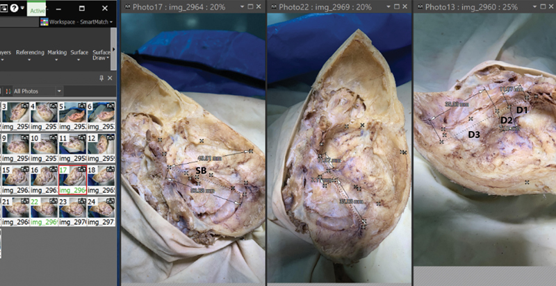
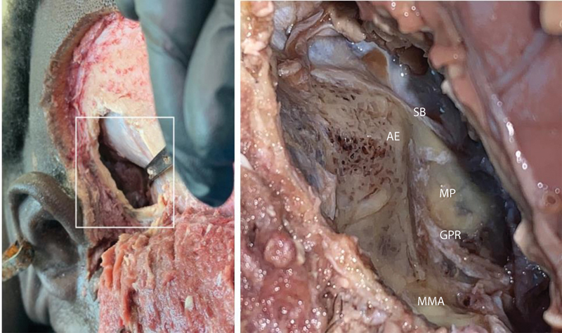
Introduction The wide range of anatomical variability of the structures of the middle cranial fossa (MCF) and the lack of reliable surgical landmarks contribute to a high level of complications in the surgical treatment of vestibular schwannomas. We hypothesized that the cranial phenotype influences the shape of the MCF, the orientation of the pyramid of the temporal bone, and the relative topography of the internal acoustic canal (IAC). Methods The skull base structures were studied on 54 embalmed cadavers and 60 magnetic resonance images of the head and neck by photo modeling, dissection, and three-dimensional analysis techniques. By the value of the cranial index, all specimens were subdivided into dolichocephalic, mesocephalic, and brachycephalic groups for comparison of variables. Results The length of the superior border of the temporal pyramid (SB), the apex to squama distance, and the width of the MCF all peaked in the brachycephalic group. The value of the angle between the SB and the axis of the acoustic canal varied from 33 to 58 degrees; it peaked in the dolichocephalic group and showed its smaller value in the brachycephalic one. The pyramid to squama angle had reversed distribution and dominated in the brachycephalic group. Conclusion The cranial phenotype influences the shape of the MCF, temporal pyramid, and IAC. Presented in this article data help specialists operating on the vestibular schwannoma to localize the IAC based on the individual shape of a skull.

Keywords: anatomical variability; cranial index; internal acoustic canal; middle cranial fossa; temporal bone; vestibular schwannoma.


Conflict of interest statement

Conflict of Interest None declared.

Figures

Fig. 1
Fig. 1
A screenshot of the three-dimensional (3D) modeling window on the PhotoModeler platform is presented in this picture. The program analyses the light angles and distances between similar points in pictures and creates a digital 3D model of the object. The obtained 3D model is scaled by the largest diameter of the studied object, and the rotation axes are set up manually. The camera underwent preliminary calibration allowing accurate measurements with a residual of less than 3 px (PhotoModeler software, 2021).
Fig. 2
Fig. 2
A screenshot of the “measurements” window of the PhotoModeler platform is presented in this picture. The superior border of the temporal pyramid (SB) is marked along the groove of the superior petrous sinus. The anterior (medial) and posterior (lateral) margins of the porus acusticus are identified and extended up perpendicular to the SB. From the middle cranial fossa view, the following distances are measured: (D1)—between the apex of the pyramid and the projection of the anterior margin of the porus acusticus; (D2)—between the projection points of the anterior and posterior margins of the porus acusticus; and (D3)—from the projection point of the posterior margin of the porus acusticus to the most lateral point of the SB (PhotoModeler software, 2021).
Fig. 3
Fig. 3
On the axial magnetic resonance image of the head (T2-weighted), the superior border of the temporal pyramid (SB) was identified as a hypointense longitudinal shadow connecting the apex of the pyramid (AP) with the shadow of the sigmoid sinus (SS). The internal carotid artery that appears at the apex of the temporal pyramid helped demarcate the precise location of the bony apex. The axis of the internal acoustic canal (IAC) was dragged through the center of the canal and extended forward and laterally reaching the lateral wall of the middle cranial fossa. Then, the axis was projected to the same level with the SB, and the angles between the SB and the axis of the IAC angle and between the SB and the temporal squama (SB angle) were measured (RadiAnt software package, 2021).
Fig. 4
Fig. 4
The image ( A ) shows the planning of the skin incision for subtemporal craniotomy on a cadaver specimen (male, mesocephalic phenotype). The line of incision begins at the level of the root of the zygomatic arch 1 cm anterior to the auricle; then, it extends upward along the line of hair growth and arches over the temporal fossa. On the image ( B ), the skin flap (SF), temporal facial, and temporal muscle (TM) are reflected anteriorly. The 4- to 5-cm bony flap (BF) is marked on the surface of the temporal squama in immediate proximity to the root of the zygomatic arch and the auricle.
Fig. 5
Fig. 5
Surgical view of the right middle cranial fossa reached via the subtemporal approach (simulation on a male cadaver; mesocephalic phenotype, cranial index = 76.4). The dura mater is elevated exposing the anterior surface of the temporal pyramid with the following anatomical landmarks: SB, superior border of the pyramid; AE, arcuate eminence; GPR, greater petrosal nerve; MP, meatal plate; MMA, middle meningeal artery (dissected). The precise localization of the internal acoustic canal, its length, and the distance from the surface of the bone are still unclear at this stage of the procedure. Note: the operative corridor is significantly smaller in females (Maina et al, 2007 6 ).
Fig. 6
Fig. 6
The axial magnetic resonance image of the head shows: ( A ) relatively small angle between the axis of the internal acoustic canal (IAC) and the superior border of the temporal pyramid (SB); ( B ) relatively large angle between the lateral wall of the middle cranial fossa (squama, S) and the SB. The cranial index of this patient is equal to 86.4 (brachycephalic phenotype).
Fig. 7
Fig. 7
The axial magnetic resonance image of the head shows: ( A ) relatively large angle between the axis of the internal acoustic canal (IAC) and the superior border of the temporal pyramid (SB); ( B ) relatively small angle between the lateral wall of the middle cranial fossa (squama, S) and the SB. The cranial index of this patient is equal to 72.3 (dolichocephalic phenotype).
Fig. 8
Fig. 8
The phenotypical variability of the middle cranial fossa (MCF), the orientation of the pyramid of the temporal bone, and orientation of the internal acoustic canal (IAC) are demonstrated on three-dimensional (3D) models of cadaveric skull bases with cranial indexes 70.67% (a), 75.10% (b), and 85.23% (c): dolichocephalic, mesocephalic, and brachycephalic specimens, respectively. The distance from the temporal squama to the projection point of the lateral border of the porus acusticus (PA) along the superior border of the pyramid (D3) increases with the laterolateral diameter of the skull. The angle between the superior border of the pyramid and the axis of the IAC (yellow arrow) is significantly large in specimens with narrow skulls. The 3D modeling technique was used to visualize the data (PhotoModeler software, 2021).

References

    1. Myrseth E, Pedersen P H, Møller P, Lund-Johansen M.Treatment of vestibular schwannomas. Why, when and how? Acta Neurochir (Wien) 200714907647–660., discussion 660 - PubMed
    1. Gonzalez F, Ferreira M, Zabramski J, Spetzler R, Deshmukh P. The middle fossa approach. Barrow Quarterly. 2000;16(04):67–85.
    1. Staecker H, Nadol J B, Jr, Ojeman R, Ronner S, McKenna M J. Hearing preservation in acoustic neuroma surgery: middle fossa versus retrosigmoid approach. Am J Otol. 2000;21(03):399–404. - PubMed
    1. Farahani R, Nooranipour M, Nikakhtar K. Anthropometry of internal acoustic meatus. Int J Morphol. 2007;5(04):861–865.
    1. Faure A, Masse H, Gayet-Delacroix M. What is the arcuate eminence? Surg Radiol Anat. 2003;25(02):99–104. - PubMed