. 2023 Jul;30(7):103710.
doi: 10.1016/j.sjbs.2023.103710. Epub 2023 Jun 19.

Fatal intracranial haemorrhage in acute promyelocytic leukemia patients with short isoform of PML-RARα: Review of molecular and radiological data

Affiliations

Fatal intracranial haemorrhage in acute promyelocytic leukemia patients with short isoform of PML-RARα: Review of molecular and radiological data

Manal Abudawood et al. Saudi J Biol Sci. 2023 Jul.

Abstract

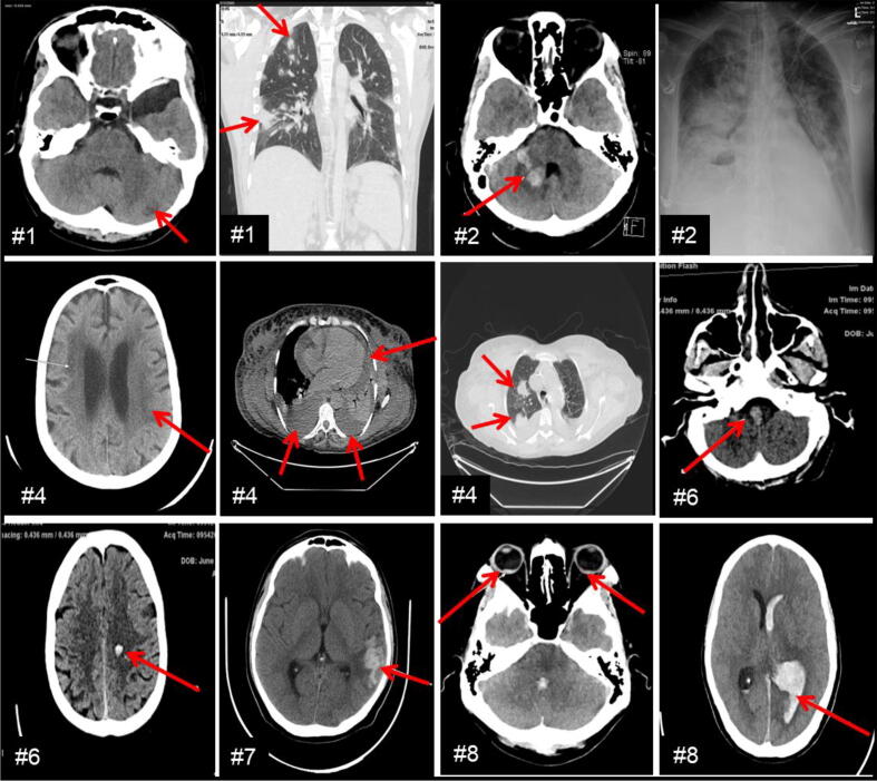
Three major PML-RARα fusion gene transcripts (long [bcr1], variant [bcr2], and short [bcr3]) are currently used in clinical laboratories for the diagnosis and treatment monitoring of APL patients. Despite highly improved outcome, relapse and intracranial haemorrhage that may lead to early death is still an unsolved complication in APL. We reviewed APL patients confirmed by qPCR for the presence of PML-RARα transcripts (n = 27) and studied their outcome in relation to the isoform expression at diagnosis and follow-up in King Fahad Medical City. Eight in twenty-seven patients showed bcr3 and nineteen patients with bcr1 as major isoforms at diagnosis. Half of the bcr3 patients (n = 4/8) showed early mortality, prolonged qPCR positivity, 4-fold higher neutrophil/lymphocyte ratio, higher creatinine levels, and significantly reduced relapse free and overall survival time compared with bcr1 patients. Radiological findings in bcr3 patients revealed CNS involvement in the form of intracranial haemorrhage and periventricular microangiopathy and no CNS involvement in bcr1 patients. In conclusion, PML-RARα isoform expression at diagnosis in selective patients influences disease course over time and may even lead to early mortality due to haemorrhage. Thus, timely reporting of the specific PML-RARα isoform by clinical laboratories and CNS assessment by radiology can prevent complications leading to death in some APL patients.

Keywords: Acute promyelocytic leukemia (APL); All-trans-retinoic acid (ATRA); Arsenic trioxide (ATO); Central nervous system (CNS); Computerized tomography (CT); Minimal residual disease (MRD); Molecular remission (MR); PML-RARα isoforms; Quantitative polymerase chain reaction (qPCR).


Conflict of interest statement

The authors declare that they have no known competing financial interests or personal relationships that could have appeared to influence the work reported in this paper.

Figures

Fig. 1
Fig. 1
Genomic browser image with exon map showing different PML-RARα isoforms and their detection by karyotyping, FISH and qPCR at diagnosis: A) upper panel-chromosome 15 and 17 map showing the breakpoints (arrow) involved in translocation t(15;17) with the regions shown (dashed red box), lower panel-detailed exon map of the PML and RARa genes on respective chromosomes with coding exons (e1, e2, e3 etc., texture boxes) and non-coding exons (solid boxes). The arrows represent the breakpoints at respective locations to generate the two isoforms of PML-RARα bcr1 (long L- isoform) and bcr3 (short S- isoform), B) representative APL case showing detection of PML-RARα at diagnosis by Karyotype (a), FISH (b) and qPCR for bcr1 (c) and bcr3 (d) on RotorGene instrument using commercially available kits (Ipsogen Qiagen, Germany). The graph shows a high positive HP (***), low positive LP (*) and abl1 gene as internal control used in each qPCR run.
Fig. 2
Fig. 2
Clinical laboratory markers WBC, PLT and NLR at presentation and at day 28 post induction treatment for bcr1 vs bcr3 APL patients: A) WBC, platelet (PLT) counts and NLR median values at presentation (day 0) for bcr3 vs bcr1 APL patients, B) WBC, platelet (PLT) counts and NLR median values post induction treatment (day 28) for bcr3 vs bcr1 APL patients. The fold changes are shown, ns means not significant changes since APL patients go into remission post induction.
Fig. 3
Fig. 3
Prolonged qPCR positivity post induction and early reappearance (time to molecular relapse) of PML-RARα in bcr3 vs bcr1 patients: A) representative APL case showing qPCR data for bcr3 at diagnosis (day 0), and post treatment monitoring at day 28, and at day 60 showing isoform detected but below level of quantification (LOQ, positivity), B) representative APL case showing qPCR data for bcr1 at diagnosis (d 0), and post treatment monitoring at d 28 showing isoform not detected (ND), Arrows show bcr3/abl1 and/or bcr1/abl1 detection, C) qPCR monitoring data over extended period of time for bcr3 vs bcr1 APL patients. # on the upper left side of the figures represent representative case numbers and numbers on the lower left side represents time for the reappearance of bcr3 and/or bcr1 isoform (time to molecular relapse). The graphs also show a high positive HP (***), low positive LP (*) and abl1 gene as internal control used in each qPCR run. An arrow also represents amplification of bcr3 and/or bcr1.
Fig. 4
Fig. 4
Reduced relapse free survival (RFS) and overall survival (OS) in bcr3 vs bcr1 APL patients: Kaplan Meier-based estimation of RFS (4A) and OS probability (4B) in bcr3 vs bcr1 patients (p < 0.05). Red lines represent bcr3 patients with reduced survival compared with green lines representing bcr1 patients. Numbers at risk for bcr1 and bcr3 groups are shown at the bottom. Note that four patients that showed early mortality were excluded in the RFS analyses.
Fig. 5
Fig. 5
Abnormal imaging findings in APL patients with bcr3 isoform of PML-RARα at presentation: A) Brain, chest, abdomen computerized tomography (CT) scans, and chest X-rays in bcr3 patients showing various abnormalities such as left cerebellar hypodensity, hepatomegaly and nodular opacities (arrows #1), right cerebellar hematoma and pulmonary edema (arrows #2) periventricular microangiopathic changes, pulmonary opacities, bilateral pleural and pericardial effusion (arrows #4), intracranial haemorrhage in right occipital and right medulla, pulmonary edema (arrows #6), left cerebral hematoma (arrows #7), left intraventricular and bilateral orbital haemorrhage (arrows #8).

References

    1. Abaza Y., Kantarjian H., Garcia-Manero G., Estey E., Borthakur G., Jabbour E., Faderl S., O’Brien S., Wierda W., Pierce S., Brandt M., McCue D., Luthra R., et al. Long-term outcome of acute promyelocytic leukemia treated with all-trans-retinoic acid, arsenic trioxide, and gemtuzumab. Blood. 2017;129:1275–1283. doi: 10.1182/blood-2016-09-736686. - DOI - PMC - PubMed
    1. Daver N., Kantarjian H., Marcucci G., et al. Clinical characteristics and outcomes in patients with acute promyelocytic leukaemia and hyperleucocytosis. British J. Haematol. 2015;168(5):646–653. - PMC - PubMed
    1. De Botton S., Dombret H., Sanz M., Miguel J.S., Caillot D., Zittoun R., Gardembas M., Stamatoulas A., Condé E., Guerci A., Gardin C., Geiser K., Makhoul D.C., Reman O., de la Serna J., Lefrere F., Chomienne C., Chastang C., Degos L., Fenaux P. Incidence, clinical features, and outcome of all trans-retinoic acid syndrome in 413 cases of newly diagnosed acute promyelocytic leukemia. The European APL Group. Blood. 1998;15;92(8):2712–8 - PubMed
    1. De Thé H., Chomienne C., Lanotte M., Degos L., Dejean A. The t(15;17) translocation of acute promyelocytic leukaemia fuses the retinoic acid receptor α gene to a novel transcribed locus. Nature. 1990;347:558–561. - PubMed
    1. Dinmohamed A.G., Visser O. Incidence of acute promyelocytic leukemia across Europe: results of RARECAREnet—a population-based study. Stem Cell Investig. 2019;6:37. doi: 10.21037/sci.2019.10.03. - DOI - PMC - PubMed

LinkOut - more resources