. 2023 Jul 1;79(Pt 7):180-192.
doi: 10.1107/S2053230X23005381. Epub 2023 Jul 5.

Tetracycline-modifying enzyme SmTetX from Stenotrophomonas maltophilia

Affiliations

Tetracycline-modifying enzyme SmTetX from Stenotrophomonas maltophilia

Martin Malý et al. Acta Crystallogr F Struct Biol Commun. .

Abstract

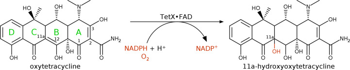
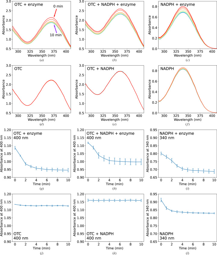
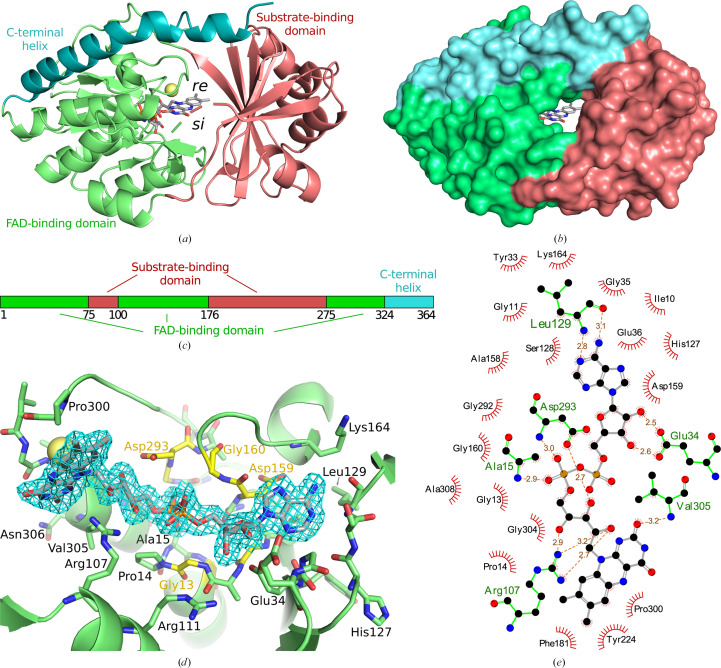
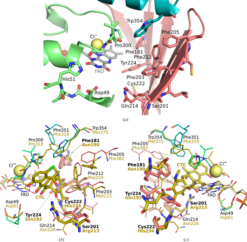
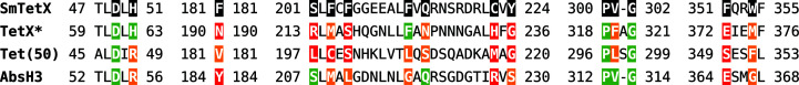
The resistance of the emerging human pathogen Stenotrophomonas maltophilia to tetracycline antibiotics mainly depends on multidrug efflux pumps and ribosomal protection enzymes. However, the genomes of several strains of this Gram-negative bacterium code for a FAD-dependent monooxygenase (SmTetX) homologous to tetracycline destructases. This protein was recombinantly produced and its structure and function were investigated. Activity assays using SmTetX showed its ability to modify oxytetracycline with a catalytic rate comparable to those of other destructases. SmTetX shares its fold with the tetracycline destructase TetX from Bacteroides thetaiotaomicron; however, its active site possesses an aromatic region that is unique in this enzyme family. A docking study confirmed tetracycline and its analogues to be the preferred binders amongst various classes of antibiotics.

Keywords: FAD-dependent monooxygenases; antibiotic resistance; tetracycline.


Figures

Figure 1
Figure 1
Simplified reaction scheme of the degradation of oxytetracycline catalyzed by the tetracycline destructase TetX. The four individual rings of the molecule are labelled in green. The C atoms susceptible to modification by tetracycline destructases are labelled with their numbers.
Figure 2
Figure 2
SmTetX causes changes in the UV–Vis spectrum of oxytetracycline (OTC) and NADPH. The initial concentrations of the reagents, which are listed in each panel, were [OTC] = 0.5 mM, [NADPH] = 0.5 mM and [enzyme] = 0.1 µM. The individual enzymatic assays were performed in parallel in 100 mM TAPS pH 8.5. (af) Absorbance scans are plotted at 1 min intervals over a time course of 10 min, represented by a rainbow colour gradient [from red through yellow to blue as shown in (a)]. Background absorbance of the buffer was subtracted. (gl) Monitoring of the decrease in absorbance of OTC (400 nm) and NADPH (340 nm); standard errors of the mean from measurements in triplicate are shown.
Figure 3
Figure 3
Structural analysis of SmTetX. The substrate-binding domain is coloured red, the FAD-binding domain green and the C-terminal helix blue. C atoms of FAD are shown in grey in stick representation and a chloride anion is shown as a yellow sphere. (a, b) Overall fold represented in secondary structure (a) and surface (b) views along the access tunnel to the re site. (c) Schematic diagram of the SmTetX domain structure; the numbers correspond to the native amino-acid sequence. (d) The binding of FAD in combined stick and secondary-structure representation: a view from the flavin si site. The 2mF oDF c electron density is contoured in blue at the 1σ level. C atoms of the residues belonging to the GXGXXG, DG and GD binding motifs are coloured yellow. (e) Interactions of FAD with the protein. Hydrogen bonds are shown in orange with distances in Å. The graphics were prepared in PyMOL 2.5 (Schrödinger) and LigPlot+ (Laskowski & Swindells, 2011 ▸).
Figure 4
Figure 4
The active site of SmTetX is located at the re site. (a) Composition of the active site in combined stick and secondary-structure representation. C atoms of the substrate-binding domain are coloured red, those of the FAD-binding domain green and those of the C-terminal helix blue. The chloride anion is shown as a yellow sphere. (b, c) Alignment of SmTetX with the TetX–chlortetracycline (CTC) complex (Volkers et al., 2011; PDB entry 2y6r) in yellow shown in stick representation. The r.m.s.d. was 3.74 Å using 284 Cα atoms of chains A. The graphics were prepared in PyMOL 2.5 (Schrödinger).
Figure 5
Figure 5
Multiple structure-based sequence alignment of SmTetX with the tetracycline destructases TetX from B. thetaiotaomicron (PDB entry 2y6r; Volkers et al., 2011 ▸) and Tet(50) (PDB entry 5tue; Park et al., 2017 ▸), and the putative reductase AbsH3 from Streptomyces sp. (PDB entry 6n04; Clinger et al., 2021 ▸). Regions significant for the re site of the enzyme are shown. The key residues in the binding pocket of SmTetX are highlighted by a black background (and shown in Fig. 4 ▸). Identity, similarity and dissimilarity of the key amino acids are marked with green, orange and red backgrounds, respectively. *The sequence and structure of this region are the same for TetX, TetX2, TetX4 and TetX7 (Cheng et al., 2022 ▸).

References

    1. Afonine, P. V., Moriarty, N. W., Mustyakimov, M., Sobolev, O. V., Terwilliger, T. C., Turk, D., Urzhumtsev, A. & Adams, P. D. (2015). Acta Cryst. D71, 646–666. - PMC - PubMed
    1. Agirre, J., Atanasova, M., Bagdonas, H., Ballard, C. B., Baslé, A., Beilsten-Edmands, J., Borges, R. J., Brown, D. G., Burgos-Mármol, J. J., Berrisford, J. M., Bond, P. S., Caballero, I., Catapano, L., Chojnowski, G., Cook, A. G., Cowtan, K. D., Croll, T. I., Debreczeni, J. É., Devenish, N. E., Dodson, E. J., Drevon, T. R., Emsley, P., Evans, G., Evans, P. R., Fando, M., Foadi, J., Fuentes-Montero, L., Garman, E. F., Gerstel, M., Gildea, R. J., Hatti, K., Hekkelman, M. L., Heuser, P., Hoh, S. W., Hough, M. A., Jenkins, H. T., Jiménez, E., Joosten, R. P., Keegan, R. M., Keep, N., Krissinel, E. B., Kolenko, P., Kovalevskiy, O., Lamzin, V. S., Lawson, D. M., Lebedev, A. A., Leslie, A. G. W., Lohkamp, B., Long, F., Malý, M., McCoy, A. J., McNicholas, S. J., Medina, A., Millán, C., Murray, J. W., Murshudov, G. N., Nicholls, R. A., Noble, M. E. M., Oeffner, R., Pannu, N. S., Parkhurst, J. M., Pearce, N., Pereira, J., Perrakis, A., Powell, H. R., Read, R. J., Rigden, D. J., Rochira, W., Sammito, M., Sánchez Rodríguez, F., Sheldrick, G. M., Shelley, K. L., Simkovic, F., Simpkin, A. J., Skubak, P., Sobolev, E., Steiner, R. A., Stevenson, K., Tews, I., Thomas, J. M. H., Thorn, A., Valls, J. T., Uski, V., Usón, I., Vagin, A., Velankar, S., Vollmar, M., Walden, H., Waterman, D., Wilson, K. S., Winn, M. D., Winter, G., Wojdyr, M. & Yamashita, K. (2023). Acta Cryst. D79, 449–461.
    1. Aleksandrov, A., Proft, J., Hinrichs, W. & Simonson, T. (2007). ChemBioChem, 8, 675–685. - PubMed
    1. Arita, M., Karsch-Mizrachi, I. & Cochrane, G. (2021). Nucleic Acids Res. 49, D121–D124. - PMC - PubMed
    1. Berman, H., Henrick, K. & Nakamura, H. (2003). Nat. Struct. Mol. Biol. 10, 980. - PubMed

MeSH terms