

Bioflavonoid luteolin prevents sFlt-1 release via HIF-1α inhibition in cultured human placenta
- PMID: 37405762
- PMCID: PMC10348062
- DOI: 10.1096/fj.202300611R
Bioflavonoid luteolin prevents sFlt-1 release via HIF-1α inhibition in cultured human placenta
Abstract
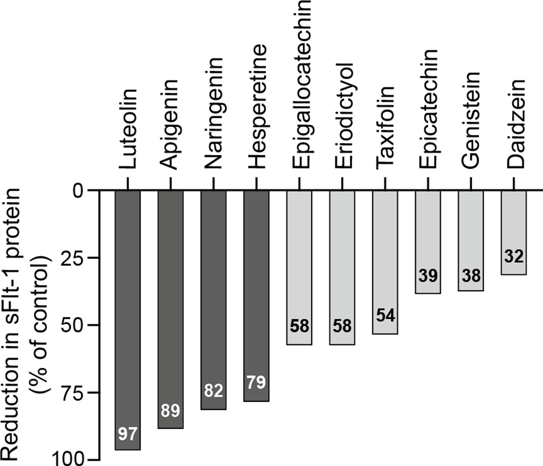
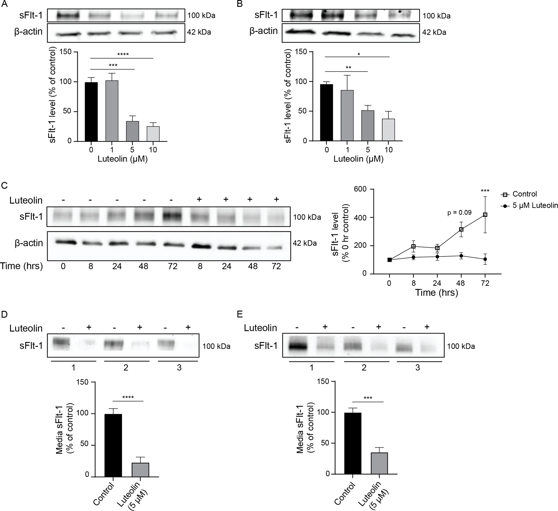
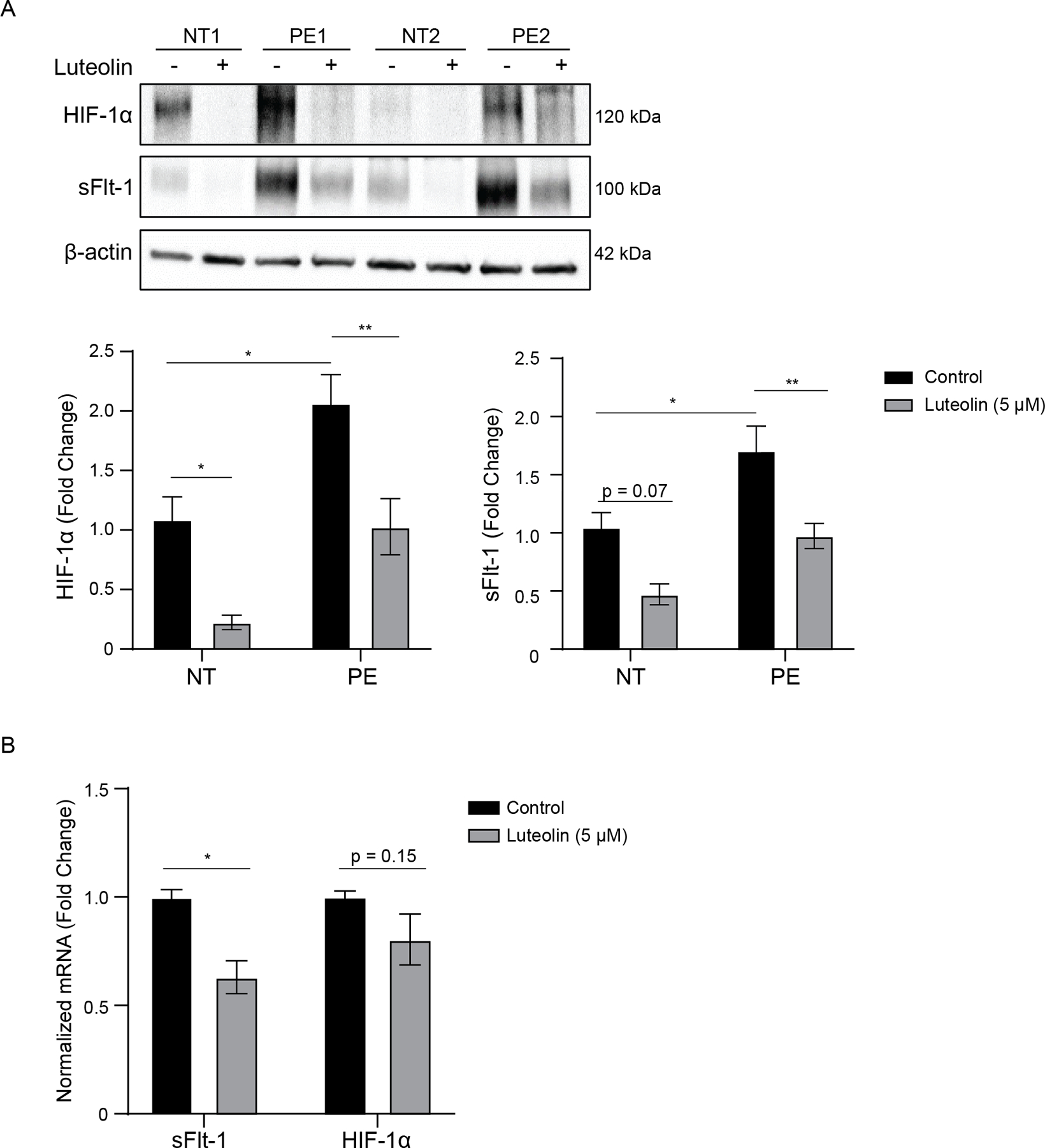
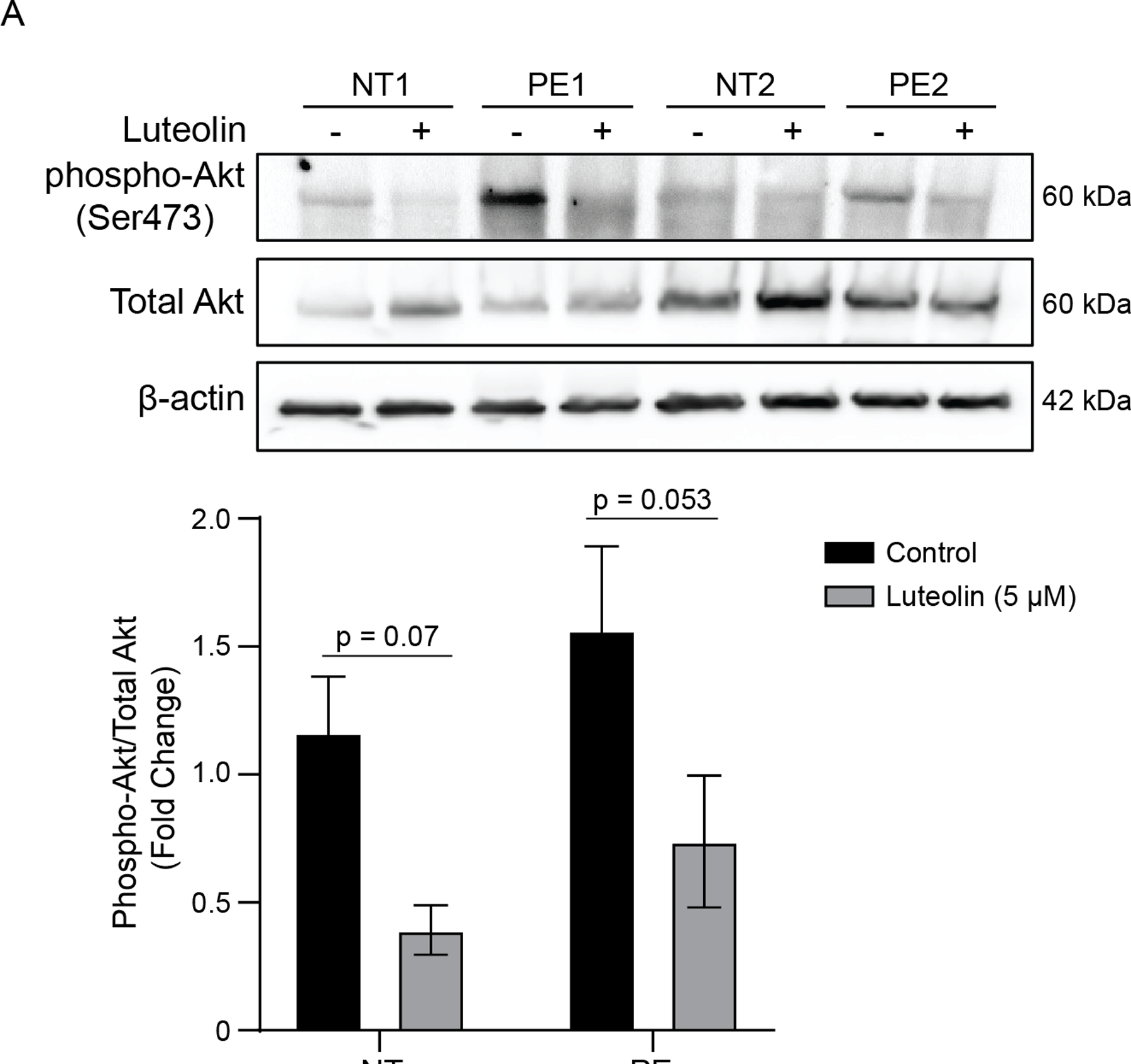
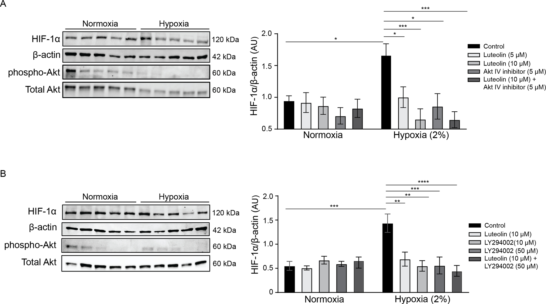
Preeclampsia (PE) is a serious hypertensive complication of pregnancy and is a leading cause of maternal death and major contributor to maternal and perinatal morbidity, including establishment of long-term complications. The continued prevalence of PE stresses the need for identification of novel treatments which can target prohypertensive factors implicated in the disease pathophysiology, such as soluble fms-like tyrosine kinase 1 (sFlt-1). We set out to identify novel compounds to reduce placental sFlt-1 and determine whether this occurs via hypoxia-inducible factor (HIF)-1α inhibition. We utilized a commercially available library of natural compounds to assess their ability to reduce sFlt-1 release from primary human placental cytotrophoblast cells (CTBs). Human placental explants from normotensive (NT) and preeclamptic (PE) pregnancies were treated with varying concentrations of luteolin. Protein and mRNA expression of sFlt-1 and upstream mediators were evaluated using ELISA, western blot, and real-time PCR. Of the natural compounds examined, luteolin showed the most potent inhibition of sFlt-1 release, with >95% reduction compared to vehicle-treated. Luteolin significantly inhibited sFlt-1 in cultured placental explants compared to vehicle-treated in a dose- and time-dependent manner. Additionally, significant decreases in HIF-1α expression were observed in luteolin-treated explants, suggesting a mechanism for sFlt-1 downregulation. The ability of luteolin to inhibit HIF-1α may be mediated through the Akt pathway, as inhibitors to Akt and its upstream regulator phosphatidylinositol-3 kinase (PI3K) resulted in significant HIF-1α reduction. Luteolin reduces anti-angiogenic sFlt-1 through inhibition of HIF-1α, making it a novel candidate for the treatment of PE.
Keywords: flavones; hypoxia; placenta; preeclampsia; sFlt-1.
© 2023 The Authors. The FASEB Journal published by Wiley Periodicals LLC on behalf of Federation of American Societies for Experimental Biology.
Conflict of interest statement
Figures
Similar articles
-
Luteolin prevents TNF-α-induced NF-κB activation and ROS production in cultured human placental explants and endothelial cells.Placenta. 2024 Jan;145:65-71. doi: 10.1016/j.placenta.2023.12.004. Epub 2023 Dec 8. Placenta. 2024. PMID: 38096686 Free PMC article.
-
Trimethylamine N-Oxide increases soluble fms-like tyrosine Kinase-1 in human placenta via NADPH oxidase dependent ROS accumulation.Placenta. 2021 Jan 1;103:134-140. doi: 10.1016/j.placenta.2020.10.021. Epub 2020 Oct 19. Placenta. 2021. PMID: 33120049
-
[Expression of hypoxia-inducible factor-1alpha, vascular endothelial growth factor and sFlt-1 in preeclampsia placenta].Zhonghua Fu Chan Ke Za Zhi. 2006 Jul;41(7):440-4. Zhonghua Fu Chan Ke Za Zhi. 2006. PMID: 17083805 Chinese.
-
Pathogenesis of Preeclampsia and Therapeutic Approaches Targeting the Placenta.Biomolecules. 2020 Jun 24;10(6):953. doi: 10.3390/biom10060953. Biomolecules. 2020. PMID: 32599856 Free PMC article. Review.
-
Placental-specific sFLT-1: role in pre-eclamptic pathophysiology and its translational possibilities for clinical prediction and diagnosis.Mol Hum Reprod. 2017 Feb 10;23(2):69-78. doi: 10.1093/molehr/gaw077. Mol Hum Reprod. 2017. PMID: 27986932 Review.
Cited by
-
Placental and Renal Pathways Underlying Pre-Eclampsia.Int J Mol Sci. 2024 Feb 27;25(5):2741. doi: 10.3390/ijms25052741. Int J Mol Sci. 2024. PMID: 38473987 Free PMC article. Review.
-
Luteolin prevents TNF-α-induced NF-κB activation and ROS production in cultured human placental explants and endothelial cells.Placenta. 2024 Jan;145:65-71. doi: 10.1016/j.placenta.2023.12.004. Epub 2023 Dec 8. Placenta. 2024. PMID: 38096686 Free PMC article.
-
Soyghurt Potentially Controls the Level of sFlt1 and PLGF in Preeclampsia Maternal Serum-Induced Placental Trophoblast Cell in vitro.J Exp Pharmacol. 2024 Mar 13;16:111-122. doi: 10.2147/JEP.S446961. eCollection 2024. J Exp Pharmacol. 2024. PMID: 38504909 Free PMC article.
-
Autophagy-related biomarkers in preeclampsia: the underlying mechanism, correlation to the immune microenvironment and drug screening.BMC Pregnancy Childbirth. 2024 Jan 2;24(1):1. doi: 10.1186/s12884-023-06211-2. BMC Pregnancy Childbirth. 2024. PMID: 38166707 Free PMC article.
References
-
- (2019)ACOG Practice Bulletin No. 202 Summary: Gestational Hypertension and Preeclampsia. Obstet. Gynecol 133, 211–214 - PubMed
-
- Hogan MC, Foreman KJ, Naghavi M, Ahn SY, Wang M, Makela SM, Lopez AD, Lozano R, and Murray CJ (2010) Maternal mortality for 181 countries, 1980–2008: a systematic analysis of progress towards Millennium Development Goal 5. Lancet 375, 1609–1623 - PubMed
-
- Kuklina EV, Ayala C, and Callaghan WM (2009) Hypertensive disorders and severe obstetric morbidity in the United States. Obstet Gynecol 113, 1299–1306 - PubMed
Publication types
MeSH terms
Substances
Grants and funding
LinkOut - more resources
Full Text Sources
Research Materials
Miscellaneous
