. 2023 Oct 24;7(20):6092-6107.
doi: 10.1182/bloodadvances.2023010045.

Somatic mutational landscape of hereditary hematopoietic malignancies caused by germline variants in RUNX1, GATA2, and DDX41

Claire C Homan  1   2 Michael W Drazer  3 Kai Yu  4 David M Lawrence  1   2   5 Jinghua Feng  2   5 Luis Arriola-Martinez  1   2 Matthew J Pozsgai  3 Kelsey E McNeely  3 Thuong Ha  1   2 Parvathy Venugopal  1   2 Peer Arts  1   2 Sarah L King-Smith  1   2 Jesse Cheah  1   2 Mark Armstrong  1   2 Paul Wang  2   5 Csaba Bödör  6 Alan B Cantor  7 Mario Cazzola  8   9 Erin Degelman  10 Courtney D DiNardo  11 Nicolas Duployez  12   13 Remi Favier  14 Stefan Fröhling  15   16 Ana Rio-Machin  17 Jeffery M Klco  18 Alwin Krämer  19 Mineo Kurokawa  20 Joanne Lee  21 Luca Malcovati  8   9 Neil V Morgan  22 Georges Natsoulis  23 Carolyn Owen  24 Keyur P Patel  11 Claude Preudhomme  12   13 Hana Raslova  25 Hugh Rienhoff  23 Tim Ripperger  26 Rachael Schulte  27 Kiran Tawana  28 Elvira Velloso  29   30 Benedict Yan  21 Erika Kim  31 Raman Sood  4 Amy P Hsu  32 Steven M Holland  32 Kerry Phillips  33 Nicola K Poplawski  33   34 Milena Babic  1   2 Andrew H Wei  35 Cecily Forsyth  36 Helen Mar Fan  37 Ian D Lewis  38 Julian Cooney  39 Rachel Susman  40 Lucy C Fox  41 Piers Blombery  41 Deepak Singhal  42 Devendra Hiwase  34   42 Belinda Phipson  43   44 Andreas W Schreiber  2   5   45 Christopher N Hahn  1   2   34 Hamish S Scott  1   2   5   34 Paul Liu  4 Lucy A Godley  3 Anna L Brown  1   2   34 NISC Comparative Sequencing Program
Affiliations

Somatic mutational landscape of hereditary hematopoietic malignancies caused by germline variants in RUNX1, GATA2, and DDX41

Claire C Homan et al. Blood Adv. .

Abstract

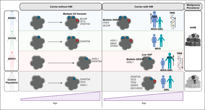
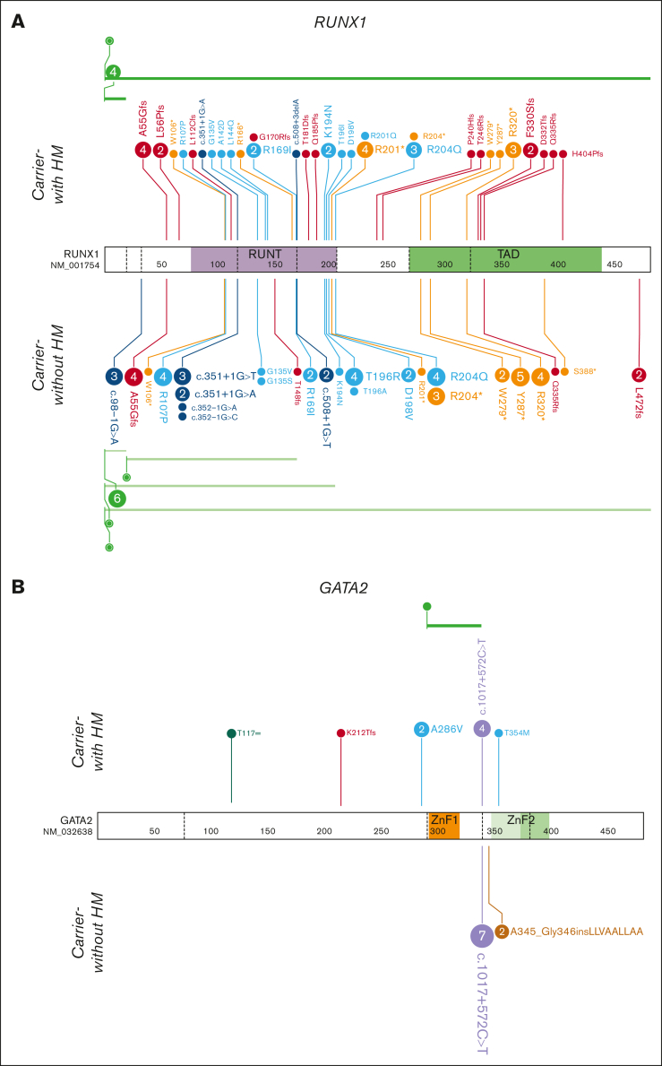
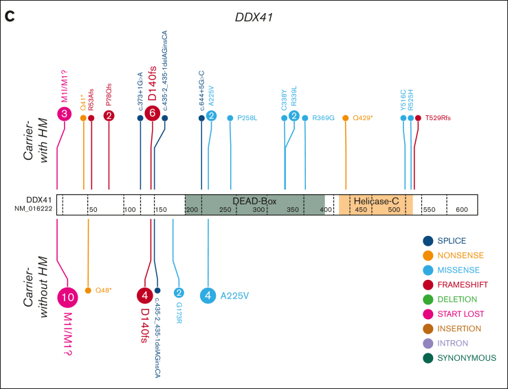
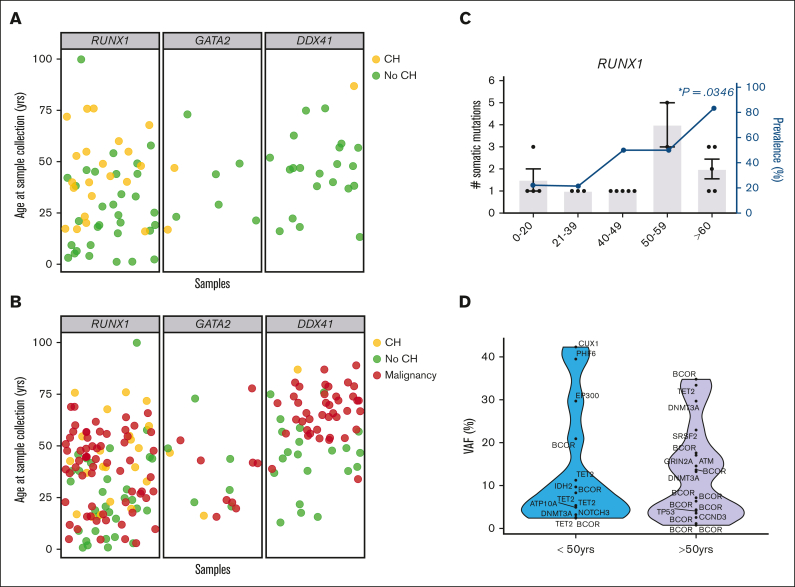
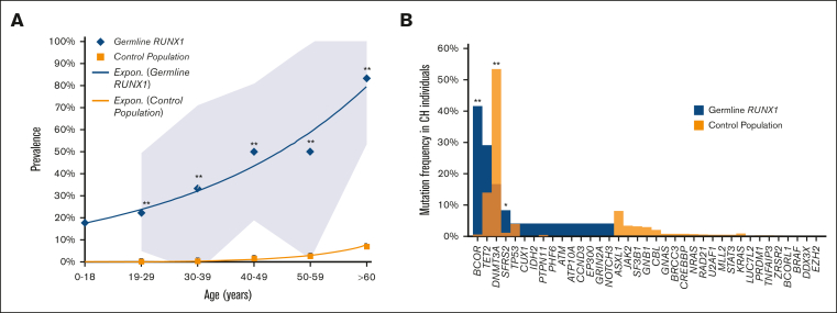
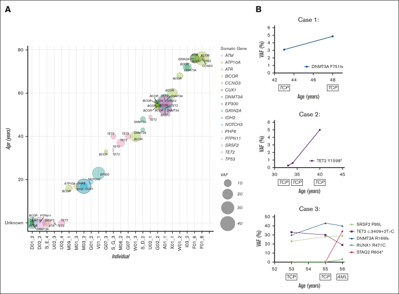
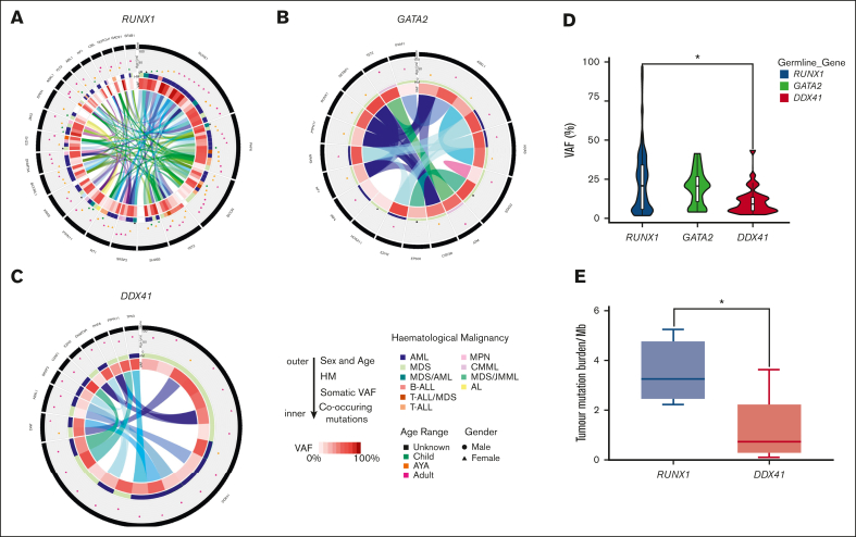
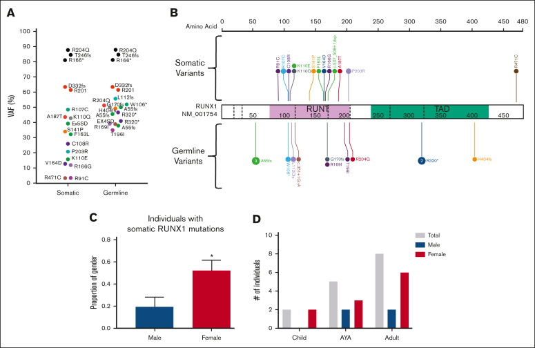
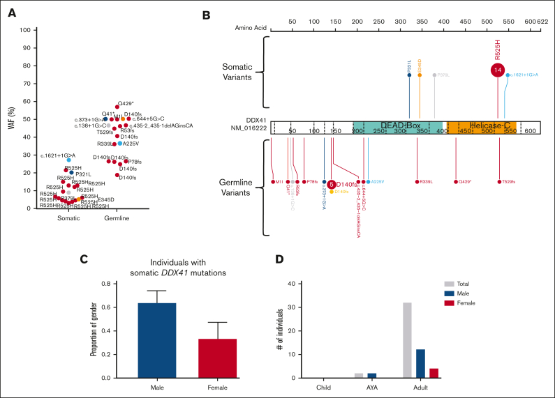
Individuals with germ line variants associated with hereditary hematopoietic malignancies (HHMs) have a highly variable risk for leukemogenesis. Gaps in our understanding of premalignant states in HHMs have hampered efforts to design effective clinical surveillance programs, provide personalized preemptive treatments, and inform appropriate counseling for patients. We used the largest known comparative international cohort of germline RUNX1, GATA2, or DDX41 variant carriers without and with hematopoietic malignancies (HMs) to identify patterns of genetic drivers that are unique to each HHM syndrome before and after leukemogenesis. These patterns included striking heterogeneity in rates of early-onset clonal hematopoiesis (CH), with a high prevalence of CH in RUNX1 and GATA2 variant carriers who did not have malignancies (carriers-without HM). We observed a paucity of CH in DDX41 carriers-without HM. In RUNX1 carriers-without HM with CH, we detected variants in TET2, PHF6, and, most frequently, BCOR. These genes were recurrently mutated in RUNX1-driven malignancies, suggesting CH is a direct precursor to malignancy in RUNX1-driven HHMs. Leukemogenesis in RUNX1 and DDX41 carriers was often driven by second hits in RUNX1 and DDX41, respectively. This study may inform the development of HHM-specific clinical trials and gene-specific approaches to clinical monitoring. For example, trials investigating the potential benefits of monitoring DDX41 carriers-without HM for low-frequency second hits in DDX41 may now be beneficial. Similarly, trials monitoring carriers-without HM with RUNX1 germ line variants for the acquisition of somatic variants in BCOR, PHF6, and TET2 and second hits in RUNX1 are warranted.


Conflict of interest statement

Conflict-of-interest disclosure: The authors declare no competing financial interests.

A complete list of the members of the NIH Intramural Sequencing Center Comparative Sequencing Program appears in “Appendix.”

Figures

None
Graphical abstract
Figure 1.
Figure 1.
HHM genomics cohorts. Germ line variants in the HHM cohorts were visualized using the ProteinPaint web application. Carriers-with HM cohorts (diagnosed with HM) are visualized above the protein. Carriers-without HM cohorts (no HM diagnosis) are below the protein. Variants (displayed as protein changes where possible) are color coded by variant type. The number of individuals with each variant is indicated within the circle when the number is greater than 1. (A) Germ line RUNX1 (66 carriers-without HM and 52 carriers-with HM individuals); (B) Germ line GATA2 (9 carriers-without HM and 13 carriers-with HM individuals); and (C) Germ line DDX41 (22 carriers-without HM and 29 carriers-with HM individuals) cohorts.
Figure 1.
Figure 1.
HHM genomics cohorts. Germ line variants in the HHM cohorts were visualized using the ProteinPaint web application. Carriers-with HM cohorts (diagnosed with HM) are visualized above the protein. Carriers-without HM cohorts (no HM diagnosis) are below the protein. Variants (displayed as protein changes where possible) are color coded by variant type. The number of individuals with each variant is indicated within the circle when the number is greater than 1. (A) Germ line RUNX1 (66 carriers-without HM and 52 carriers-with HM individuals); (B) Germ line GATA2 (9 carriers-without HM and 13 carriers-with HM individuals); and (C) Germ line DDX41 (22 carriers-without HM and 29 carriers-with HM individuals) cohorts.
Figure 2.
Figure 2.
Defining the spectrum of CH in germline GATA2, RUNX1, and DDX41 carriers-without HM. (A) Each individual in the carriers-without HM cohort was defined as having CH (yellow) or no identifiable CH (no-CH, green), based on the identification of somatic clinically relevant variants, driver somatic variants. The age of the individual at the time of sample collection is indicated. (B) Correlation of the ages of malignancy development (HM, red) observed in the germ line malignancy cohorts with the carriers-without HM cohort, with (yellow) and without CH (green). (C) Demographics of individuals with CH variants in the RUNX1 germ line carriers-without HM cohorts. Column graph shows the number of somatic variants identified in individuals with CH. Error bars show the standard error of the mean. Line graphs show the prevalence of CH in the carriers-without HM germ line cohort in different age groups. ∗P < .05, logistic regression model. (D) Violin plots showing the distribution of VAFs of driver somatic variants (shown in panel A) in the germ line RUNX1 carriers-without HM cohort in individuals under the age of 50 years or >50 years old. VAFs for X-chromosome genes were normalized in male individuals to compensate for ploidy, enabling comparison with autosomal genes. HM, hematologic malignancy.
Figure 3.
Figure 3.
Spectrum of CH in age-related CH compared with the germ line RUNX1 carriers-without HM cohort. (A) Prevalence of CH in the control population compared with the germ line RUNX1 carriers-without HM cohort. The control population includes cohorts from Jaiswal et al32 and Genovese et al33 (n = 27 783). The 0 to 18 age group is only available for the RUNX1 cohort. (B) Mutational spectrum of CH in the control population compared with the germ line RUNX1 carriers-without HM cohort. Graph shows the frequency of variants in individuals with CH in each cohort. The control population includes cohorts from Desai et al,36 Abelson et al,35 and Jaiswal et al.32P < .05, ∗∗P < .001, 2-proportions Z test, approximate to normal distribution, ±95% confidence interval.
Figure 4.
Figure 4.
Molecular monitoring of germ line RUNX1 carriers with CH. (A) Driver somatic variants identified in RUNX1 carriers-without HM individuals across age. Circle size = increasing VAF, color = gene. Individuals with unknown age were given the value 0 (D01_2, D02_2, S_E_4, U02_3, U02_4). (B) Longitudinal case studies with the VAF of detected driver somatic variants plotted across years. Clinical diagnosis at the time of monitoring is indicated below the age of the individual at which the sample was collected. AML, acute myeloid leukemia; TCP, thrombocytopenia.
Figure 5.
Figure 5.
Clinically relevant somatic variants identified in the germ line carriers-with HM cohorts. Distribution of the clinically relevant somatic variants, driver somatic variants, identified in the carriers-with HM cohorts. From outside to inside: 1. Gene with the somatic variant. The length of the black bar indicates the frequency of variant within the germ line carriers-with HM cohort. 2. The age and sex of the individual with the somatic variant. Triangle = female, circle = male. Age groups are indicated by colors (green = child [≤14 years], orange = AYA [15-39 years], adult = pink [≥40 years], black = the age of the individual is unknown). 3. The type of HM is indicated by the color of the bar. 4. VAF of the somatic variant in the sample as represented on a sliding scale (darker = high VAF, lighter = low VAF). 5. The inner ring depicts the association of different somatic variants in the sample. The colored ribbon depicts a unique sample and the associated somatic variants observed in the sample. (A) Germ line RUNX1 carriers-with HM cohort. Only shown are the genes that are identified as somatically mutated in 2 or more individuals. (B) Germ line GATA2 carriers-with HM cohort, showing all driver somatic variants and (C) Germ line DDX41 carriers-with HM cohort showing all driver somatic variants. (D) Violin plot displaying the distribution of driver somatic variant VAFs observed in the germ line carriers-with HM cohorts. Boxes represent the 25th and 75th percentiles, with the horizontal line in the middle indicating the median, and the vertical lines representing the 95th percentile cohorts. ∗P < .05, 1-way analysis of variance of log-transformed values, with Tukey multiple comparison test. (E) TMB in germ line RUNX1 and DDX41 carriers-with HM cohorts. TMB is the number of SNV and INDELs divided by 38Mb coding region. Only malignancy samples where we had available a matched germ line control tissue were used for analysis. Boxes represent the 25th and 75th percentiles, with the horizontal line in the middle indicating the median, and the vertical lines representing the max and min values. ∗P < .05 nonparametric Mann-Whitney U test. AYA, adolescents and young adults; AML, acute myeloid leukemia; AL, acute leukemia; B-ALL, B-cell acute lymphoblastic leukemia; CML, chronic myeloid leukemia; CMML, chronic myelomonocytic leukemia; JMML, juvenile myelomonocytic leukemia; MPN, myeloproliferative neoplasms; T-ALL, T-cell acute lymphoblastic leukemia.
Figure 6.
Figure 6.
Somatic variants in RUNX1 are the most common event in the germ line RUNX1 carriers-with HM cohort. (A) Plot of acquired somatic RUNX1 variants and associated germ line RUNX1 variants. Data points are colored according to the somatic and associated germ line variant observed in the patient. VAF of more than 60% indicates a copy neutral loss of heterozygosity (CNLOH) or Trisomy 21. (B) Somatic and germ line RUNX1 variants are visualized using the ProteinPaint web application. Variants are colored according to the somatic and associated germ line variant observed in the patient. The number of probands for each variant is indicated within the circle where the number is greater than 1. All variants are annotated to RUNX1c; NM_001754.4; LRG_ 482. (C) The proportion of male and females harboring a somatic RUNX1 variant is significantly different. ∗P < .05. (D) Sex and age distribution of individuals with a somatic RUNX1 variant; adult ≥ 40 years, AYA = 15 to 39 years, children ≤14 years. Data points are colored according to the somatic and associated germ line variant observed in the patient. AYA, adolescents and young adults.
Figure 7.
Figure 7.
A somatic variant in DDX41 is the most common event in the germ line DDX41 carriers-with HM cohort. (A) Plot of acquired somatic DDX41 variants and associated germ line DDX41 variants. Data points are colored according to the somatic and associated germ line variant observed in the patient. (B) Somatic and germ line DDX41 variants are visualized using the ProteinPaint web application. Variants are colored according to the somatic variant and associated germ line variant observed in the patient. The number of probands for each variant is indicated within the circle where the number is greater than 1. All variants are annotated to DDX41; NM_016222.4; LRG_ 1386. (C) The proportion of male and females harboring a somatic DDX41 variant shows no significant difference. (D) Sex and age distribution of individuals with a somatic DDX41; adult ≥40 years, AYA = 15 to 39 years, children ≤14 years. AYA, adolescents and young adults.

References

    1. Tawana K, Brown AL, Churpek JE. Integrating germline variant assessment into routine clinical practice for myelodysplastic syndrome and acute myeloid leukaemia: current strategies and challenges. Br J Haematol. 2022;196(6):1293–1310. - PubMed
    1. Arber DA, Orazi A, Hasserjian R, et al. The 2016 revision to the World Health Organization classification of myeloid neoplasms and acute leukemia. Blood. 2016;127(20):2391–2405. - PubMed
    1. Feurstein S, Drazer MW, Godley LA. Genetic predisposition to leukemia and other hematologic malignancies. Semin Oncol. 2016;43(5):598–608. - PubMed
    1. Polprasert C, Schulze I, Sekeres MA, et al. Inherited and somatic defects in DDX41 in myeloid neoplasms. Cancer Cell. 2015;27(5):658–670. - PMC - PubMed
    1. Abou Dalle I, Kantarjian H, Bannon SA, et al. Successful lenalidomide treatment in high risk myelodysplastic syndrome with germline DDX41 mutation. Am J Hematol. 2020;95(2):227–229. - PubMed

Publication types