. 2023 Sep 1;435(17):168193.
doi: 10.1016/j.jmb.2023.168193. Epub 2023 Jul 3.

The Dysferlin C2A Domain Binds PI(4,5)P2 and Penetrates Membranes

Affiliations

The Dysferlin C2A Domain Binds PI(4,5)P2 and Penetrates Membranes

Ethiene Kwok et al. J Mol Biol. .

Abstract

Dysferlin is a large membrane protein found most prominently in striated muscle. Loss of dysferlin activity is associated with reduced exocytosis, abnormal intracellular Ca2+ and the muscle diseases limb-girdle muscular dystrophy and Miyoshi myopathy. The cytosolic region of dysferlin consists of seven C2 domains with mutations in the C2A domain at the N-terminus resulting in pathology. Despite the importance of Ca2+ and membrane binding activities of the C2A domain for dysferlin function, the mechanism of the domain remains poorly characterized. In this study we find that the C2A domain preferentially binds membranes containing PI(4,5)P2 through an interaction mediated by residues Y23, K32, K33, and R77 on the concave face of the domain. We also found that subsequent to membrane binding, the C2A domain inserts residues on the Ca2+ binding loops into the membrane. Analysis of solution NMR measurements indicate that the domain inhabits two distinct structural states, with Ca2+ shifting the population between states towards a more rigid structure with greater affinity for PI(4,5)P2. Based on our results, we propose a mechanism where Ca2+ converts C2A from a structurally dynamic, low PI(4,5)P2 affinity state to a high affinity state that targets dysferlin to PI(4,5)P2 enriched membranes through interaction with Tyr23, K32, K33, and R77. Binding also involves changes in lipid packing and insertion by the third Ca2+ binding loop of the C2 domain into the membrane, which would contribute to dysferlin function in exocytosis and Ca2+ regulation.

Keywords: C2 domain; CEST; calcium; dysferlin; genetic code expansion.


Conflict of interest statement

Declaration of Competing Interest The authors declare that they have no known competing financial interests or personal relationships that could have appeared to influence the work reported in this paper.

Figures

Figure 1:
Figure 1:. Dysferlin.
(Top) Diagram of dysf showing the location of the 7 C2 domains (C2A-C2G), FerA domain, dysf domain, and single pass transmembrane domain (TMD). (Bottom) Dysferlin structure predicted by AlphaFold. The C2A domain is highlighted in blue.
Figure 2:
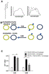
Figure 2:. PI(4,5)P2 recruits dysf C2A.
(A) Structure of laurdan probe (left) and diagram of changes in laurdan emission spectra upon protein interaction with laurdan-containing liposomes . Arrows denote the changes in wavelength emission associated with C2 domain binding. (B) Diagram illustrating recruitment of C2A to PC:PS:PI(4,5)P2:laurdan (69:25:5:1) containing liposomes preferentially over PC:PS (75:25) liposomes. C2B distributes more evenly between the two sets of liposomes. (C) Mean change in GP values for listed liposome sample. (N = 3; error = ± standard deviation; *P < 0.01).
Figure3:
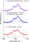
Figure3:. PI(4,5)P2 interaction influences the C2A-membrane binding geometry.
VSF spectra of C2A bound to the 3:1 DPPC:DPPS (red circles), 95:5 DPPC:PI(4,5)P2 (blue squares), and 70:25:5 DPPC:DPPS:PI(4,5)P2 (purple triangles) lipid monolayers. Solid lines are spectral fits to the data.
Figure 4:
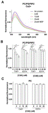
Figure 4:. PI(4,5)P2-TopFluor quenches in the presence of C2A.
(A) Representative titration of C2A mixed at the given concentration with liposomes composed of PC:PS:PI(4,5)P2:PI(4,5)P2-TopFluor (70:25:4.5:0.5). (B) Quantitation of PI(4,5)P2-TopFluor fluorescence intensity for C2A or C2B at the listed concentration in the presence of 250 μM Ca2+ or 3 mM EDTA. (C) Quantitation of PC-TopFluor fluorescence intensity for C2A at the listed concentration in the presence of 250 μM Ca2+ or 3 mM EDTA. (N = 3; error = ± standard deviation; *P < 0.05).
Figure 5:
Figure 5:. The Ca2+ binding loops of C2A insert into membranes.
(A) Structure of dysf C2A (PDB 4IHB) highlighting residues T17 (green) and M75 (red) that were converted to TAG sites for Acd incorporation to generate Acd17 and Acd75. (B) Representative Acd75 emission spectra when mixed with liposomes composed of PC:PS:PI(4,5)P2 (72.5 : 25 : 2.5 ratio), as well as PC:PS:PI(4,5)P2 liposomes with 1 mol % 5-doxyl phosphocholine or 12-doxyl phosphocholine. (C, D) Quantitation of (C) Acd75 and (D) Acd17 fluorescence in samples with conditions listed in (C) in the presence of 5 mM EDTA (white) or 1 mM Ca2+ (black). (N = 3; error = ± standard deviation; *P < 0.01).
Figure 6:
Figure 6:. Titration of IP3 into Dysferlin C2A at high and low Ca2+.
A. Representative titrations are shown for three residues, two in the basic patch and one in the Ca2+ binding loop. 5 mM Ca2+ data is shown in closed black circles, 5 mM EDTA data is shown in open squares. Fits to determine the Kd’s are shown in black and gray respectively. B. Overlay of 15N-TROSY spectra collected at increasing concentrations (light orange to dark orange) of IP3 in the presence of 5 mM CaCl2. C. Overlay of 15N-TROSY spectra collected at increasing concentrations (light blue to dark blue) of IP3 in the presence of 5 mM EDTA (low Ca2+). D. Table of IP3 binding affinities determined from the NMR titrations at high and low Ca2+. Error is reported as +/− the value shown. The structure of dysferlin C2A (PDB 4IHB) with chemical shift perturbation ranging from the largest perturbation 0.439 (blue) to the 1σ cut-off of 0.052 ppm (yellow) is shown on the right.
Figure 7:
Figure 7:
a) Residues exchanging at 3 mM CaCl2 mapped onto the structure (4IHB). b) Selected CEST traces showing increased exchange at lower Ca2+ concentrations. Data (circles) and fits (lines) for 50 (dark), 25 (medium), and 10 (light) Hz saturation field strengths. Solid vertical lines indicate the 15N chemical shift for the major state, dotted vertical lines indicate the 15N chemical shift for the minor state, with error indicated by grey shading.
Figure 8.
Figure 8.
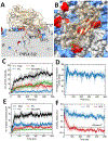
MD simulations of C2A domain (pdb 7JOF) with PC:PS:PI(4,5)P2 (70:25:5) membranes. A) Representative cross section of membrane showing the interaction of C2A domain with a PI(4,5)P2 lipid. B) Representative top-down orientation of C2A domain interacting with the membrane. PS lipids colored in blue and PIP(4,5)2 lipids colored in red. C) Number of H-bonds between C2A domain and neighboring lipids. D) Tilt angle of C2A domain relative to the membrane normal (see Methods). E) Number of lipid molecules that contact the C2A domain. F) Insertion depth for residues T17 and M75 relative to the depth of the glycerol phosphorous atom in the lipid membrane.

References

    1. Bulankina AV, Thoms S (2020) Functions of Vertebrate Ferlins. Cells 9(3). doi: 10.3390/cells9030534. - DOI - PMC - PubMed
    1. Lek A, Evesson FJ, Sutton RB, North KN, Cooper ST (2012) Ferlins: Regulators of Vesicle Fusion for Auditory Neurotransmission, Receptor Trafficking and Membrane Repair. Traffic 13(2):185–194. - PubMed
    1. Han R, Rader EP, Levy JR, Bansal D, Campbell KP (2011) Dystrophin deficiency exacerbates skeletal muscle pathology in dysferlin-null mice. Skelet Muscle 1(1):35. - PMC - PubMed
    1. Kerr JP, et al. (2013) Dysferlin stabilizes stress-induced Ca2+ signaling in the transverse tubule membrane. Proc Natl Acad Sci U S A 110(51):20831–20836. - PMC - PubMed
    1. Bansal D, Campbell KP (2004) Dysferlin and the plasma membrane repair in muscular dystrophy. Trends Cell Biol 14(4):206–213. - PubMed

Publication types

MeSH terms

LinkOut - more resources